



Phospho-MEK1/2(Ser217/221) Antibody (YA5835)
Based on 1 publication(s) in Google Scholar
Phospho-MEK1/2(Ser217/221) Antibody (YA5835) is a Rabbit-derived and non-conjugated IgG monoclonal antibody, targeting to Phospho-MEK1/2(Ser217/221).
-
Host:
Rabbit
-
Isotype:
IgG/Kappa
-
Application:
WB, IHC-P, ICC/IF, IP, ELISA
-
Reactivity :
Human, Mouse, Rat
-
Formulation:
Supplied in PBS, 50% glycerol, 0.05% Proclin 300, 0.05%BSA
-
Conjugation:
Non-conjugated


To place orders, for customer services and technical support, please contact: MedChemExpress USA
Tel: 609-228-6898 E-mail: [email protected] [email protected]



Publications Citing Use of MedChemExpress (MCE) Phospho-MEK1/2(Ser217/221) Antibody (YA5835)
More

Applications
| Application |
IHC-P

IHC-P: Immunohistochemistry-Paraffin
|
WB
WB: Western Blot
|
ICC/IF
ICC/IF: Immunocytochemistry/
Immunofluorescence |
ELISA
ELISA: Enzyme Linked Immunosorbent Assay
|
IP
IP: Immunoprecipitation
|
|---|---|---|---|---|---|
| Dilution Ratio | 1:1000-1:5000 | 1:2000-1:10000 | 1:200-1:1000 | 1:5000-1:20000 | 1:50-1:200 |


Product Details
Phospho-MEK1/2(Ser217/221) Antibody (YA5835) is a Rabbit-derived and non-conjugated IgG monoclonal antibody, targeting to Phospho-MEK1/2(Ser217/221).
-
Host Rabbit

-
Clonality Monoclonal

-
Species ReactivityHuman, Mouse, Rat
-
Observed Molecular WeightObserved band size: 44 kDa
Note: Due to possible protein modifications or aggregation, the molecular weight should be confirmed by actual measurement, and the predicted value is for reference only.

-
Calculated Molecular Weight Predicted band size: 44 kDa

Protein A
Non-conjugated
Phosphorylated
IgG/Kappa


Product Properties
-
Appearance
Liquid
-
Formulation
Supplied in PBS, 50% glycerol, 0.05% Proclin 300, 0.05%BSA
-
Concentration
Batch-dependent, Please check the COA for the concentration of each lot. Check Lot Concentration
-
Storage & Stability
Stored at -20°C for 1 year. Avoid repeated freeze / thaw cycles.
-
Shipping
Shipping with blue ice.


Publications (1)
-
Journal Impact Factor
-
Most Recent


Verification Images
-
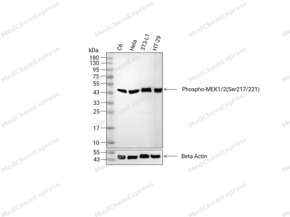 Western blot analysis of extracts from C6(lane2(20μg), Hela(20μg), 3T3-L1(lane4(20μg) and HT-29(lane5(20μg) using Phospho-MEK1/2(Ser217/221) Antibody (YA5835)(HY-P86143). Proteins were transferred to a PVDF membrane and blocked with 5% non-fat milk in TBST at 4°C overnight. The primary antibody (1/5000) and Loading control antibody (Beta Actin, HY-P80993, 1/10,000) was used in 5% non-fat milk in TBST for 2 hour at room temperature. Goat Anti-Rabbit IgG-HRP Secondary Antibody (HY-P8001, 1/10,000) was used for 1 hour at room temperature.
 -
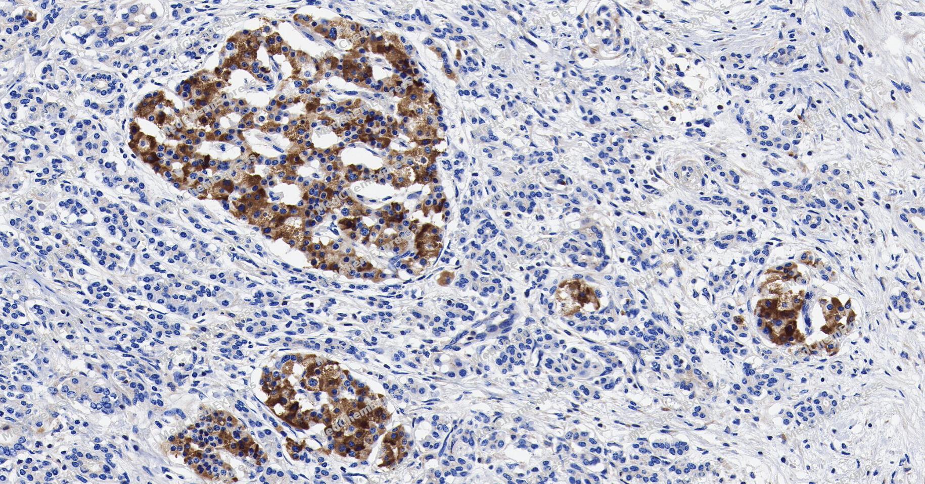 Immunohistochemical analysis of paraffin-embedded human pancreatic cancer tissue using Phospho-MEK1/2(Ser217/221) Antibody (YA5835). The section was pre-treated using heat mediated antigen retrieval with Tris/EDTA buffer (pH 9.0) for 20 minutes. The tissues were probed with the primary antibody (HY-P86143, 1/100) overnight at 4℃. The detection was performed using an HRP conjugated compact polymer system. DAB was used as the chromogen. Tissues were counterstained with hematoxylin and mounted with neutral balsam.
 -
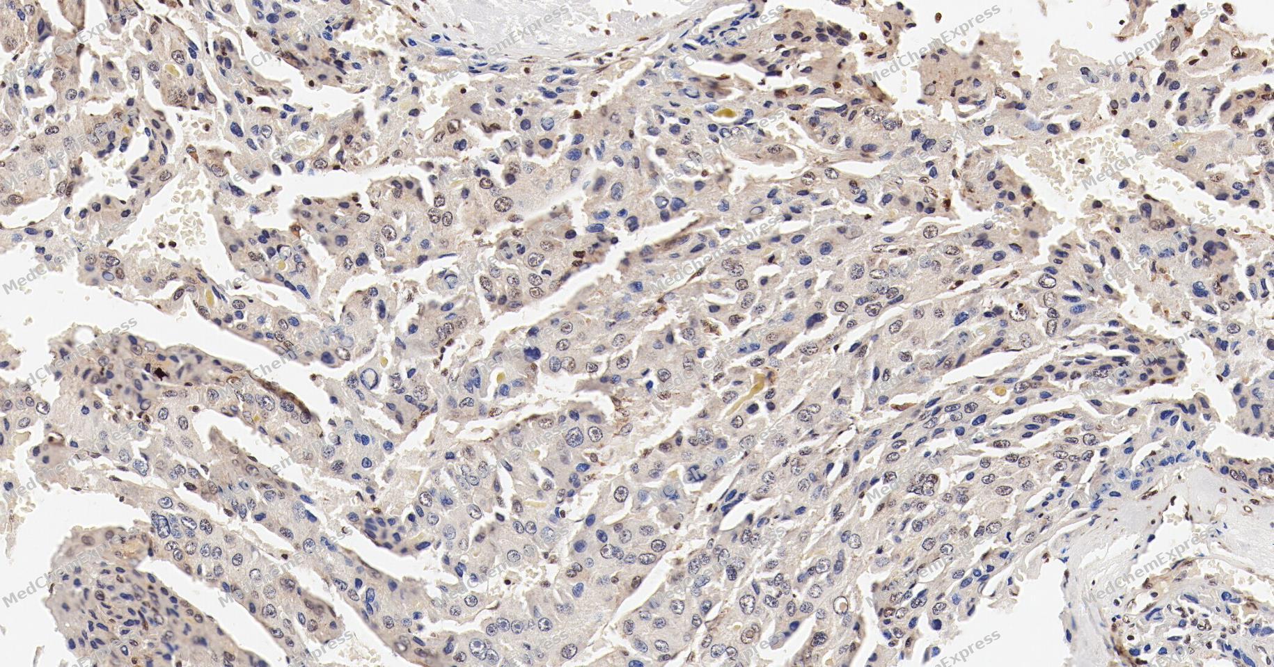 Immunohistochemical analysis of paraffin-embedded human hepatocellular carcinoma tissue using Phospho-MEK1/2(Ser217/221) Antibody (YA5835). The section was pre-treated using heat mediated antigen retrieval with Tris/EDTA buffer (pH 9.0) for 20 minutes. The tissues were probed with the primary antibody (HY-P86143, 1/100) overnight at 4℃. The detection was performed using an HRP conjugated compact polymer system. DAB was used as the chromogen. Tissues were counterstained with hematoxylin and mounted with neutral balsam.
 -
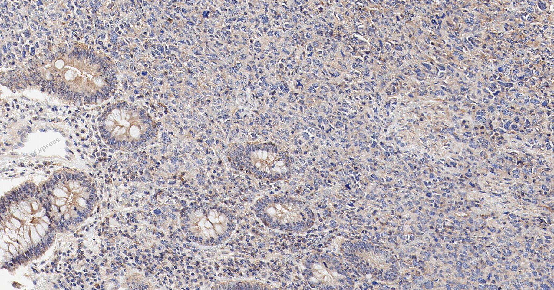 Immunohistochemical analysis of paraffin-embedded human colon cancer tissue using Phospho-MEK1/2(Ser217/221) Antibody (YA5835). The section was pre-treated using heat mediated antigen retrieval with Tris/EDTA buffer (pH 9.0) for 20 minutes. The tissues were probed with the primary antibody (HY-P86143, 1/100) overnight at 4℃. The detection was performed using an HRP conjugated compact polymer system. DAB was used as the chromogen. Tissues were counterstained with hematoxylin and mounted with neutral balsam.
 -
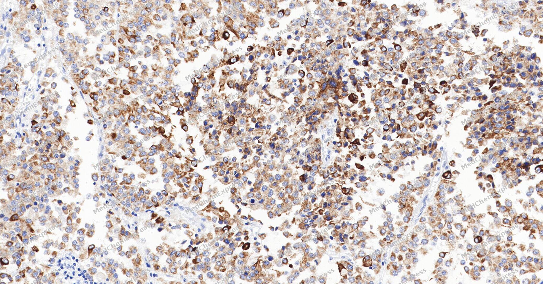 Immunohistochemical analysis of paraffin-embedded human malignant melanoma tissue using Phospho-MEK1/2(Ser217/221) Antibody (YA5835). The section was pre-treated using heat mediated antigen retrieval with Tris/EDTA buffer (pH 9.0) for 20 minutes. The tissues were probed with the primary antibody (HY-P86143, 1/100) overnight at 4℃. The detection was performed using an HRP conjugated compact polymer system. DAB was used as the chromogen. Tissues were counterstained with hematoxylin and mounted with neutral balsam.
 -
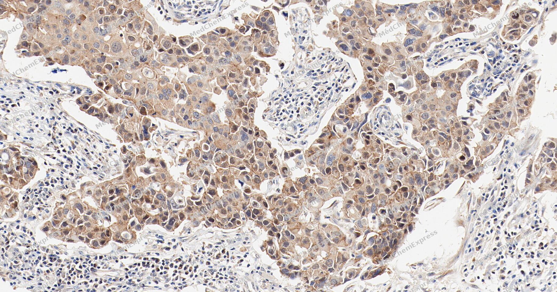 Immunohistochemical analysis of paraffin-embedded human breast cancer tissue using Phospho-MEK1/2(Ser217/221) Antibody (YA5835). The section was pre-treated using heat mediated antigen retrieval with Tris/EDTA buffer (pH 9.0) for 20 minutes. The tissues were probed with the primary antibody (HY-P86143, 1/100) overnight at 4℃. The detection was performed using an HRP conjugated compact polymer system. DAB was used as the chromogen. Tissues were counterstained with hematoxylin and mounted with neutral balsam.
 -
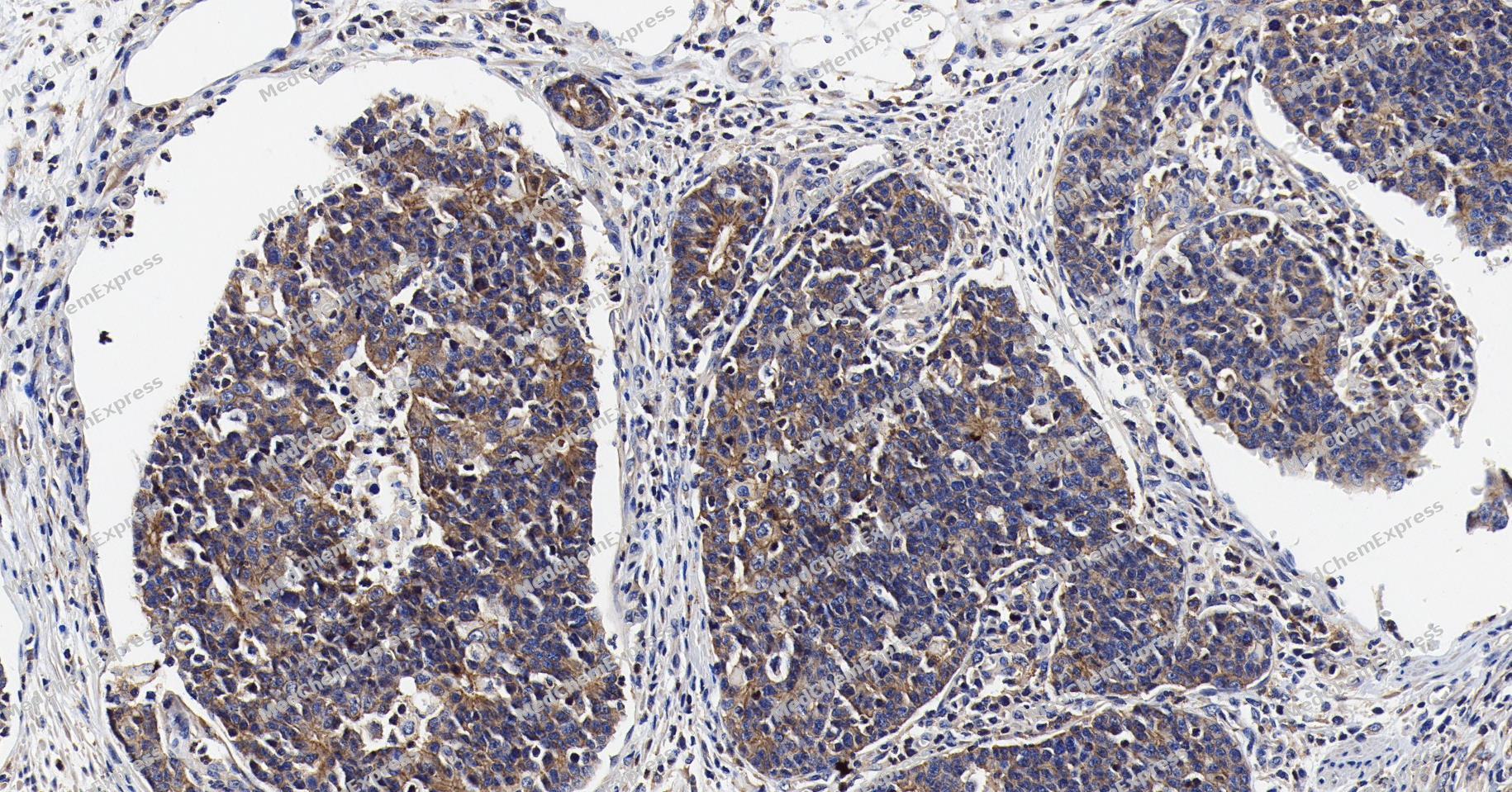 Immunohistochemical analysis of paraffin-embedded human gastric cancer tissue using Phospho-MEK1/2(Ser217/221) Antibody (YA5835). The section was pre-treated using heat mediated antigen retrieval with Tris/EDTA buffer (pH 9.0) for 20 minutes. The tissues were probed with the primary antibody (HY-P86143, 1/100) overnight at 4℃. The detection was performed using an HRP conjugated compact polymer system. DAB was used as the chromogen. Tissues were counterstained with hematoxylin and mounted with neutral balsam.
 -
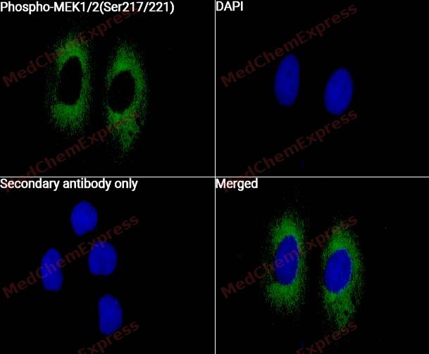 Immunocytochemistry analysis of Hela cells labelingPhospho-MEK1/2(Ser217/221) With Phospho-MEK1/2(Ser217/221) antibody (HY-P86143) at 1/300 dilution. Cells were fixed in 4% paraformaldehyde for 15 minutes at room temperature, permeabilized with 0.1% Triton X-100 in PBS for 15 minutes at room temperature, then blocked with quick block buffer for 10 minutes at room temperature. Cells were then incubated with Phospho-MEK1/2(Ser217/221) antibody (HY-P86143) at 1/300 dilution in quick block buffer overnight at 4 ℃. AF488-conjugated Goat Anti-Rabbit IgG H&L(HY-P8002, Green) was used as the secondary antibody at 1/1,000 dilution. PBS instead of the primary antibody was used as the secondary antibody only control. The Nuclear counterstain was DAPI (Blue).
 -
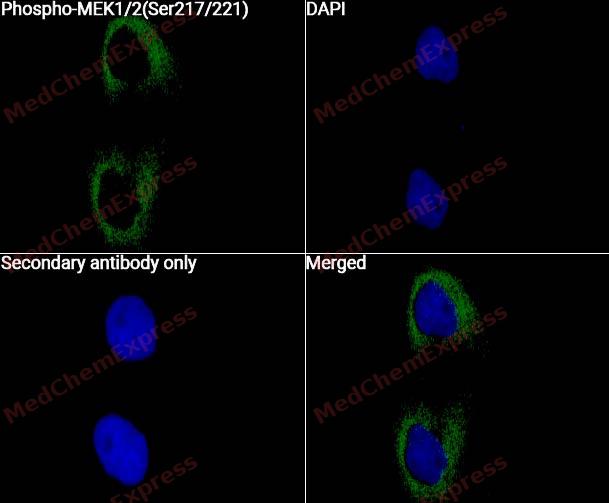 Immunocytochemistry analysis of Hela cells labeling Phospho-MEK1/2(Ser217/221) With Phospho-MEK1/2(Ser217/221) antibody (HY-P86143) at 1/500 dilution. Cells were fixed in 4% paraformaldehyde for 15 minutes at room temperature, permeabilized with 0.1% Triton X-100 in PBS for 15 minutes at room temperature, then blocked with quick block buffer for 10 minutes at room temperature. Cells were then incubated with Phospho-MEK1/2(Ser217/221) antibody (HY-P86143) at 1/500 dilution in quick block buffer overnight at 4 ℃. AF488-conjugated Goat Anti-Rabbit IgG H&L(HY-P8002, Green) was used as the secondary antibody at 1/1,000 dilution. PBS instead of the primary antibody was used as the secondary antibody only control. The Nuclear counterstain was DAPI (Blue).


Background
-
Function
MEK1/2 is a P36507: Catalyzes the concomitant phosphorylation of a threonine and a tyrosine residue in a Thr-Glu-Tyr sequence located in MAP kinases. Activates the ERK1 and ERK2 MAP kinases. Activates BRAF in a KSR1 or KSR2-dependent manner; by binding to KSR1 or KSR2 releases the inhibitory intramolecular interaction between KSR1 or KSR2 protein kinase and N-terminal domains which promotes KSR1 or KSR2-BRAF dimerization and BRAF activation
Q02750: Dual specificity protein kinase which acts as an essential component of the MAP kinase signal transduction pathway. Binding of extracellular ligands such as growth factors, cytokines and hormones to their cell-surface receptors activates RAS and this initiates RAF1 activation. RAF1 then further activates the dual-specificity protein kinases MAP2K1/MEK1 and MAP2K2/MEK2. Both MAP2K1/MEK1 and MAP2K2/MEK2 function specifically in the MAPK/ERK cascade, and catalyze the concomitant phosphorylation of a threonine and a tyrosine residue in a Thr-Glu-Tyr sequence located in the extracellular signal-regulated kinases MAPK3/ERK1 and MAPK1/ERK2, leading to their activation and further transduction of the signal within the MAPK/ERK cascade. Activates BRAF in a KSR1 or KSR2-dependent manner; by binding to KSR1 or KSR2 releases the inhibitory intramolecular interaction between KSR1 or KSR2 protein kinase and N-terminal domains which promotes KSR1 or KSR2-BRAF dimerization and BRAF activation. Depending on the cellular context, this pathway mediates diverse biological functions such as cell growth, adhesion, survival and differentiation, predominantly through the regulation of transcription, metabolism and cytoskeletal rearrangements. One target of the MAPK/ERK cascade is peroxisome proliferator-activated receptor gamma (PPARG), a nuclear receptor that promotes differentiation and apoptosis. MAP2K1/MEK1 has been shown to export PPARG from the nucleus. The MAPK/ERK cascade is also involved in the regulation of endosomal dynamics, including lysosome processing and endosome cycling through the perinuclear recycling compartment (PNRC), as well as in the fragmentation of the Golgi apparatus during mitosis
[1]. -
Subcellular Localization
P36507: Cytoplasm; Membrane; Peripheral membrane protein
Q02750: Cytoplasm, cytoskeleton, microtubule organizing center, centrosome; Cytoplasm, cytoskeleton, microtubule organizing center, spindle pole body; Cytoplasm; Nucleus; Membrane; Peripheral membrane protein -
Expression
Tissue_specificity: Q02750: Widely expressed, with extremely low levels in brain -
Isoforms & Post-Translational Modification
Q02750 has 2 isomers: Q02750-1: 43439 Da (predicted); Q02750-2: 40764 Da (predicted).
Phosphorylation at Ser-218 and Ser-222 by MAP kinase kinase kinases (BRAF or MEKK1) positively regulates kinase activity (PubMed:29433126, PubMed:8131746). Also phosphorylated at Thr-292 by MAPK1/ERK2 and at Ser-298 by PAK (PubMed:16129686). MAPK1/ERK2 phosphorylation of Thr-292 occurs in response to cellular adhesion and leads to inhibition of Ser-298 phosphorylation by PAK (PubMed:16129686). Autophosphorylated at Ser-218 and Ser-222, autophosphosphorylation is promoted by NEK10 following UV irradiation (PubMed:20956560);(Microbial infection) Acetylation by Yersinia YopJ prevents phosphorylation and activation, thus blocking the MAPK signaling pathway -
Subunit
P36507: Interacts with MORG1 (By similarity). Interacts with SGK1 (PubMed:19447520). Interacts with KSR1 (PubMed:10409742). Interacts with KSR1 and BRAF; the interaction with KSR1 mediates KSR1-BRAF dimerization (PubMed:29433126). Interacts with GLS (PubMed:22538822)
Q02750: Found in a complex with at least BRAF, HRAS, MAP2K1, MAPK3/ERK1 and RGS14 (By similarity). Forms a heterodimer with MAP2K2/MEK2 (By similarity). Forms heterodimers with KSR2 which further dimerize to form tetramers (By similarity).
Documentation
-
Data Sheet (235 KB)
-
SDS (252 KB)
- English - EN (252 KB)
- Français - FR (252 KB)
- Deutsch - DE (252 KB)
- Norwegian - NO (252 KB)
- Español - ES (252 KB)
- Swedish - SV (252 KB)
- Italian - IT (252 KB)
- Korean - KR (252 KB)
- Portuguese - PT (252 KB)
-
User Guide for Antibodies (1077 KB)

 Powered by Bioz
