1. Resources
  2. Articles
  3. What is autophagy and how to unlock autophagy detection
What is autophagy and how to unlock autophagy detection
Background of Autophagy

Autophagy, derived from the Greek meaning "eating of self", is critical in life sciences. Autophagy is a highly conserved eukaryotic cellular recycling process. Unhealthy cells (such as lack of nutrients) will initiate the autophagy program to save themselves and obtain essential amino acids, fatty acids, and other nutrients by degrading proteins and organelles to maintain basic life activities. Autophagy plays an indispensable role in maintaining homeostasis and autophagy dysfunction is implicated in the pathogenesis of various human diseases.

p27 regulates autophagy

On August 17, 2020, the team led by Arnaud Besson at the University of Toulouse published an anticle on autophagy (Figure 1), which clarified the new mechanism of autophagy and cell cycle regulation.

Figure 1. p27 regulates the autophagy-lysosomal pathway to coordinate cell cycle and growth
Background

p27Kip1 (p27) is considered an inhibitor of cyclin CDKs with the ability to induce cell cycle arrest. Nutrient deprivation is often the primary inducer of autophagy, and studies have shown that p27 in the cytoplasm is a positive regulator of autophagy (under conditions of glucose deprivation or serum deprivation), which can protect cells from stress-induced cells apoptosis. However, the specific molecular mechanism by how p27 regulates autophagy remains unknown. This article,will provide an in-depth interpretation of the autophagy-related article (Fig. 1) published by Arnaud Besson et al. in Nature cell biology in 2020, helping to understand the new mechanism of autophagy and cell cycle regulation.

p27 regulates autophagy

Previous studies have shown that p27 in the cytoplasm is a positive regulator of autophagy in the cytoplasm (under glucose deprivation or serum deprivation), which can protect cells from stress-induced apoptosis. However, the specific molecular mechanism by which p27 regulates autophagy remains unknown. Studying the changes in p27+/+ MEFs and p27-/- MEFs cells in response to amino acid deprivation, Arnaud’s team demonstrated that p27 promotes autophagy in amino acid-deficient cells.

Cells:

p27+/+ MEFs: p27 wild-type (p27+/+) mouse embryonic fibroblasts (MEFs), derived from p27+/+ embryos.
p27-/- MEFs: p27-/- MEFs, derived from p27-/- embryos.

1.p27 promotes autophagy

LC3B-II, a marker for autophagosomes, is located in the autophagosome membrane and is degraded upon fusion with lysosomes. As shown in Figure 2a, for short periods of amino acid deprivation, the expression levels of LC3B-II were similar in p27+/+ and p27-/- mouse embryonic fibroblasts. With prolonged amino acid deprivation, LC3B-II expression levels in p27-/- were significantly increased and showed a statistically significant difference at 48 hours (Fig. 2b). In the subsequent experiments, 48h was chosen as the induction time for prolonged amino acid deprivation. In Figure 2c, LC3B immunofluorescence staining also echoes this observation (red arrow). This suggests that p27 promotes autophagy in amino acid-deprived cells[1].

Figure 2. Expression of LC3B, LC3B-I and LC3B-II in cells at different time of amino acid deprivation[1].
a-b: Expression and quantification of LC3B-I and LC3B-II of amino acid-deprived p27+/+ and p27-/- MEFs for the indicated time periods; c-d: Immunofluorescence staining and quantitative analysis of LC3B of p27+/+ and p27-/- MEFs at 0 h and 48h following amino acid starvation(green fluorescence).

2.Detection of p27-regulated autophagy flux

Autophagy is a multi-step dynamic process. In response to nutrient deprivation stress (such as amino acid or glucose deprivation), cells receive autophagy-inducing signals and subsequently form flattened and double-membrane phagophores in the cytoplasm. Then, the phagophores continues to extend, wrapping some cellular elements or autophagic cargo and forming a closed spherical autophagosome. Autophagosomes are fused with lysosomes to form autolysosomes. Lysosomal enzymes degrade the inner autophagosomal membrane and sequestered material, resulting in degradation of nutrients that are then reused by cells. During the whole process, cells phagocytose autophagic cargos into autophagosomes, and then, autolysosomes are formed and degrade autophagic cargos[2]. This process is called autophagic flux (Fig. 3). If any problem occurs in the autophagic flux, autophagy will not complete its biological function.

3.p27- regulated autophagy flux detection

Autophagy is a multi-step dynamic process. In response to nutrient deprivation stress, such as amino acid or glucose deprivation, cells receive autophagy-inducing signals; subsequently, they form flattened phagocytes (Phagophores) that feature a double membrane structure in the cytoplasm. The phagocytic vesicles extend continuously to wrap cellular elements or autophagic cargo, followed byforming a closed spherical autophagosome (Autophagosome). And then, autophagosomes are fused with lysosomes to form autolysosomes. Lysosomal enzymes degrade the inner autophagosomal membrane and sequestered materials, and the nutrients produced by the degradation will be reused by cells.

This multiple-step process, also known as autophagic flux, encompasses all stages of autophagy in that cells phagocytose autophagic cargos into autophagosomes, autolysosomes are formed and degrade autophagic cargos (Fig. 3)[2]. Autophagy cannot complete its function if any step of autophagic flux is impaired.

Figure 3. Schematic diagram of autophagy in response to nutrient deprivation stress[2] Phagophore-Autophagosome-Autolysosome

As mentioned above, as the marker of autophagosomes, LC3B-II levels in mammalian cells usually keep balanced. The fluctuation only occurs in the mutual conversion between LC3B-I and LC3B-II.

If LC3B-II expression is increased, it may be caused by the increasing autophagosomes after early-stage autophagy activation. Or it may be caused by a later-stage accumulation of autophagolysosomes due to the clearing problem. Therefore, changes in the expression of LC3B-II at a single time point do not reflect changes in autophagic flux.

The Arnaud team studied autophagic flux in p27+/+ and p27-/- MEFs through WB for protein checking and the mCherry-eGFP-LC3B dual-fluorescence system for immunofluorescence.

(1) Detection of LC3B-II changes in autophagic flux using chloroquine-treated cells

It is known that Chloroquine (CQ) increases lysosomal pH by accumulating within lysosomes as a deprotonated weak base[3]. In acidic lysosomes, CQ increases the pH and results in the inactivation of acid hydrolase in lysosomes, thereby inhibiting the fusion and degradation of intracellular autophagolysosomes. Previous research has shown that CQ treatment of cells led to LC3B-II accumulation. Therefore, changes in LC3B-II observed at this moment only represented the number changing in autophagosomes.

In the article, after CQ treatment, autophagic fluxes were similar in both cells under short-term amino acid deprivation (0-4 h) (Figure 4a,b). However, the LC3B-II amount was significantly reduced in p27-/- MEFs compared with p27+/+ MEFs after prolonged amino acid deprivation treatment (Fig. 4c, d). This suggests that p27 promotes autophagic flux in amino acid-deficient cells[1].

Figure 4. Chloroquine affects the expression of cellular LC3B-II[1]
a-b: Quantitative analysis of LC3B-II expression and changes in autophagic flux with or without CQ following 0 h and 48h of amino acid starvation; c-d: Quantitative analysis of LC3B-I and LC3B-II expression and changes in autophagic flux following 0 h and 48h of amino acid starvation

(2) Accumulation of p62/SQSTM1 in cells

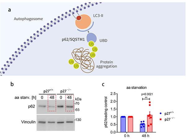
P62 (also known as SQSTM1 protein) is a regulator of autophagosome formation withsubstrate specificity, and acts as a bridge between to -be degraded ubiquitinated substrates and LC3B-II. In general, p62 binds ubiquitinated proteins and then gets into autophagosomes, eventually fuses with lysosomes to form autolysosomes for clearance (Fig. 5a). When the autophagic flux is suppressed, p62/SQSTM1 will accumulate in the impaired autophagic flux cells. And the overall P62 expression level in the cells was negatively correlated with autophagic activity. As shown in Figure 5b, p62 expression was higher in amino acids deprived p27-/- cells over time. Correspondingly, autophagic flux was impaired in the p27-/- cells. This further explains that p27 can promote autophagic flux activation[1].

Figure 5. p62 accumulates in cells with impaired autophagic flux
Figure 5. p62 accumulates in cells with impaired autophagic flux[1][4]
a: p62 is involved in the formation of autophagosomes[4]; b: WB of p62 and Vinculin (Loading control) following 0 h and 48 h amino acid starvation; c: Quantitative analysis of p62 normalization.

(3) Detection of autophagosomes and autolysosomes by mCherry-eGFP-LC3B dual fluorescence system.

The mCherry (red light)-eGFP (green light)-LC3B tandem fluorescent protein can detect the fusion proteins during autophagic flux. It exploits the pH difference between acidic autolysosomes and neutral autophagosomes and the sensitivity of different fluorescein to pH to monitor the progression from autophagosome to autolysosome (Figure 6a).

Figure 6. Tandem fluorescent probes and fluorescent colocalization to detect autophagic flux[1][5]
a: Schematic diagram and mechanism of mCherry-eGFP-LC3B tandem fluorescent probes[5]; b: The formation of autophagosomes and autolysosomes in p27+/+ and p27−/− MEFs expressing mCherry–eGFP–LC3B following amino acid starvation for 48h with or without CQ for 2h; c: Quantitative analysis of autophagosomes (yellow) and autolysosomes (red)

As shown in Figure 6a-b, mCherry fluorophore is more tolerable to low pH than GFP . The GFP signal will be quenched after entering the lysosome due to the drop in pH, while the mCherry fluorophore can still produce red fluorescence after entering the lysosome.Therefore, if yellow fluorescence appears due to the co-localization ofgreen and red fluorescence in cells,it indicates that the mCherry-eGFP-LC3B fusion protein did not fuse with the lysosome, which means the autophagic flux was blocked. On the contrary, if only red fluorescence appeared in cells without GFP, it suggests that the mCherry-eGFP-LC3B fusion protein was localized in lysosomes or autophagolysosome and autophagic flux was activated.

The proportion of autophagic lysosomes was decreased in p27-/- cells compared to p27+/+ cells (72% vs. 95%), further suggesting that p27 promotes autophagic flux (Figure 6b-c).

(4) p27 promotes autophagosome maturation

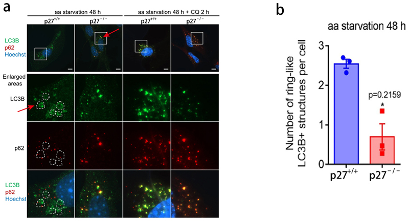
Figure 7. p27 promotes the formation of intermediates during autophagosome maturation
Figure 7. p27 promotes the formation of intermediates during autophagosome maturation[1]
a: Immunostaining of LC3B and p62 in p27+/+ and p27-/- MEFs following 0 h and 48h amino acid starvation (dotted lines depict LC3B-positive ring structures); b: Quantification of LC3B cyclic aggregates in p27+/+ and p27-/- MEFs.
Previous research has shown that during autophagosome maturation, LC3B-positive autophagic vesicles aggregate around the nucleus to form a ring-shaped aggregated structure, which is considered an intermediate in autophagosome maturation[6]. As shown in Figure 7a, ring-shaped aggregates were evident in p27+/+ cells but were rarely observed in p27-/- cells. Instead, small vesicular structures were observed. These datasuggest that p27 promotes the autophagosome maturation process.
Conclusion

1. p27 could promote autophagic flux activation in chronically amino acid deficient cells.compared with p27-/- cells, following long-term amino acid deprivation in p27+/+ MEFs showedup-regulated LC3B-II, decreased p62 expression,the increased proportion of autophagy-lysosomes, and the formation of cyclic aggregates during autophagosome maturation.

2. Autophagy flux is a dynamic multiple-step cellular metabolic process, in which every step is indispensable. Up to date, no single method can detect autophagy with absolute accuracy and specificity. Therefore, a combination of experimental procedures should be used to assess autophagy activity, such as autophagy-related tools and drugs, WB for autophagy-related protein detection, immunofluorescence colocalization, and mCherry-eGFP-LC3B double fluorescence detection, etc.

Related compound

Chloroquine

An antimalarial and anti-inflammatory agent widely used to treat malaria and rheumatoid arthritis. Chloroquine is an autophagy and toll-like receptors (TLRs) inhibitor.

Bafilomycin A1

A macrolide antibiotic, is also used as an autophagy inhibitor at the late stage.

E64d

a cell-permeable and irreversible broad-spectrum cysteine protease inhibitor.

Autophagy Compound Library

1,200+ autophagy pathway-related compounds that is a useful tool for the research of autophagy-related regulation and diseases.