1. Resources
  2. Product Guides
  3. Exosomes: isolation, labeling and identification
Exosomes: isolation, labeling and identification
Exosomes are nano-sized biovesicles released into surrounding body fluids after the fusion of multivesicular bodies with the plasma membrane. They carry cell-specific cargos of proteins, lipids, and genetic materials, and can selectively be taken up by neighboring or distant cells reprogramming the recipient cells’ biology[1]. They play a role in the body’s physiological and pathological processes and have been discovered in almost all body fluids, including blood, urine, saliva, breast milk, cerebrospinal fluid, semen, amniotic fluid, and ascites.
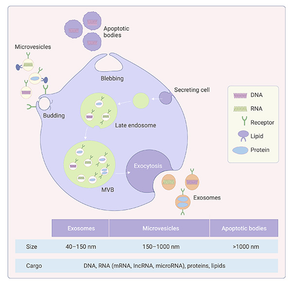
Figure 1. Classification of extracellular vesicles
Figure 1. Classification of extracellular vesicles[2]

Exosomes are the best defined extracellular vesicles (EVs) so far and their biogenesis, release pathways, size, content, and function are different from microvesicles and apoptotic bodies[3].

Feature Exosomes Microvesicles Apoptotic bodies
Size 40-150 nm 150-1000 nm ﹥1000 nm
Density 1.11-1.19 g/mL 1.02-1.22 g/mL 1.16-1.28 g/mL
Origin Multivesicular bodies fusion with plasmatic membrane Direct outward budding or blebbing from the plasma membrane Cellular debris, plasma membrane blebbing during cell apoptosis
Markers Alix, TSG101, HSC70, CD63, CD81, CD9, flotillin-1 Selectins, integrins, CD40, MMPs, Annexin A1 Annexin V, DNA, histones
Membrane permeability Membrane impermeable (PI negative) Membrane impermeable (PI negative) Membrane permeable (PI positive)
Functions Intercellular communication Intercellular communication Physiological and pathological regulation
Table 1. Characteristics of exosomes, microvesicles, and apoptotic bodies

Exosomes were first discovered in the cultured supernatant of sheep erythrocytes in 1983. Three pioneer scientists won the Nobel Prize in Physiology or Medicine in 2013 for their discoveries of machinery regulating vesicle traffic, a major transport system in our cells.

Recently, exosomes received a lot of attention due to their emerging role as intercellular messengers and their potential in disease diagnosis. Exosomes contain proteins, lipids, and RNAs that are specific to their cell origin and can deliver cargo to both nearby and distant cells[4]. Since exosomes are easy to purify/extract and detect, hence offering opportunities for disease diagnosis and treatment.

Engineering transformation and modification of exosomes can optimize their natural targeting and delivery to a specific location, hence broadening their use in clinical treatment with low immunogenicity, high biocompatibility and high efficacy.

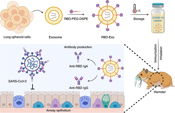
A recent study in Nature Biomedical Engineering reported a novel inhalable SARS-CoV-2 vaccine candidate consisting of a recombinant SARS-CoV-2 receptor-binding domain (RBD) conjugated to lung-derived exosomes. Since lung spheroid cells- (LSCs)derived exosomes (LSC-Exo) share surface proteins and receptors with membrane features found among airway epithelia, hence these exosomes are more distributed and retained for longer time in both the mucus-lined respiratory airway and in lung parenchyma and have enhanced internalization by antigen presenting cells (APCs) in the lung, providing a more targeted delivery carrier than commonly used liposomes. In hamsters, two doses of the vaccine attenuated severe pneumonia and reduced inflammatory infiltrates after exposure to live SARS-CoV-2[5].

Figure 2. Schematic representation of the fabrication of the RBD-Exo vaccine
The vaccine is delivered into the lungs via inhalation. RBD-Exo induces mucosal immunity and systemic immunity with the generation of RBD-specific IgA and IgG antibodies against SARS-CoV-2 infection in hamsters.
Isolation and purification of Exosomes

Cumulative evidence has revealed that exosomes can play an exceptional role in diagnostics and therapeutics of multiple diseases. An efficient, simple, and affordable method to isolate intact and pure exosomes is crucial to carrying out relevant pathways or disease research.

Herein, we summarized the commonly used exosome isolation techniques.

Strategy Principle Advantages Disadvantages
Density gradient centrifugation Components with imparity of size and density Possess various sediment speed High purity, avoiding exosomal damage Labor-intensive, Preliminary preparation and Cumbersome operation
Ultrafiltration Particles with various size and molecular weight Convenient, Without special equipment and reagents Clogging on filtering membrane, Loss of exosomes of small particle diameter
Immunoaffinity Based on interaction between antibodies and specific membrane proteins of exosomes High specificity for exosome subtypes isolation Expensive, depending on specificity of the antibody
Polymer precipitation The influence of exosomal solubility or dispersibility under the high hydrophilic polymers Imple operation, suitable for large-volume samples Potential contaminants (co-purifying protein aggregates or residuary polymers)
Size exclusion chromatography (SEC) Particles with various size and molecular weight Simple, economical, maintain the biological function and structure Special columns and packing are required, lipoprotein contamination
Table 2. Comparison of exosome isolation techniques[6]
Furthermore, highly pure exosomes can be easily obtained with exosome isolation kit
Figure 3. Exosome isolation procedures
Labeling and Identification of Exosomes

In 2014,the International Society for Extracellular Vesicles (ISEV) provided researchers with a minimal set of biochemical, biophysical and functional standards that should be used to attribute any specific biological cargo or function to extracellular vesicles (EVs). ISEV suggests a format of characteristics of EVs that should be analyzed and at least 2 different technologies should be used to characterize individual EVs.

Detection of exosome-enriched proteins

Analytic techniques to detect exosome-enriched proteins include Western blot (WB), high resolution flow cytometry (FACS) or global proteomic analysis using mass spectrometry to identify exosome transporting-related transmembrane proteins (CD63/CD81/CD9), heat shock proteins (HSP60/HSP70/HSPA5/CCT2/HSP90) and some cell-specific proteins that are enriched in exosomes membrane. Among them, CD63 and TSG101 are the most commonly used exosome markers.

Identification of exosome morphology with TEM

Exosomes morphology can be directly identified under transmission electron microscopy (TEM) with high resolution to provide an indication of the heterogeneity of the samples.

Size distribution and concentration measurements with NTA

NTA (Nanoparticle Tracking Analysis) technology can quickly and accurately analyze the particle size distribution and concentration of large number of exosomes in the sample.

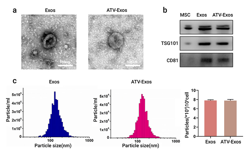
A study by Muyu Yu et al. focused on whether exosomes derived from the bone marrow MSC (BMSC) pretreated with atorvastatin (ATV) could exhibit better pro-angiogenic ability. As shown in Fig 4, they isolated exosomes from non-pretreated BMSC (Exos) and ATV pretreated BMSC (ATV-Exos) and evaluated their characterization by transmission electron microscopy (TEM), nanoparticle tracking analysis (NTA) and Western blotting[7].

Figure 8. Comparison of different linkers
Figure 4. The characterization of BMSC-derived exosomes[7]
a. The morphology of Exos and ATV-Exos was examined by TEM. b. The specific surface markers (Alix, TSG101, CD81) of Exos and ATV-Exos were assessed by Western blotting. c. The diameter and particle concentration of Exos and ATV-Exos were detected via NTA.

Labeling and tracing of exosomes

In vitro labeling or in vivo tracing of isolated exosomes is helpful for further studies on the functions and applications of exosomes. There are many methods for labeling and tracing of exosomes, mainly including lipophilic tracers and membrane-permeable compound tracers.

Lipophilic Tracers

Lipophilic dyes (DiO, DiI, DiD, DiR) can label exosomes well, and are also commonly used dyes in exosome research.

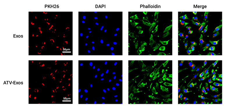
In vitro tracing: Exosomes can be traced in vitro by using PKH26 dye and scanning it by confocal microscope. As shown in Fig. 5, PKH26-labeled exosomes (red) were localized in the perinuclear region of HUVEC by using laser scanning confocal microscopy, confirming the internalization of exosomes by endothelial cells[7].

Figure 5. The uptake verification of Exos and ATV-Exos by HUVEC
Figure 5. The uptake verification of Exos and ATV-Exos by HUVEC[7]
Exosomes, cytoskeleton, and cell nucleus were stained red, green, and blue, respectively

In vivo tracing:

In vivo tracing can be done by using DIR dye and scanning by IVIS 200 Optical Imaging System.

Figure 6. In vivo imaging of DIR-labeled 4T1 exosomes in mouse mammary fat pads
Figure 6. In vivo imaging of DIR-labeled 4T1 exosomes in mouse mammary fat pads[8]
Intravenous injection of 4T1 exosomes into Balb/c mice with 4T1 tumors. Exosomes are labeled with the lipophilic fluorescent tracer DIR. A scale of the radiance efficiency is presented to the right of each live mouse image

Membrane permeable compound tracers

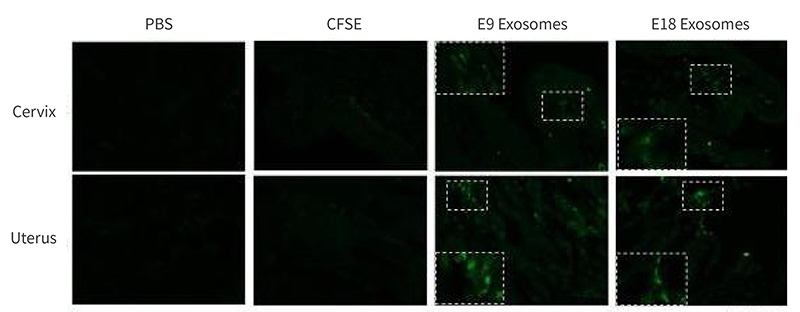
The membrane-permeable carboxyfluorescein diacetate succimidyl ester (CFSE) is a lipophilic fluorescent dye that can passively diffuses into cells, which is also commonly used for effective tracing of exosomes. It is colorless and non-fluorescent until the acetate groups are cleaved by intracellular esterases to yield highly fluorescent carboxyfluorescein succinimidyl ester. The succinimidyl ester group reacts with intracellular amines forming fluorescent conjugates that are well retained by cells.

Figure 6. In vivo imaging of DIR-labeled 4T1 exosomes in mouse mammary fat pads
Figure 7. Confocal images of CFSE-labeled embryonic tissue exosomes[9]
MCE has launched multiple exosome-related products. Please check our website for specific product information.
Related Products

All MCE products are only used for scientific researches or drug registration applications, we do not provide products and services for any personal use.

References
[1]. Zhang Y, Liu Y, Liu H, Tang WH. Exosomes: biogenesis, biologic function and clinical potential. Cell Biosci. 2019 Feb 15;9:19.
[2]. Naveed Akbar, Valerio Azzimato, Robin P Choudhury, Myriam Aouadi, et al. Extracellular vesicles in metabolic disease. Diabetologia. 2019 Dec;62(12): 2179-2187.
[3]. Doyle LM, Wang MZ. Overview of Extracellular Vesicles, Their Origin, Composition, Purpose, and Methods for Exosome Isolation and Analysis. Cells. 2019 Jul 15;8(7):727.
[4]. Li X, Corbett AL, Taatizadeh E, Tasnim N, Little JP, Garnis C, Daugaard M, Guns E, Hoorfar M, Li ITS. Challenges and opportunities in exosome research-Perspectives from biology, engineering, and cancer therapy. APL Bioeng. 2019 Mar 27;3(1):011503
[5]. Zhenzhen Wang, Kristen D Popowski, et al. Exosomes decorated with a recombinant SARS-CoV-2 receptor-binding domain as an inhalable COVID-19 vaccine. Nat Biomed Eng. 2022 Jul 4
[6]. Chen J, Li P, Zhang T, Xu Z, Huang X, Wang R, Du L. Review on Strategies and Technologies for Exosome Isolation and Purification. Front Bioeng Biotechnol. 2022 Jan 5;9:811971
[7]. Muyu Yu, Wei Liu, Junxian Li, et al. Exosomes derived from atorvastatin-pretreated MSC accelerate diabetic wound repair by enhancing angiogenesis via AKT/eNOS pathway. Stem Cell Res Ther. 2020 Aug 12;11(1):350.
[8]. Tyson Smyth, Max Kullberg, et al. Biodistribution and delivery efficiency of unmodified tumor-derived exosomes. J Control Release. 2015 Feb 10; 199: 145-55.
[9]. Samantha Sheller-Miller, Jayshil Trivedi, et al. Exosomes Cause Preterm Birth in Mice: Evidence for Paracrine Signaling in Pregnancy. Sci Rep. 2019 Jan 24;9(1):608.