



PKH 26
Based on 52 publication(s) in Google Scholar
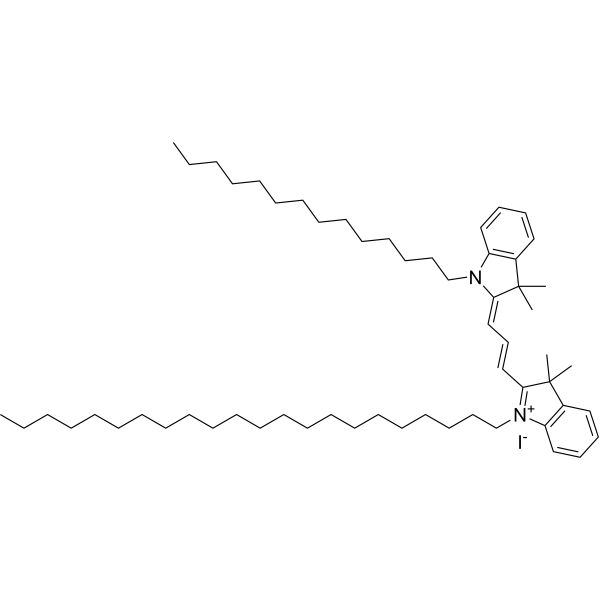
PKH 26 is a red fluorescent dye, PKH 26 can stably bind to the lipid region of cell membrane and emit red fluorescence (Ex/Em=551/567 nm), which is mainly used for in vitro cell labeling, in vitro cell proliferation studies and in vivo and in vitro cell tracing studies.
For research use only. We do not sell to patients.
- Purity : 99.94%
- CAS No.: 154214-55-8
- Formula: C59H97IN2
- Molecular Weight:961.32
-
Storage:
-20°C, protect from light, stored under nitrogen
* In solvent : -80°C, 6 months; -20°C, 1 month (protect from light, stored under nitrogen)

To place orders, for customer services and technical support, please contact: MedChemExpress USA
Tel: 609-228-6898 E-mail: [email protected] [email protected]


-
 Biological Activity
-
Chemical Information
-
Solvent & Solubility
- Protocols
- Purity & Documentation
- References
-
 Help & FAQs


-
HY-D1048
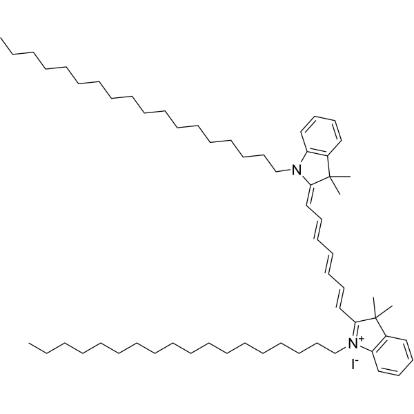
DiRMembrane Dye -
HY-D0969
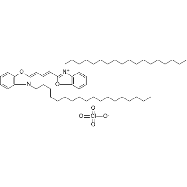
DiOFluorescent Probe -

HY-D1421
PKH 67Green Fluorescent Dye -

HY-18739
Phorbol 12-myristate 13-acetatePKC Activator -
HY-D1783
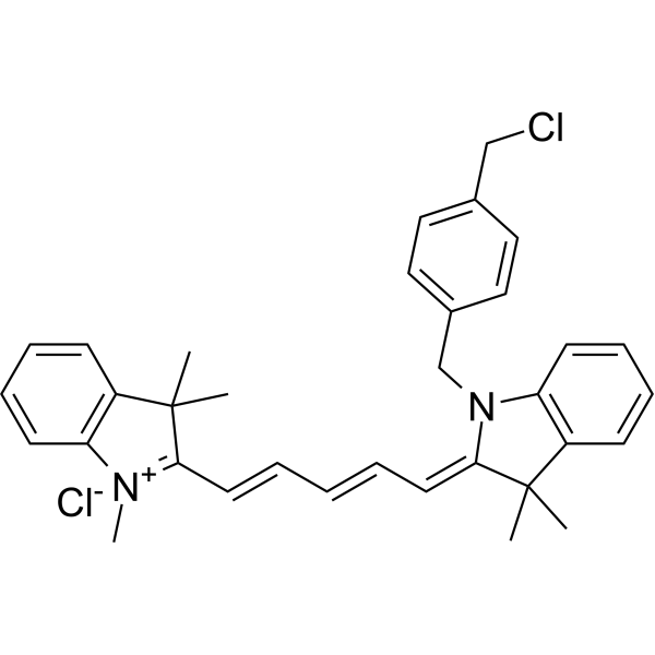
MitoTracker Deep Red FMRed Fluorescent Dye -

HY-10583
Y-27632 dihydrochlorideROCK Inhibitor -
HY-19363
GW4869N-SMase Inhibitor -

HY-DY1011
PKH 26 (solution)Fluorescent -

HY-13259
MG-132Proteasome Inhibitor -
HY-D0940
H2DCFDAROS-Sensitive Probe -
HY-D1434
FM1-43Track Endocytosis/Exocytosis -
HY-12318
IBMXPDE Inhibitor
Publications Citing Use of MedChemExpress (MCE) PKH 26
More- Cancer Cell. 2026 May 7:S1535-6108(26)00215-1.
- Cancer Cell. 2026 Jan 8:S1535-6108(25)00545-8. [Abstract]
- Mol Cancer. 2025 May 26;24(1):151. [Abstract]
- Mol Cancer. 2025 Apr 16;24(1):116. [Abstract]
- Adv Mater. 2023 Nov;35(48):e2302503. [Abstract]
- Nat Commun. 2025 Mar 20;16(1):2743. [Abstract]
- J Nanobiotechnology. 2026 Jul 1.
- J Nanobiotechnology. 2025 Dec 23;23(1):782. [Abstract]
- J Nanobiotechnology. 2025 Jul 22;23(1):535. [Abstract]
- J Nanobiotechnology. 2025 Jul 16;23(1):522. [Abstract]
- J Nanobiotechnology. 2024 May 22;22(1):276. [Abstract]
- Theranostics. 2025 Jan 1;15(1):86-102. [Abstract]
- MedComm (2020). 2026 Jun 13;7(6):e70828.
- Adv Sci (Weinh). 2025 Sep 24:e05921. [Abstract]
- Adv Sci (Weinh). 2025 May;12(19):e2501772. [Abstract]
- Biomaterials. 2026 May 28:335:124345. [Abstract]
- Chem Eng J. 2025 Jul 15.
- Mater Today Bio. 2026 May 6:38:103185. [Abstract]
- Mater Today Bio. 2026 Feb 25:37:102955. [Abstract]
- Journal of Future Foods. 2026 Jun 2.
- J Cachexia Sarcopenia Muscle. 2025 Dec;16(6):e70159. [Abstract]
- Nano Res. 2024 Aug 13;17:9898–9907.
- Oncogene. 2025 Mar 28. [Abstract]
- Int J Surg. 2025 Dec 17. [Abstract]
- Antioxidants (Basel). 2026 Feb 27;15(3):300. [Abstract]
- Cell Rep. 2026 Jun 25;45(7):117606. [Abstract]
- Pharmaceutics. 2023 Oct 13;15(10):2456. [Abstract]
- Neurosci Bull. 2026 Aug 8.
- CNS Neurosci Ther. 2025 Sep;31(9):e70597. [Abstract]
- Organogenesis. 2023 Dec 31;19(1):2285836. [Abstract]
- Cells. 2026 Aug 4;15(15):1408.
- Int Immunopharmacol. 2026 Oct 1:186:117107.
- Int J Mol Sci. 2024 Mar 29;25(7):3854. [Abstract]
- J Ovarian Res. 2024 Nov 14;17(1):224. [Abstract]
- Toxicology. 2025 Nov:517:154214. [Abstract]
- Cell Signal. 2025 Oct:134:111963. [Abstract]
- Mol Cell Biochem. 2025 Jun;480(6):3873-3888. [Abstract]
- J Fungi (Basel). 2026 Aug 11;12(8):597.
- FASEB J. 2026 Aug 31;40(16):e72157.
- FASEB J. 2023 Mar;37(3):e22821. [Abstract]
- Cell Stress Chaperones. 2025 Jul;30(4):100081. [Abstract]
- Cardiovasc Drugs Ther. 2026 Jun 18. [Abstract]
- J Neuroimmunol. 2026 Apr:413:578875. [Abstract]
- Immunobiology. 2025 Jul 16;230(4):153103. [Abstract]
- Anim Reprod Sci. 2026 Jul:290:108180. [Abstract]
- Oncol Lett. 2023 Nov 16;27(1):24. [Abstract]
- bioRxiv. 2026 Jul 26.
- bioRxiv. 2026 May 18:2026.05.14.725149. [Abstract]
- bioRxiv. 2025 Nov 4.
- Heliyon. 2025 Jan 14;11(2):e41960. [Abstract]
- Research Square Preprint. 2023 Apr 12.
- Research Square Print. 2022 Aug.
 Customer Validation & Images

-
Cell Imaging/Staining
-
Cell Imaging/Staining
-
Cell Imaging/Staining
-
Cell Imaging/Staining
-
Cell Imaging/Staining


Biological Activity
Description
In Vitro
Guide (The following is the experimental plan we recommend. This plan serves only as a reference guide. The specific operations should be adjusted according to your actual needs.)
1. Preparation of PKH 26 working solution
1.1 Preparation of stock solution
Prepare a 1 mM stock solution using DMSO
1.2 Preparation of PKH 26 working solution
Dilute the stock solution in serum-free cell culture medium or PBS to obtain a working solution of 5-10 μM.
Note: Please adjust the concentration of the PKH 26 working solution as needed
2. Cell staining
2.1 Suspended cells (6-well plate)
a. Centrifuge at 4°C for 1000g for 3-5 minutes, discard the supernatant, wash twice with PBS, each time for 5 minutes. The cell density is 1×10⁶/mL.
b. Add 1 mL of the working solution, incubate at room temperature for 10-45 minutes.
c. Centrifuge at 400g for 4-5 minutes at 4°C, discard the supernatant.
d. Wash twice with PBS, each time for 5 minutes.
e. Resuspend the cells in serum-free cell culture medium or PBS. Observe with a fluorescence microscope or flow cytometer.
2.2 Attached cells
a. Cultivate attached cells on a sterile cover glass.
b. Remove the cover glass from the culture medium and aspirate the excess medium.
c. Add 100 μL of the working solution, gently shake to fully cover the cells, incubate at room temperature for 5-30 minutes.
d. Wash twice with the culture medium, each time for 5 minutes. Observe with a fluorescence microscope or flow cytometer.
MedChemExpress (MCE) has not independently confirmed the accuracy of these methods. They are for reference only.
- BrdU Incorporation Assay
- Somatic Cell Culture
- CFSE Dye Dilution Proliferation Assay
- Dye-dilution cell tracking and proliferation staining
- CCK-8/WST-8 Cell Proliferation Assay
- Protocol for Cell Cycle
- Cell Counting-Based Growth Curve Assay
- Colony Formation (Clonogenic) Assay
- Cell Viability Determination by MTT Colorimetric Assay
- EdU Incorporation Assay (Click Chemistry-Based DNA Synthesis Measurement)
- Ki-67 Immunostaining Proliferation Assay
- PCNA Immunodetection Proliferation Assay
- Protocol for Cell Counting and Cell Density Analysis
- MTT Cell Proliferation Assay
Emission (Em)
567
Excitation (Ex)
551


Chemical Information
-
CAS No. 154214-55-8
-
Appearance Solid
-
Molecular Weight 961.32
-
Formula C59H97IN2
-
Color Brown to reddish brown
-
SMILES
CC1(C)C(/C=C/C=C2N(CCCCCCCCCCCCCC)C3=C(C=CC=C3)C/2(C)C)=[N+](CCCCCCCCCCCCCCCCCCCCCC)C4=C1C=CC=C4.[I-]
-
Shipping
Room temperature in continental US; may vary elsewhere.
-
Storage
-20°C, protect from light, stored under nitrogen
* In solvent : -80°C, 6 months; -20°C, 1 month (protect from light, stored under nitrogen)


Publications (52)
-
Journal Impact Factor
-
Most Recent
-
-
Cancer Cell
2026 Jan 8:S1535-6108(25)00545-8. PMID: 41512868 -
Mol Cancer
GP73-dependent regulation of exosome biogenesis promotes colorectal cancer liver metastasis. [Abstract]2025 May 26;24(1):151. PMID: 40414849 -
Mol Cancer
ADAR1-high tumor-associated macrophages induce drug resistance and are therapeutic targets in colorectal cancer. [Abstract]2025 Apr 16;24(1):116. PMID: 40241135 -
Adv Mater
A Targeted Exosome Therapeutic Confers Both Cfdna Scavenging and Macrophage Polarization for Ameliorating Rheumatoid Arthritis. [Abstract]2023 Nov;35(48):e2302503. PMID: 37681753 -
Nat Commun
Adipose progenitor cell-derived extracellular vesicles suppress macrophage M1 program to alleviate midlife obesity. [Abstract]2025 Mar 20;16(1):2743. PMID: 40113754
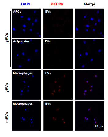
PKH 26 purchased from MedChemExpress. Usage Cited in: Nat Commun. 2025 Mar 20;16(1):2743. [Abstract]
The engulfment of PKH26-labeled EVs (red) by APCs, adipocytes and macrophages was detected by confocal microscopy.
-
-
J Nanobiotechnology
CAT/SOD-Enriched Achyranthes bidentata nanovesicles mitigate TMJOA via ROS scavenging and JNK/FOXO1 pathway Inhibition. [Abstract]2025 Dec 23;23(1):782. PMID: 41430692
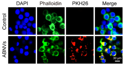
PKH 26 purchased from MedChemExpress. Usage Cited in: J Nanobiotechnology. 2025 Dec 23;23(1):782. [Abstract]
Macrophage uptake of PKH26 (24 h)-labeled ABNVs (red).
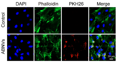
PKH 26 purchased from MedChemExpress. Usage Cited in: J Nanobiotechnology. 2025 Dec 23;23(1):782. [Abstract]
PKH26 (24 h)-labeled ABNVs were efficiently internalized by chondrocytes.
-
J Nanobiotechnology
A fibrin gel-loaded Gouqi-derived nanovesicle (GqDNV) repairs the heart after myocardial infarction by inhibiting p38 MAPK/NF-κB p65 pathway. [Abstract]2025 Jul 22;23(1):535. PMID: 40696398 -
J Nanobiotechnology
Exosomes derived from platelet-rich plasma alleviate synovial inflammation by enhancing synovial lymphatic function. [Abstract]2025 Jul 16;23(1):522. PMID: 40671121 -
J Nanobiotechnology
Gouqi-derived nanovesicles (GqDNVs) inhibited dexamethasone-induced muscle atrophy associating with AMPK/SIRT1/PGC1α signaling pathway. [Abstract]2024 May 22;22(1):276. PMID: 38778385 -
Theranostics
iRGD-TRP-PK1-modified red blood cell membrane vesicles as a new chemotherapeutic drug delivery and targeting system in head and neck cancer. [Abstract]2025 Jan 1;15(1):86-102. PMID: 39744235 -
-
Adv Sci (Weinh)
MicroRNA-Enriched Plant-Derived Exosomes Alleviate Colitis by Modulating Systemic Immunity, Metabolic Homeostasis, and Gut Microbiota. [Abstract]2025 Sep 24:e05921. PMID: 40994128
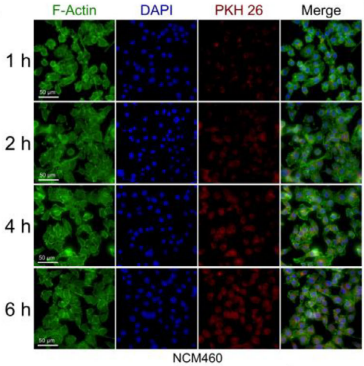
PKH 26 purchased from MedChemExpress. Usage Cited in: Adv Sci (Weinh). 2025 Sep 24:e05921. [Abstract]
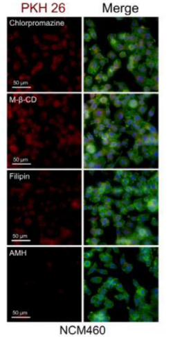
Representative fluorescence microscopy images depicting the time‐dependent cellular uptake of CAEs in NCM460 cells at 1‐, 2‐, 4‐, and 6‐h incubation (These histogram bars served as a negative reference for gating PKH 26‐positive cells).
PKH 26 purchased from MedChemExpress. Usage Cited in: Adv Sci (Weinh). 2025 Sep 24:e05921. [Abstract]
NCM460 cells after 6‐h treatment.
-
Adv Sci (Weinh)
Rational Design and Organoid-Based Evaluation of a Cocktail CAR-γδ T Cell Therapy for Heterogeneous Glioblastoma. [Abstract]2025 May;12(19):e2501772. PMID: 40112194 -
Biomaterials
Stealth red blood cells with broad-spectrum antigenic shielding for transfusion therapy in antibody-mediated hemolytic anemia. [Abstract]2026 May 28:335:124345. PMID: 42229299 -
-
Mater Today Bio
MRI-guided diagnosis and dual-pathway neuroprotection in ischemic stroke via PDA-engineered apple exosome-like nanoparticles. [Abstract]2026 May 6:38:103185. PMID: 42182870 -
Mater Today Bio
Targeted inhibition of PARP-1 in pulmonary epithelial cells and macrophages via SPA-functionalized microparticles attenuates sepsis-induced lung injury. [Abstract]2026 Feb 25:37:102955. PMID: 41809387 -
-
J Cachexia Sarcopenia Muscle
C2C12-Derived ApoVs Promote Skeletal Muscle Development and Ameliorate Age-Related Muscle Loss Through Igf1r/PI3K/AKT/mTOR Pathway. [Abstract]2025 Dec;16(6):e70159. PMID: 41344911 -
-
Oncogene
sEV-mediated intercellular transformation from MGAT4AHigh to MGAT4ALow tumor cells via the HOTAIRM1/miR-196b-5p axis promotes apoptosis resistance in CTCL. [Abstract]2025 Mar 28. PMID: 40155530 -
Int J Surg
Apoptotic extracellular vesicles from peripancreatic adipose-derived mesenchymal stem cells ameliorate severe acute pancreatitis through the transcription factor EB-mediated autophagy-lysosomal pathway: an experimental study. [Abstract]2025 Dec 17. PMID: 41403290 -
Antioxidants (Basel)
Kynurenic Acid/GPR35 Signaling Protects the Infarcted Heart by Suppressing Macrophage mtDNA-Triggered cGAS-STING Activation. [Abstract]2026 Feb 27;15(3):300. PMID: 41897447 -
Cell Rep
The TNRC6B/circTNRC6B axis drives lymphovascular invasion and metastasis through an intragenic self-degradation loop. [Abstract]2026 Jun 25;45(7):117606. PMID: 42360875 -
Pharmaceutics
Small Molecule Cocktails Promote Fibroblast-to-Leydig-like Cell Conversion for Hypogonadism Therapy. [Abstract]2023 Oct 13;15(10):2456. PMID: 37896216 -
-
CNS Neurosci Ther
Dual-Functionalized Extracellular Vesicles Promote Brain Repair and Remodeling Following Ischemic Stroke in Mice. [Abstract]2025 Sep;31(9):e70597. PMID: 40955109 -
Organogenesis
Regulation of Interferon-β-Modified Human Umbilical Cord Mesenchymal Stem Cell-Derived Exosomes in Proliferation and Apoptosis of Prostate Cancer Cells. [Abstract]2023 Dec 31;19(1):2285836. PMID: 38031805 -
-
-
Int J Mol Sci
2024 Mar 29;25(7):3854. PMID: 38612664 -
J Ovarian Res
Human umbilical cord mesenchymal stem cell-derived extracellular vesicles harboring IGF-1 improve ovarian function of mice with premature ovarian insufficiency through the Nrf2/HO-1 pathway. [Abstract]2024 Nov 14;17(1):224. PMID: 39543679 -
Toxicology
Role of neuroglia cell-derived microvesicles in cobalt chloride-induced mitochondrial autophagy in neurons. [Abstract]2025 Nov:517:154214. PMID: 40490208 -
Cell Signal
Exosomal miR-382-5p prevents pre-metastatic niche formation by inhibiting GPR176/GNAS-CXCR1/CXCR2 axis in colorectal cancer liver metastasis. [Abstract]2025 Oct:134:111963. PMID: 40578589 -
Mol Cell Biochem
Upregulation of YY1 in M2 macrophages promotes secretion of exosomes containing hsa-circ-0000326 via super-enhancers to facilitate prostate cancer progression. [Abstract]2025 Jun;480(6):3873-3888. PMID: 39960585 -
-
-
FASEB J
GSK-3β/β-catenin pathway plays crucial roles in the regulation of NK cell cytotoxicity against myeloma cells. [Abstract]2023 Mar;37(3):e22821. PMID: 36794671 -
Cell Stress Chaperones
COL5A2-mediated endoplasmic reticulum stress promotes macrophage M2 polarization in lung adenocarcinoma. [Abstract]2025 Jul;30(4):100081. PMID: 40345640 -
Cardiovasc Drugs Ther
Shenfu Injection Inhibits Cardiac Fibroblast Activation and Improves Myocardial Fibrosis by Down-regulating M1 Macrophage-Derived Exosomal miR-155-5p. [Abstract]2026 Jun 18. PMID: 42313305 -
J Neuroimmunol
Exosomes derived from neural stem cells contribute to cerebral ischemia/reperfusion injury via inhibiting autophagy in rats. [Abstract]2026 Apr:413:578875. PMID: 41619451 -
Immunobiology
Exosomes derived from baicalin-pretreated bone marrow mesenchymal stem cells inactivate the TLR4/MyD88/NF-kB pathway to improve asthma. [Abstract]2025 Jul 16;230(4):153103. PMID: 40695068 -
Anim Reprod Sci
Cryopreservation and xenotransplantation of spotted sea bass (Lateolabrax maculatus) spermatogonia. [Abstract]2026 Jul:290:108180. PMID: 41955652 -
Oncol Lett
Exosome‑delivered miR‑486‑3p inhibits the progression of osteosarcoma via sponging CircKEAP1/MARCH1 axis components. [Abstract]2023 Nov 16;27(1):24. PMID: 38058466 -
-
bioRxiv
BLOC1S1 regulates autolysosomal and exosomal dynamics during CD4+ T cell differentiation. [Abstract]2026 May 18:2026.05.14.725149. PMID: 42239123 -
-
Heliyon
Small extracellular vesicles derived from miRNA-486 overexpressed dental pulp stem cells mitigate high altitude pulmonary edema through PTEN/PI3K/AKT/eNOS pathway. [Abstract]2025 Jan 14;11(2):e41960. PMID: 39906863 -
-


Solvent & Solubility
In Vitro:
DMSO : 1.67 mg/mL (1.74 mM; ultrasonic and warming and heat to 60°C; Hygroscopic DMSO has a significant impact on the solubility of product, please use newly opened DMSO)
Please refer to the solubility information to select the appropriate solvent. Once prepared, please aliquot and store the solution to prevent product inactivation from repeated freeze-thaw cycles.
Storage method and period of stock solution: -80°C, 6 months; -20°C, 1 month (protect from light, stored under nitrogen). When stored at -80°C, please use it within 6 months. When stored at -20°C, please use it within 1 month.
Please refer to the solubility information to select the appropriate solvent. Once prepared, please aliquot and store the solution to prevent product inactivation from repeated freeze-thaw cycles.
Storage method and period of stock solution: -80°C, 6 months; -20°C, 1 month (protect from light, stored under nitrogen). When stored at -80°C, please use it within 6 months. When stored at -20°C, please use it within 1 month.
Concentration (start) × Volume (start) = Concentration (final) × Volume (final)


Purity & Documentation
-
Data Sheet (271 KB)
-
SDS (252 KB)
- English - EN (252 KB)
- Français - FR (252 KB)
- Deutsch - DE (252 KB)
- Norwegian - NO (252 KB)
- Español - ES (252 KB)
- Swedish - SV (252 KB)
- Italian - IT (252 KB)
- Korean - KR (252 KB)
- Portuguese - PT (252 KB)
-
Handling Instructions (2659 KB)
References


Complete Stock Solution Preparation Table
Please refer to the solubility information to select the appropriate solvent. Once prepared, please aliquot and store the solution to prevent product inactivation from repeated freeze-thaw cycles.
Storage method and period of stock solution: -80°C, 6 months; -20°C, 1 month (protect from light, stored under nitrogen). When stored at -80°C, please use it within 6 months. When stored at -20°C, please use it within 1 month.
| Optional Solvent | Concentration Solvent Mass | 1 mg | 5 mg | 10 mg | 25 mg |
|---|---|---|---|---|---|
| DMSO | 1 mM | 1.0402 mL | 5.2012 mL | 10.4024 mL | 26.0059 mL |


PKH 26 Related Classifications
HY-D1451 Related Classifications
 Powered by Bioz