



GMP EGF Protein, Human
Based on 5 publication(s) in Google Scholar
EGF proteins act as potent stimulators of growth in a variety of epidermal and epithelial tissues in both in vivo and in vitro settings, while promoting the proliferation of specific fibroblasts in cell culture. This multifaceted protein also acts as a magnesium stimulating hormone, driving magnesium reabsorption in the renal distal tubule through engagement of EGFR and activation of the magnesium channel TRPM6. GMP EGF Protein, Human is the recombinant human-derived EGF protein, expressed by E. coli , with tag free.
- Species: Human
- Source: E. coli
-
Storage:Stored at -20°C for 2 years from date of receipt. After reconstitution, it is stable at 4°C for 1 week or -20°C for longer (with carrier protein). It is recommended to freeze aliquots at -20°C or -80°C for extended storage.

To place orders, for customer services and technical support, please contact: MedChemExpress USA
Tel: 609-228-6898 E-mail: [email protected] [email protected]


-
 Biological Activity
-
Technical Parameters
-
Product Properties
- Documentation
- References
- Help & FAQs


-

HY-10182
LaduviglusibGSK-3 Inhibitor -
HY-N0583
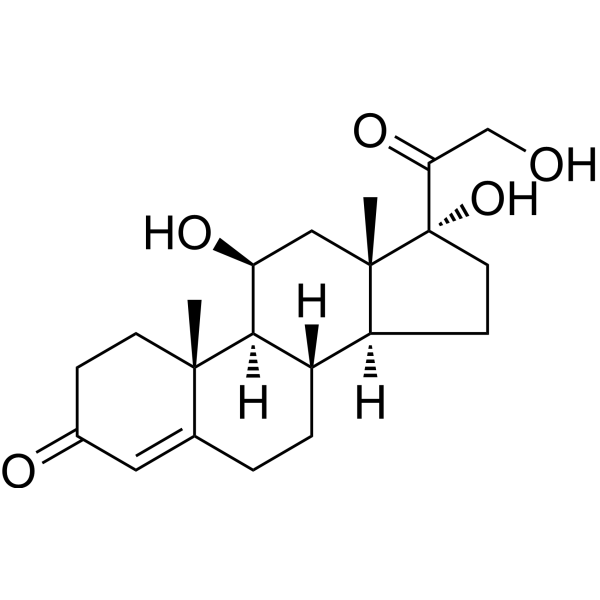
HydrocortisoneGlucocorticoid Receptor Agonist -
HY-B0272
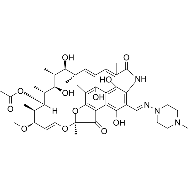
RifampicinAntivirus Agent -

HY-P701242
Heat Stable FGF basic/bFGF Protein, Human -
HY-B0764
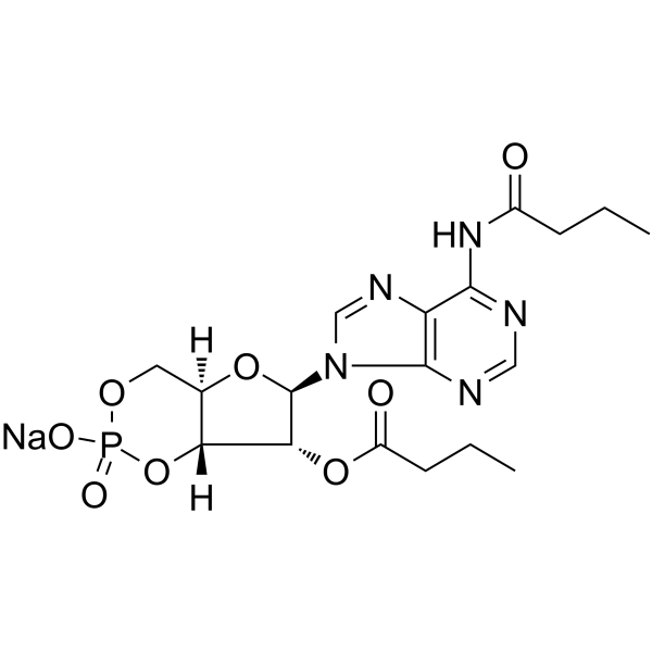
Bucladesine sodiumcAMP Analog -

HY-10431
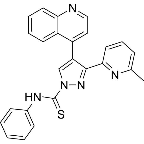
SB-431542TGF-β Receptor Kinase Inhibitor -
HY-10432
A 83-01ALK4/5/7 Inhibitor -
HY-P7004
FGF basic/bFGF Protein, Human (146a.a) -
HY-P70311
Activin A Protein, Human/Mouse/Rat (HEK293) -
HY-P7055
PDGF-BB Protein, Human -
HY-P7014
FGF-4 Protein, Human -

HY-K0301
Cell Counting Kit-8


Biological Activity
Description
EGF proteins act as potent stimulators of growth in a variety of epidermal and epithelial tissues in both in vivo and in vitro settings, while promoting the proliferation of specific fibroblasts in cell culture. This multifaceted protein also acts as a magnesium stimulating hormone, driving magnesium reabsorption in the renal distal tubule through engagement of EGFR and activation of the magnesium channel TRPM6. GMP EGF Protein, Human is the recombinant human-derived EGF protein, expressed by E. coli , with tag free.
Background
GMP EGF Protein emerges as a potent stimulator of the growth of diverse epidermal and epithelial tissues in both in vivo and in vitro environments, along with fostering the proliferation of specific fibroblasts in cell culture. This multifaceted protein also acts as a magnesiotropic hormone, driving magnesium reabsorption in the renal distal convoluted tubule through the engagement of EGFR and activation of the magnesium channel TRPM6. Notably, GMP EGF Protein possesses the capability to induce neurite outgrowth in motoneurons of the pond snail Lymnaea stagnalis in vitro. Its molecular interactions include binding to EGFR, thereby promoting EGFR dimerization, and interacting with RHBDF2, suggesting involvement in diverse cellular processes. Furthermore, GMP EGF Protein engages with RHBDF1, potentially influencing the retention of EGF in the endoplasmic reticulum and regulating its degradation through endoplasmic reticulum-associated degradation (ERAD).
In Vivo
EGF Protein (Human) (10 μg/g, cream, a single dose for 7 days) reduces inflammatory infiltrate and results in wound re-epithelialization in rats with full thicknessskin wounds[1].
EGF Protein (Human) (50 μg/g, cream, every 24 h for 7 days) significantly stimulates re-epithelialization at high levels, preferably in combination with a protease inhibitor in pigs with thickness wounds on the back[1].
EGF Protein (Human) (30 μg/kg, s.c., daily for 3 weeks) attenuates the oesophageal damage normally caused by the sclerotherapy (surgical dilation of the oesophagus) procedure in Gottingen minipigs[1].
Verified Bioactivity
The specific activity is > 1×10⁶ IU/mg.


Publications (5)
-
Journal Impact Factor
-
Most Recent
-
Oncogene
TRIM21-mediated ubiquitination of SIX2 attenuates breast cancer stemness via LGSN suppression. [Abstract]2025 Sep 15. PMID: 40954199 -
J Agric Food Chem
2024 Nov 6;72(44):24387-24399. PMID: 39435975 -
EMBO Rep
Noncanonical Wnt signaling promotes colon tumor growth, chemoresistance and tumor fibroblast activation. [Abstract]2023 Apr 5;24(4):e54895. PMID: 36704936 -
Chem Biol Drug Des
Artesunate Enhances Sensitivity of Renal Cancer Cells to Sunitnib by Mediating Tripartite Motif Containing 24-Induced Ubiquitination of Paired Box 6. [Abstract]2025 May;105(5):e70116. PMID: 40346910 -
Biochem Biophys Res Commun
CENPA-driven transcriptional activation of ECT2 enhances EGFR inhibitor resistance in lung adenocarcinoma. [Abstract]2025 Sep 1:777:152287. PMID: 40618443


Technical Parameters
-
Species Human
-
Source E. coli
-
Tag Tag Free
-
Accession
P01133-1 (N971-R1023)
-
Molecular Construction
-
N-term
-
GMP EGF (N971-R1023)
Accession # P01133-1 -
C-term
-
-
Protein Length
Full Length of EGF
-
Synonyms
EGF; Alternative Protein EGF; Epidermal Growth Factor; Beta-Urogastrone; Pro-Epidermal Growth Factor; HOMG4; Epidermal Growth Factor (Beta-Urogastrone); URG
-
AA Sequence
NSDSECPLSHDGYCLHDGVCMYIEALDKYACNCVVGYIGERCQYRDLKWWELR
-
Predicted Molecular Mass
6.2 kDa
-
Molecular Weight
Approximately 11 kDa, based on SDS-PAGE under reducing conditions.
-
Purity
≥ 95%, as determined by reducing SDS-PAGE.


Product Properties
Lyophilized powder.
Lyophilized from a 0.22 μm filtered solution of 20 mM Tris, 200 mM NaCl, pH 8.0.
Note: For SPR assay, please replace the buffer. Primary amine components (e.g., Tris, imidazole) can affect protein-coupled chips.
<0.01 EU/μg, determined by LAL method.
It is not recommended to reconstitute to a concentration less than 100 μg/mL in injection water.
Stored at -20°C for 2 years from date of receipt. After reconstitution, it is stable at 4°C for 1 week or -20°C for longer (with carrier protein). It is recommended to freeze aliquots at -20°C or -80°C for extended storage.
Room temperature in continental US; may vary elsewhere.


Documentation
-
Data Sheet (261 KB)
-
SDS (252 KB)
- English - EN (252 KB)
- Français - FR (252 KB)
- Deutsch - DE (252 KB)
- Norwegian - NO (252 KB)
- Español - ES (252 KB)
- Swedish - SV (252 KB)
- Italian - IT (252 KB)
- Korean - KR (252 KB)
- Portuguese - PT (252 KB)
-
Handling Instructions (2659 KB)


References
[1]. Wong WR, et al. Applications, and efficient large-scale production, of recombinant human epidermal growth factor. Biotechnol Genet Eng Rev. 2001;18:51-71. [Content Brief]
[2]. Lao G, et al. Controlled release of epidermal growth factor from hydrogels accelerates wound healing in diabetic rats. J Am Podiatr Med Assoc. 2012 Mar-Apr;102(2):89-98. [Content Brief]
[3]. Lao G, et al. Controlled release of epidermal growth factor from hydrogels accelerates wound healing in diabetic rats. J Am Podiatr Med Assoc. 2012 Mar-Apr;102(2):89-98. [Content Brief]

Calculators
Concentration (start) × Volume (start) = Concentration (final) × Volume (final)


 Powered by Bioz

