



N-Cadherin Protein, Mouse (HEK293)

Based on 1 Customer Validation
N-Cadherin Protein is a calcium-dependent cell adhesion protein that mediates homotypic cell-cell adhesion by dimerizing with a CDH2 chain from another cell. N-Cadherin Protein plays a role in cell adhesion, cell sorting, and neural development. N-Cadherin Protein, Mouse (HEK293) is the recombinant mouse-derived N-Cadherin protein, expressed by HEK293 , with tag free.
- Species: Mouse
- Source: HEK293
-
Storage:Stored at -80°C for 1 year from date of receipt. It is stable at -20°C for 3 months after opening. It is recommended to freeze aliquots at -80°C for extended storage. Avoid repeated freeze-thaw cycles.


To place orders, for customer services and technical support, please contact: MedChemExpress USA
Tel: 609-228-6898 E-mail: [email protected] [email protected]


-
 Biological Activity
-
Technical Parameters
-
Product Properties
- Documentation
- Help & FAQs




Biological Activity
Description
N-Cadherin Protein is a calcium-dependent cell adhesion protein that mediates homotypic cell-cell adhesion by dimerizing with a CDH2 chain from another cell. N-Cadherin Protein plays a role in cell adhesion, cell sorting, and neural development. N-Cadherin Protein, Mouse (HEK293) is the recombinant mouse-derived N-Cadherin protein, expressed by HEK293 , with tag free.
Background
N-Cadherin, a calcium-dependent cell adhesion protein, primarily facilitates homotypic cell-cell adhesion through dimerization with a CDH2 chain from neighboring cells. This property contributes to the sorting of heterogeneous cell types. Additionally, N-Cadherin serves as a regulator of neural stem cell quiescence by anchoring these cells to ependymocytes in the adult subependymal zone; MMP24-mediated cleavage of N-Cadherin affects this anchorage, modulating neural stem cell quiescence. In pancreatic beta cells, N-Cadherin plays a crucial role in forming cell-to-cell junctions with neural crest stem cells, promoting the extension of processes by the latter. The protein is essential for proper neurite branching, pre- and postsynaptic organization, and may be involved in neuronal recognition mechanisms. In hippocampal neurons, N-Cadherin potentially regulates dendritic spine density. Functionally, N-Cadherin forms homodimers via its extracellular region and can also form heterodimers with other cadherins in trans, involving a cadherin chain from another cell. Interactions with CDCP1, PCDH8, and OBSCN further underscore the complexity of N-Cadherin's cellular activities, with potential internalization through the TAOK2/p38 MAPK pathway. Moreover, it is identified in a complex containing FGFR4, NCAM1, CDH2, PLCG1, FRS2, SRC, SHC1, GAP43, and CTTN, highlighting its association with diverse cellular components.
Verified Bioactivity
Measured by the ability of the immobilized protein to support the adhesion of U-118-MG cells. The ED50 for this effect is 0.1447 μg/mL, corresponding to a specific activity is 6910.85 units/mg.
MCE Validation Data
-
Bioactivity - Cell-Based Assay
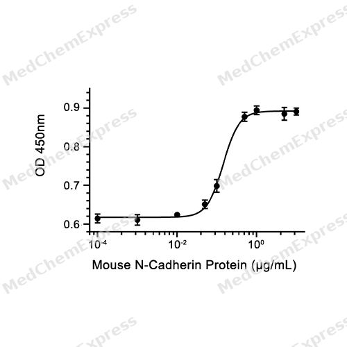
Bioactivity - Cell-Based Assay


Technical Parameters
-
Species Mouse
-
Source HEK293
-
Tag Tag Free
-
Accession
P15116 (S26-A724)
-
Molecular Construction
-
N-term
-
N-Cadherin (S26-A724)
Accession # P15116 -
C-term
-
-
Protein Length
Partial
-
Synonyms
CDH2; N-Cadherin; Prev. NCAD; Calcium-Dependent Adhesion Protein, Neuronal; CDHN; CD325 Antigen; Neural Cadherin; N-Cadherin 1; Cadherin-2; ARVD14; CD325; ADHD8; Cadherin 2, Type 1, N-Cadherin (Neuronal); ACOGS; Cadherin 2; CDw325
-
AA Sequence
SGEIALCKTGFPEDVYSAVLPKDVHEGQPLLNVKFSNCNRKRKVQYESSEPADFKVDEDGTVYAVRSFPLTAEQAKFLIYAQDKETQEKWQVAVNLSREPTLTEEPMKEPHEIEEIVFPRQLAKHSGALQRQKRDWVIPPINLPENSRGPFPQELVRIRSDRDKNLSLRYSVTGPGADQPPTGIFIINPISGQLSVTKPLDRELIARFHLRAHAVDINGNQVENPIDIVINVIDMNDNRPEFLHQVWNGSVPEGSKPGTYVMTVTAIDADDPNALNGMLRYRILSQAPSTPSPNMFTINNETGDIITVAAGLDREKVQQYTLIIQATDMEGNPTYGLSNTATAVITVTDVNDNPPEFTAMTFYGEVPENRVDVIVANLTVTDKDQPHTPAWNAAYRISGGDPTGRFAILTDPNSNDGLVTVVKPIDFETNRMFVLTVAAENQVPLAKGIQHPPQSTATVSVTVIDVNENPYFAPNPKIIRQEEGLHAGTMLTTLTAQDPDRYMQQNIRYTKLSDPANWLKIDPVNGQITTIAVLDRESPNVKNNIYNATFLASDNGIPPMSGTGTLQIYLLDINDNAPQVLPQEAETCETPEPNSINITALDYDIDPNAGPFAFDLPLSPVTIKRNWTINRLNGDFAQLNLKIKFLEAGIYEVPIIITDSGNPPKSNISILRVKVCQCDSNGDCTDVDRIVGAGLGTGA
-
Predicted Molecular Mass
77.8 kDa
-
Molecular Weight
Approximately 91 kDa & 114 kDa, based on SDS-PAGE under reducing conditions, due to the glycosylation.
-
Glycosylation
Yes
-
Purity
≥ 90%, as determined by reducing SDS-PAGE.


Product Properties
Solution
Supplied as a 0.22 μm filtered solution of PBS, pH 7.4.
<1 EU/μg, determined by LAL method.
Stored at -80°C for 1 year from date of receipt. It is stable at -20°C for 3 months after opening. It is recommended to freeze aliquots at -80°C for extended storage. Avoid repeated freeze-thaw cycles.
Shipping with dry ice.


Documentation
-
Data Sheet (240 KB)
-
SDS (252 KB)
- English - EN (252 KB)
- Français - FR (252 KB)
- Deutsch - DE (252 KB)
- Norwegian - NO (252 KB)
- Español - ES (252 KB)
- Swedish - SV (252 KB)
- Italian - IT (252 KB)
- Korean - KR (252 KB)
- Portuguese - PT (252 KB)
-
Handling Instructions (2659 KB)

Calculators
Concentration (start) × Volume (start) = Concentration (final) × Volume (final)


 Powered by Bioz

