



PD-L2 Protein, Rat (HEK293, His)
Based on 1 Customer Validation
Programmed cell death 1 ligand 2 (Pdcd1lg2, Pd-l2, B7-DC) is a member of B7 family and a ligand of programmed cell death 1. Pdcd1lg2 negatively regulates activated T cell proliferation, interferon-gamma production and interleukin-10 production in a PDCD1-independent manner, or inhibits T-cell proliferation by blocking cell cycle progression and cytokine production by interacts with PDCD1, and consequently causes cancer growth and suppresses the immune system. PD-L2 Protein, Rat (HEK293, His) is the recombinant rat-derived PD-L2 protein, expressed by HEK293 , with C-His labeled tag.
- Species: Rat
- Source: HEK293
-
Storage:Stored at -20°C for 2 years from date of receipt. After reconstitution, it is stable at 4°C for 1 week or -20°C for longer (with carrier protein). It is recommended to freeze aliquots at -20°C or -80°C for extended storage.


To place orders, for customer services and technical support, please contact: MedChemExpress USA
Tel: 609-228-6898 E-mail: [email protected] [email protected]


-
 Biological Activity
-
Technical Parameters
-
Product Properties
- Documentation
- References
- Help & FAQs




Biological Activity
Description
Programmed cell death 1 ligand 2 (Pdcd1lg2, Pd-l2, B7-DC) is a member of B7 family and a ligand of programmed cell death 1. Pdcd1lg2 negatively regulates activated T cell proliferation, interferon-gamma production and interleukin-10 production in a PDCD1-independent manner, or inhibits T-cell proliferation by blocking cell cycle progression and cytokine production by interacts with PDCD1, and consequently causes cancer growth and suppresses the immune system. PD-L2 Protein, Rat (HEK293, His) is the recombinant rat-derived PD-L2 protein, expressed by HEK293 , with C-His labeled tag.
Background
Programmed cell death 1 ligand 2 (Pdcd1lg2, Pd-l2, B7-DC) is a member of B7 family and a ligand of programmed cell death 1. Pdcd1lg2 is consist of one IgV and one IgC domain, and is involved in the costimulatory signal, negatively regulates activated T cell proliferation, interferon-gamma production and interleukin-10 production in a PDCD1-independent manner. Interaction of Pdcd1lg2 with PDCD1 inhibits T-cell proliferation by blocking cell cycle progression and cytokine production. The interaction of Pdcd1lg2 with PD-1 boosts tolerance of T-cells, induces an inhibitory effect on T-cell activation/proliferation, increases the conversion of T helper cells into Foxp3+ Treg cells, and prevents cytolysis of T cell in cancerous cells, and consequently causes cancer growth and suppresses the immune system. Pdcd1lg2 expresses chiefly on APCs such as non-hematopoietic tissues, myeloid dendritic cells, and macrophages, while Pdcd1lg is active in external side of plasma membrane and is a biomarker of pulmonary tuberculosis[1][2][3][4].
Verified Bioactivity
1.Measured by its binding ability in a functional ELISA.Immobilized rat PDCD1LG2-His at 10 μg/mL (100 μl/well) can bind rat PDCD1-Fc, The EC50 of rat PDCD1-Fc is 15-35 ng/mL.
2.Measured by its binding ability in a functional ELISA. When Recombinant Rat PD-L2 is coated at 1 μg/mL (100 μL/well), the concentration of Recombinant Rat PD-1 that produces 50% optimal binding response is 0.2136 μg/mL.
MCE Validation Data
-
Purity - SDS-PAGE
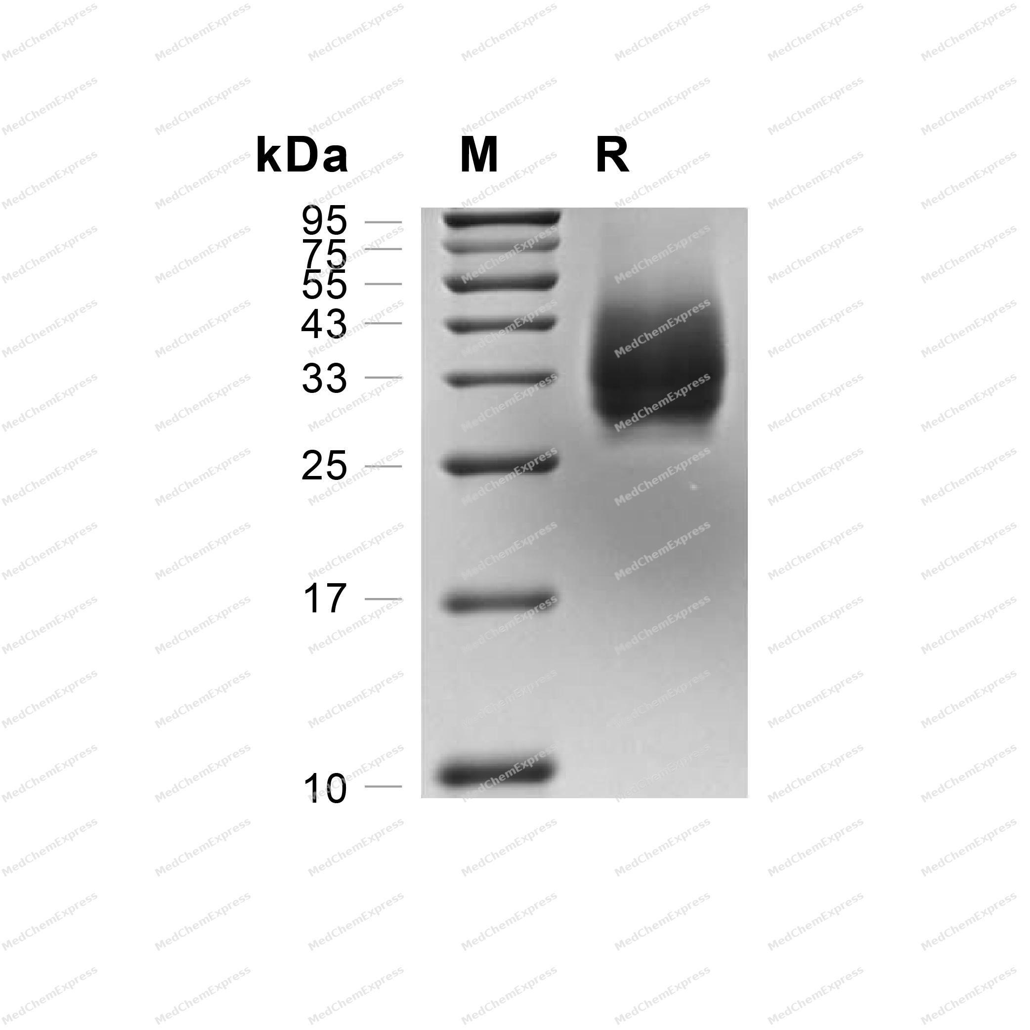
Purity - SDS-PAGE
-
Bioactivity - ELISA
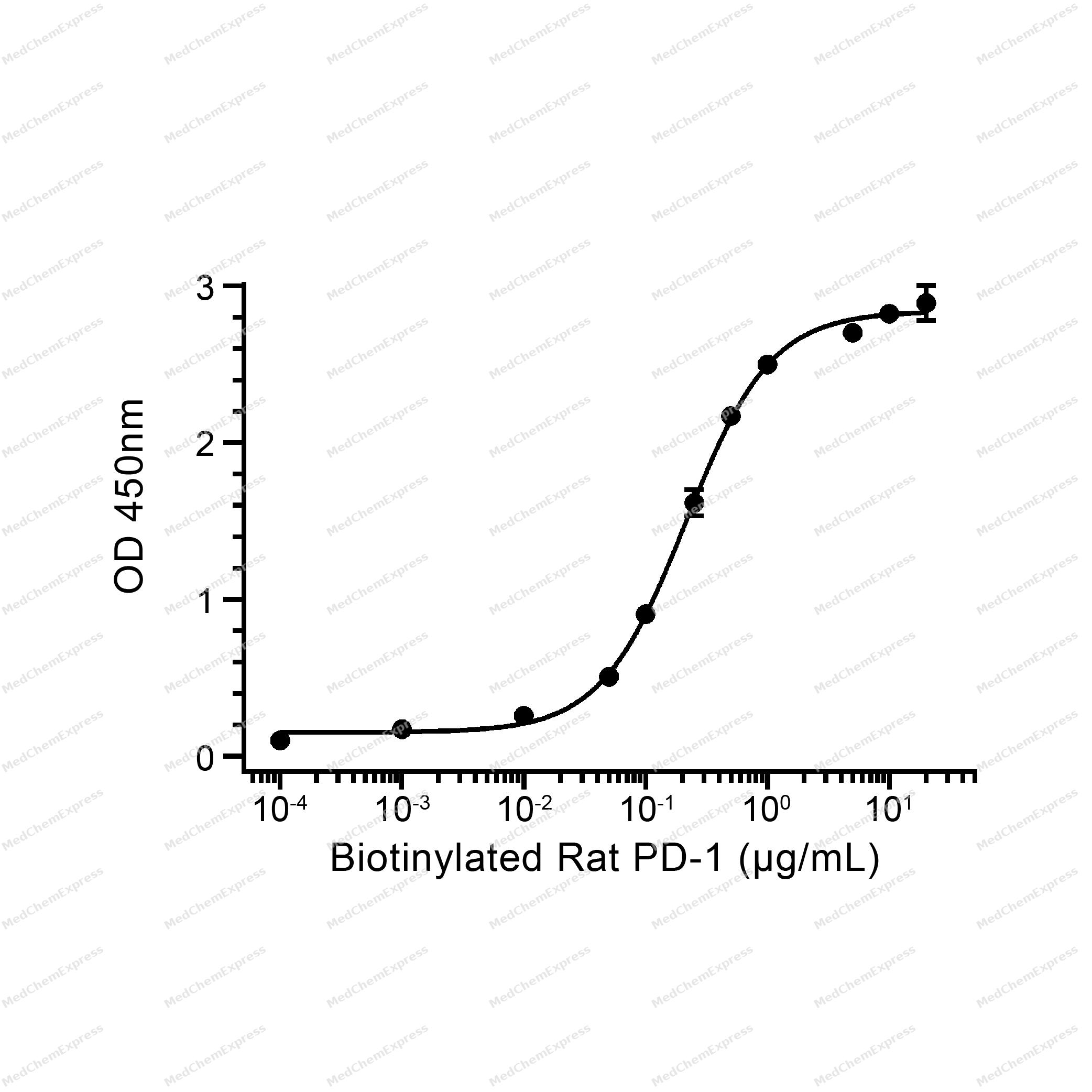
Bioactivity - ELISA


Technical Parameters
-
Species Rat
-
Source HEK293
-
Tag C-His
-
Accession
D4AAV6 (L20-R219)
-
Molecular Construction
-
N-term
-
PD-L2 (L20-R219)
Accession # D4AAV6 -
His
-
C-term
-
-
Protein Length
Partial
-
Synonyms
PDCD1LG2; B7 Dendritic Cell Molecule; Programmed Cell Death 1 Ligand 2; Programmed Death Ligand 2; PD-L2; Butyrophilin B7-DC; CD273; PDCD1 Ligand 2; B7-DC; PDCD1L2; PDL2; PD-1-Ligand 2; B7DC; CD273 Antigen; BA574F11.2; PD-1 Ligand 2; Btdc
-
AA Sequence
LFTVTAPKEVYTVDFGSSVSLECDFDRRECTELEGIRASLQKVENDTSSQSQRATLLEELLPLGKASFHIPSVQVRDSGQYRCLVICGAAWDYKYLTVKVKASYVRIDTGILEVPGTGEVQLICQARGYPLAEVSWQNVSVPANTSHIRTPEGLYQVTSVLRLKPQPNRNFSCMFWNAHMKELTSAIIDPLSWMEPKVPR
-
Molecular Weight
Approximately 36 kDa, based on SDS-PAGE under reducing conditions.
-
Purity
≥ 95%, as determined by reducing SDS-PAGE.


Product Properties
Lyophilized powder
Lyophilized from a 0.2 μm filtered solution of PBS, pH 7.4.
<1 EU/μg, determined by LAL method.
It is not recommended to reconstitute to a concentration less than 100 μg/mL in ddH2O. For long term storage it is recommended to add a carrier protein (0.1% BSA, 5% HSA, 10% FBS or 5% Trehalose).
Stored at -20°C for 2 years from date of receipt. After reconstitution, it is stable at 4°C for 1 week or -20°C for longer (with carrier protein). It is recommended to freeze aliquots at -20°C or -80°C for extended storage.
Room temperature in continental US; may vary elsewhere.


Documentation
-
Data Sheet (264 KB)
-
SDS (251 KB)
- English - EN (251 KB)
- Français - FR (251 KB)
- Deutsch - DE (251 KB)
- Norwegian - NO (251 KB)
- Español - ES (251 KB)
- Swedish - SV (251 KB)
- Italian - IT (251 KB)
- Korean - KR (251 KB)
- Portuguese - PT (251 KB)
-
Handling Instructions (2659 KB)


References
[1]. Bolandi N, et al. The Positive and Negative Immunoregulatory Role of B7 Family: Promising Novel Targets in Gastric Cancer Treatment. Int J Mol Sci. 2021 Oct 3;22(19):10719. [Content Brief]
[2]. Latchman Y, et al. PD-L2 is a second ligand for PD-1 and inhibits T cell activation. Nat Immunol. 2001 Mar;2(3):261-8. [Content Brief]
[3]. Tseng SY, et al. B7-DC, a new dendritic cell molecule with potent costimulatory properties for T cells. J Exp Med. 2001 Apr 2;193(7):839-46. [Content Brief]
[4]. Wang S, et al. Molecular modeling and functional mapping of B7-H1 and B7-DC uncouple costimulatory function from PD-1 interaction. J Exp Med. 2003 May 5;197(9):1083-91. [Content Brief]

Calculators
Concentration (start) × Volume (start) = Concentration (final) × Volume (final)


 Powered by Bioz

