



SDF-1 alpha/CXCL12 Protein, Rat
Based on 2 publication(s) in Google Scholar
SDF-1 alpha (Stromal Cell-Derived Factor-1α, SDF-1α) is a member of the chemokine α subfamily that lack the ELR domain. SDF-1α works as a chemoattractant for T- and B-lymphocytes and monocytes. SDF-1α is a ligand for CXCR4. The SDF-1α/CXCR4 signaling mediates many physiological processes including cell trafficking, angiogenesis, embryogenesis, tumor invasion and metastatic. It also controls the chemotaxis of hematopoietic stem cells homing to the bone marrow. SDF-1 alpha/CXCL12 Protein, Rat is produced in E. coli.
- Species: Rat
- Source: E. coli
-
Storage:Stored at -20°C for 2 years from date of receipt. After reconstitution, it is stable at 4°C for 1 week or -20°C for longer (with carrier protein). It is recommended to freeze aliquots at -20°C or -80°C for extended storage.


To place orders, for customer services and technical support, please contact: MedChemExpress USA
Tel: 609-228-6898 E-mail: [email protected] [email protected]


-
 Biological Activity
-
Technical Parameters
-
Product Properties
- Documentation
- References
- Help & FAQs




Biological Activity
Description
SDF-1 alpha (Stromal Cell-Derived Factor-1α, SDF-1α) is a member of the chemokine α subfamily that lack the ELR domain. SDF-1α works as a chemoattractant for T- and B-lymphocytes and monocytes. SDF-1α is a ligand for CXCR4. The SDF-1α/CXCR4 signaling mediates many physiological processes including cell trafficking, angiogenesis, embryogenesis, tumor invasion and metastatic. It also controls the chemotaxis of hematopoietic stem cells homing to the bone marrow[1][2]. SDF-1 alpha/CXCL12 Protein, Rat is produced in E. coli.
Background
Stromal cell-derived factor-1 (SDF-1), an important member of the chemokine family, is expressed in two subtypes, SDF-1α and SDF-1β, with SDF-1α being the main subtype. SDF-1α is widely present in many tissues and organs of the human body, such as the lymph nodes, bone marrow, liver, lung, muscle, small intestine, kidney, and brain, and can sustainably exist in these organs and tissues. Studies have shown that SDF-1α plays an important role in the physiological mfunctions of migration, distribution, development, differentiation, and apoptosis of various cells. Moreover, SDF-1α plays a key role in the pathological process of some diseases, such as inflammation, tumor formation and metastasis, pathogen infection, and wound repair[1][3].
SDF-1 has three isoforms, α, β, and γ, which are different at the splicing level, not at the transcriptional level. The analysis of the genomic structure of SDF-1 in human and mouse revealed two isoforms, SDF-1α and SDF-1β, which are encoded by a single gene and result from alternative splicing. SDF-1α comprises 3 exons and encodes a protein of 89 amino acids whereas SDF-1β consists of 4 exons and encodes a protein of 93 amino acids. Both isoforms are highly similar regarding their sequences with the only difference of 4 additional amino acids at the C-terminus of SDF1β. In adult rat brain, SDF-1α is the predominant one, present in astrocytes, microglia, as well as in neurons. SDF-1α is found positive in normal cholinergic neurons, such as in the medial septum and substantia innominata, and in dopaminergic neurons, such as in the substantia nigra (SN) pars compacta and the ventral tegmental area. SDF-1α is the only known ligand for CXCR4. CXCR4 is also a target for human immunodeficiency virus (HIV) binding[1][2].
In vitro and in vivo studies using ischemic reperfusion models and a pretreatment with SDF-1α results in decreased infarct size and increases resistance to hypoxic damage and apoptotic cell death via activation of ERK-1/2 and AKT phosphorylation[1]. The SDF-1α/CXCR4 signaling maintains central nervous system homeostasis through the interaction with the neurotransmitter and neuropeptide systems, the neuroendocrine systems[2]. An increasing number of animal experiments have shown that SDF-1α can enhance the migration of BMSCs, mobilize BMSCs to diseased areas, and promote their proliferation and differentiation[3].
In Vitro
Recombinant rat SDF-1α (0, 5, 10, 30, 50 or 100 ng/mL) activates PI-3K/Akt signaling, and consequently promotes the proliferation and chemotaxis in primary rat vascular smooth muscle cells (VSMCs) [4].
In Vivo
Recombinant rat SDF-1α (4 μg/4 μL; intracranial injection) ameliorates TBI-induced brain edema and blood-brain barrier disruption in Sprague-Dawley rats underwent traumatic brain injury (TBI) model. SDF-1α repressed inflammatory responses by inhibiting the expression of pro-inflammatory cytokines, such as TNF-α, IL-1β, and IL-6. SDF-1α attenuated the brain lesion by affecting the ERK and NF-κB signaling pathways after mechanical head trauma in rats[5].
Verified Bioactivity
1. Full biological activity determined by a chemotaxis bioassay using human peripheral blood monocytes is in a concentration range of 50-100 ng/ml.
2. Measured by its ability to inhibit proliferation of Jurkat cells. The ED50 this effect is 3.323 ng/mL, corresponding to a specific activity is 3.01×10⁵ units/mg.
3. Measured by its ability to chemoattract THP-1 human acute monocytic leukemia cells. The ED50 for this effect is ≤71.82 ng/mL, corresponding to a specific activity is ≥1.392×10⁴ U/mg.
MCE Validation Data
-
Purity - SDS-PAGE
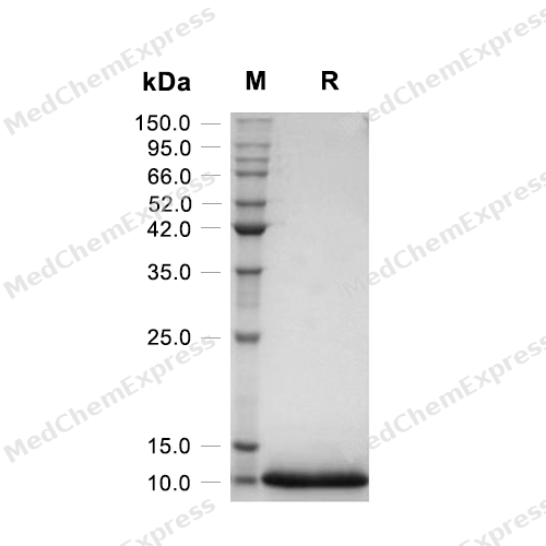
Purity - SDS-PAGE


Publications (2)
-
Journal Impact Factor
-
Most Recent
-
Oxid Med Cell Longev
Rh-CXCL-12 Attenuates Neuronal Pyroptosis after Subarachnoid Hemorrhage in Rats via Regulating the CXCR4/NLRP1 Pathway. [Abstract]2021 Nov 9:2021:6966394. PMID: 34795842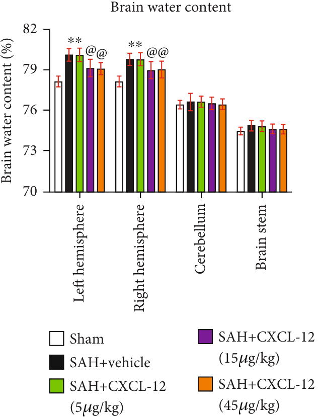
SDF-1 alpha/CXCL12 Protein, Rat purchased from MedChemExpress. Usage Cited in: Oxid Med Cell Longev. 2021 Nov 9:2021:6966394. [Abstract]
SDF-1 alpha/CXCL12 (CXCL-12) (5-45 μg/kg; intranasal administration; total volume 20 μL; 5 μL per nostril alternately every 5 min) Quantification of brain water content in the left hemisphere, right hemisphere, cerebellum, and brain stem at 24 h after SAH.
SDF-1 alpha/CXCL12 Protein, Rat purchased from MedChemExpress. Usage Cited in: Oxid Med Cell Longev. 2021 Nov 9:2021:6966394. [Abstract]
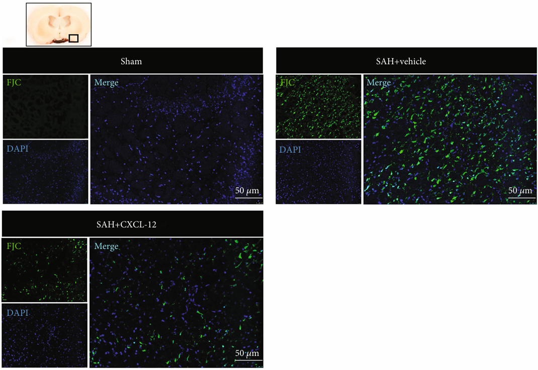
Intranasal administration of rh-CXCL-12 (SDF-1 alpha/CXCL12) (5-45 μg/kg; intranasal administration; total volume 20 μL; 5 μL per nostril alternately every 5 min) attenuated neuronal degeneration at 24 h after SAH. Representative immunofluorescence staining of FJC-positive cells in the ipsilateral basal cortex at 24 h after SAH. Green indicated FJC-positive staining, and blue indicated DAPI-positive nuclear staining. A small black square within the coronal section of the brain indicated the location of where the immunofluorescence staining images were taken.
SDF-1 alpha/CXCL12 Protein, Rat purchased from MedChemExpress. Usage Cited in: Oxid Med Cell Longev. 2021 Nov 9:2021:6966394. [Abstract]
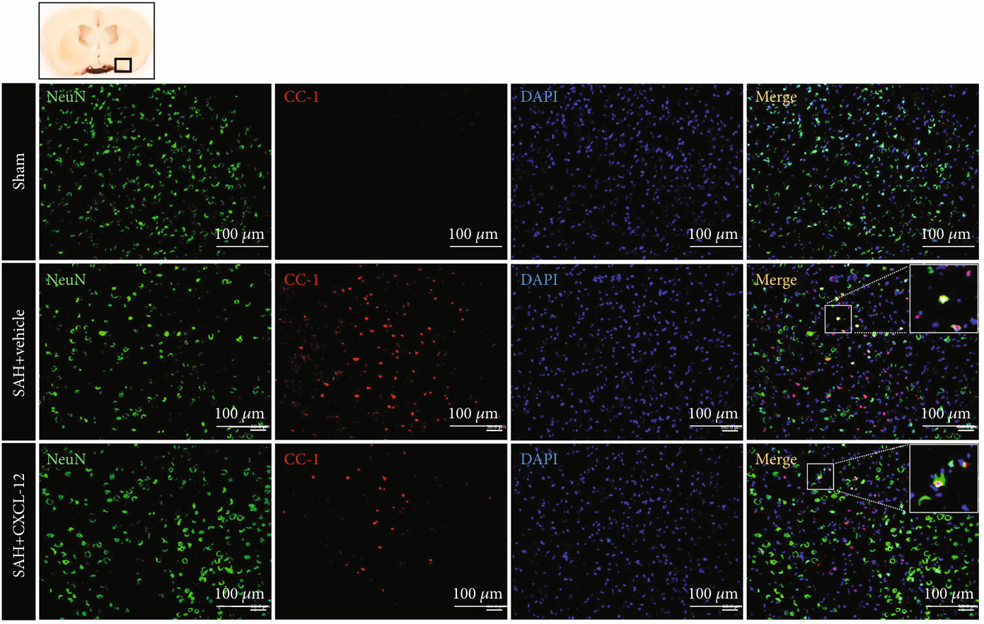
Intranasal administration of rh-CXCL-12 (SDF-1 alpha/CXCL12) (5-45 μg/kg; intranasal administration; total volume 20 μL; 5 μL per nostril alternately every 5 min) attenuated CC-1-positive neurons after SAH. Representative immunofluorescence staining of CC-1-positive neurons in the ipsilateral basal cortex at 24 h after SAH. Green indicated CC1-positive staining, red indicated neurons, and blue indicated DAPI-positive nuclear staining. A small black square within the coronal section of the brain indicated the location of where the immunofluorescence staining images were taken.
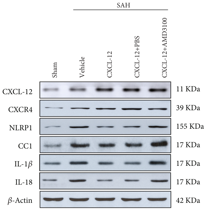
SDF-1 alpha/CXCL12 Protein, Rat purchased from MedChemExpress. Usage Cited in: Oxid Med Cell Longev. 2021 Nov 9:2021:6966394. [Abstract]
Effects of CXCR4 inhibitor on downstream proteins in the proposed signaling pathway with rh-CXCL-12 (SDF-1 alpha/CXCL12) (5-45 μg/kg; intranasal administration; total volume 20 μL; 5 μL per nostril alternately every 5 min) treatment at 24 h after SAH. Representative Western blot bands of the proteins CXCL-12, CXCR4, NLRP1, IL-1β, CC-1, and IL-18.
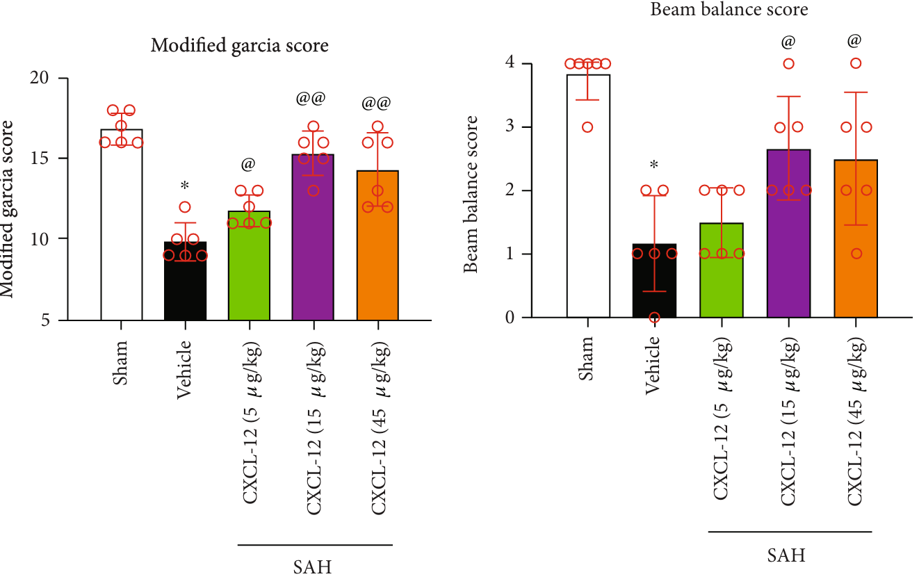
SDF-1 alpha/CXCL12 Protein, Rat purchased from MedChemExpress. Usage Cited in: Oxid Med Cell Longev. 2021 Nov 9:2021:6966394. [Abstract]
Intranasal administration of rh-CXCL-12 (SDF-1 alpha/CXCL12) (5-45 μg/kg; intranasal administration; total volume 20 μL; 5 μL per nostril alternately every 5 min) improved neurological performance on the modified Garcia and beam balance test at 24 h after SAH.
-


Technical Parameters
-
Species Rat
-
Source E. coli
-
Tag Tag Free
-
Accession
Q9QZD1/Q80YV8 (K22-K89)
-
Molecular Construction
-
N-term
-
CXCL12 (K22-K89)
Accession # Q9QZD1 -
C-term
-
-
Protein Length
Full Length of Mature Protein
-
Synonyms
CXCL12; Intercrine Reduced In Hepatomas; Prev. SDF1; SDF-1b; Prev. SDF1A; TLSF-A; Prev. SDF1B; TLSF-B; PBSF; IRH; Stromal Cell-Derived Factor 1; C-X-C Motif Chemokine 12; SCYB12; HSDF-1; TPAR1; SDF-1; Pre-B Cell Growth-Stimulating Factor; TLSF; Chemokine
-
AA Sequence
KPVSLSYRCPCRFFESHVARANVKHLKILNTPNCALQIVARLKSNNRQVCIDPKLKWIQEYLDKALNK
-
Predicted Molecular Mass
7.9 kDa
-
Molecular Weight
Approximately 8 kDa, based on SDS-PAGE under reducing conditions.
-
Purity
≥ 95%, as determined by reducing SDS-PAGE.


Product Properties
Lyophilized powder.
1.Lyophilized from a 0.22 μm filtered solution of 20 mM PB, 150 mM NaCl, pH 7.4.
2.Lyophilized from a 0.22 μm filtered solution of PBS, pH 7.0.
Please refer to the lot-specific COA for specific buffer information.
<1 EU/μg, determined by LAL method.
It is not recommended to reconstitute to a concentration less than 100 μg/mL in ddH2O. For long term storage it is recommended to add a carrier protein (0.1% BSA, 5% HSA, 10% FBS or 5% Trehalose).
Stored at -20°C for 2 years from date of receipt. After reconstitution, it is stable at 4°C for 1 week or -20°C for longer (with carrier protein). It is recommended to freeze aliquots at -20°C or -80°C for extended storage.
Room temperature in continental US; may vary elsewhere.


Documentation
-
Data Sheet (266 KB)
-
SDS (252 KB)
- English - EN (252 KB)
- Français - FR (252 KB)
- Deutsch - DE (252 KB)
- Norwegian - NO (252 KB)
- Español - ES (252 KB)
- Swedish - SV (252 KB)
- Italian - IT (252 KB)
- Korean - KR (252 KB)
- Portuguese - PT (252 KB)
-
Handling Instructions (2659 KB)


References
[1]. Santhosh K Ghadge, et al. SDF-1α as a therapeutic stem cell homing factor in myocardial infarction. Pharmacol Ther. 2011 Jan;129(1):97-108. [Content Brief]
[2]. Kryczek I, et al. Stroma-derived factor (SDF-1/CXCL12) and human tumor pathogenesis. Am J Physiol Cell Physiol. 2007 Mar;292(3):C987-95. [Content Brief]
[3]. Zheng Jiang, et al. Contribution of SDF-1α/CXCR4 signaling to brain development and glioma progression. Neurosignals. 2013;21(3-4):240-58. [Content Brief]
[4]. Zhiqiang Meng, et al. SDF Factor-1α Promotes the Migration, Proliferation, and Osteogenic Differentiation of Mouse Bone Marrow Mesenchymal Stem Cells Through the Wnt/β-Catenin Pathway. Stem Cells Dev. 2021 Jan 15;30(2):106-117. [Content Brief]
[5]. Wei Jie, et al. SDF-1α/CXCR4 axis is involved in glucose-potentiated proliferation and chemotaxis in rat vascular smooth muscle cells. Int J Exp Pathol. 2010 Oct;91(5):436-44. [Content Brief]
[6]. Weifeng Sun, et al. Intracranial injection of recombinant stromal-derived factor-1 alpha (SDF-1α) attenuates traumatic brain injury in rats. Inflamm Res. 2014 Apr;63(4):287-97. [Content Brief]

Calculators
Concentration (start) × Volume (start) = Concentration (final) × Volume (final)


 Powered by Bioz

