



TTT 3002
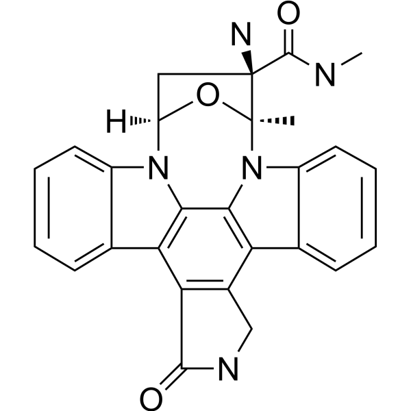
TTT 3002 is a potent and orally active FLT3 inhibitor. TTT 3002 potently inhibits FLT3 phosphorylation by activating mutations at residue D835, with an IC50 of 0.2 nM. TTT 3002 can be used for AML (acute myeloid leukemia) research.
For research use only. We do not sell to patients.
- CAS No.: 871037-95-5
- Formula: C27H23N5O3
- Molecular Weight:465.50
-
Storage:
Please store the product under the recommended conditions in the Certificate of Analysis.

To place orders, for customer services and technical support, please contact: MedChemExpress USA
Tel: 609-228-6898 E-mail: [email protected] [email protected]


-
 Biological Activity
-
Chemical Information
- Protocols
- Purity & Documentation
- References
-
 Help & FAQs


-
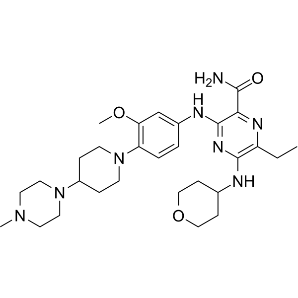
HY-12432
GilteritinibFLT3/AXL Inhibitor -
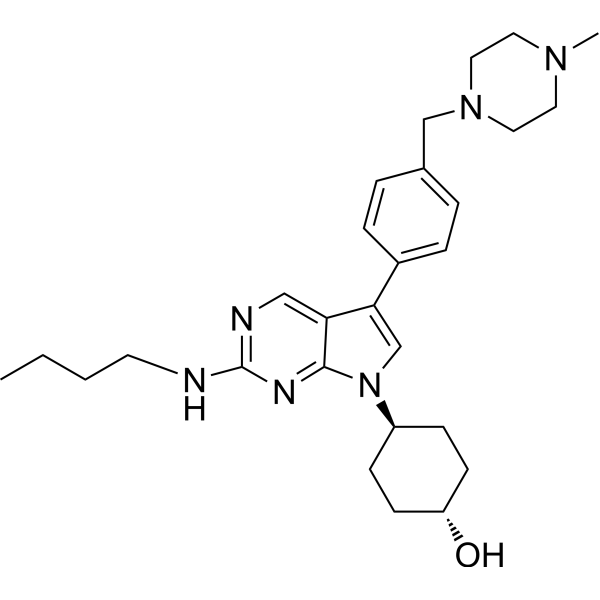
HY-12344
UNC2025MER/FLT3 Inhibitor -
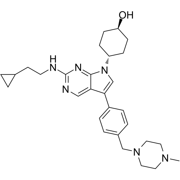
HY-101549
MRX-2843MERTK/FLT3 Inhibitor -

HY-12432R
Gilteritinib (Standard)FLT3/AXL Inhibitor -
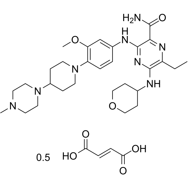
HY-12432A
Gilteritinib hemifumarateFLT3/AXL Inhibitor -
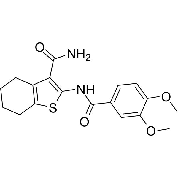
HY-13907
TCS 359FLT3 Inhibitor -
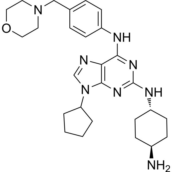
HY-112145
FLT3-IN-3FLT3 Inhibitor -
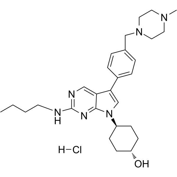
HY-12344A
UNC2025 hydrochlorideMER/FLT3 Inhibitor -
HY-19897
4SC-203Multikinase Inhibitor -
HY-128571
FLT3-IN-4FLT3 Inhibitor -
HY-110026
GTP-14564ITD-FLT3 Inhibitor -
HY-13001
QuizartinibFLT3 Inhibitor


Biological Activity
Description
Cellular Effect
|
Cell Line

|
Type | Value | Description | References |
|---|---|---|---|---|
| MV4-11 | IC50 |
<0.25 nM
Compound: TTT-3002
|
Antiproliferative activity against human MV4-11 cells assessed as reduction in cell proliferation measured after 24 hrs by MTT assay
Antiproliferative activity against human MV4-11 cells assessed as reduction in cell proliferation measured after 24 hrs by MTT assay
|
[PMID: 32659083] |
In Vitro
TTT 3002 downregulates FLT3 phosphorylation (pFLT3) in Molm14 and MV4-11 cells[1].
TTT 3002 induces cell cycle arrest followed by marked induction of apoptosis[1].
MedChemExpress (MCE) has not independently confirmed the accuracy of these methods. They are for reference only. Further protocols information, click here.
-
Cell Line:Molm14 and MV4-11 cells
-
Concentration:0, 0.25, 0.5, 1, 2 nM
-
Incubation Time:1 h
-
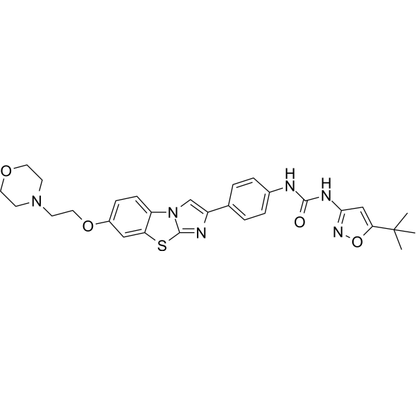
Result:Downregulated FLT3 phosphorylation (pFLT3) in Molm14 and MV4-11 cells in a dose-dependent manner. The IC50 for FLT3 phosphorylation in both cell lines was six- to seven fold lower for TTT 3002 compared with Quizartinib (HY-13001) at 0.2 vs 1.3 nM, respectively.
-
Cell Line:Molm14 and MV4-11 cells
-
Concentration:0, 1, 2, 5, 10 nM
-
Incubation Time:24 h
-
Result:Showed cell cycle arrest followed by marked induction of apoptosis, along with concurrent activation of caspase 3 and poly ADP ribose polymerase cleavage.
In Vivo
TTT 3002 (6 mg/kg, Oral gavage, single) is rapidly absorbed with a biphasic maximum serum concentration (Cmax) followed by a monoexponential decay[1].
MedChemExpress (MCE) has not independently confirmed the accuracy of these methods. They are for reference only.
-
Animal Model:BALB/C mice (female, age 6 to 8 weeks, received Ba/F3-ITD Luc+ cells by tail vein injection on day 0)[1]
-
Dosage:6 mg/kg
-
Administration:Oral gavage, twice per day, for 2 to 4 weeks
-
Result:Showed no significant changes in animal weight and was sufficient to eliminate the presence of Ba/F3-ITD Luc+ cells by day 17 (10 days of treatment).
-
Animal Model:Leukemic engrafted mice (female, age 6 to 8 weeks)[1]
-
Dosage:6 mg/kg
-
Administration:Oral gavage, single (Pharmacokinetic Analysis)
-
Result:After oral administration, TTT 3002 was rapidly absorbed with a biphasic maximum serum concentration (Cmax) followed by a monoexponential decay. The Cmax and area under the concentration-time curve from time 0 to infinity (AUC0→∞) were 613 nM and 3127 nM⋅h, respectively. The half-life, apparent volume of distribution, and apparent clearance were 3.6 hours, 21 L/kg, and 4.1 L/h per kilogram, respectively.


Chemical Information
-
CAS No. 871037-95-5
-
Molecular Weight 465.50
-
Formula C27H23N5O3
-
SMILES
C[C@]12N3C4=C(C5=CC=CC=C53)C(CNC6=O)=C6C7=C4N([C@](C[C@]2(N)C(NC)=O)([H])O1)C8=CC=CC=C78
-
Shipping
Room temperature in continental US; may vary elsewhere.
-
Storage
Please store the product under the recommended conditions in the Certificate of Analysis.

Protocols
-
Kinase activity and phosphorylation assays
Kinase activity assays measure the ability of kinases to transfer phosphate groups from ATP to specific substrates, while phosphorylation assays detect the presence and levels of phosphorylated proteins. Common methods include radiolabeled ATP incorporation (e. g. ,), ADP release detection via bioluminescence (e. g. ,[3]), enzyme-linked immunosorbent assays (ELISA) for phospho-specific epitopes (e. g. ,[6]), and microtiter-based formats for high-throughput screening (e. g. ,[8]). The ADP-Glo assay quantifies kinase activity by measuring ADP produced during phosphorylation using a luciferase-based system. Radiometric assays involve autoradiography or scintillation counting after incorporation of 32P-labeled ATP into substrate proteins. ELISA-based approaches rely on phospho-specific antibodies to detect activated kinases in cell lysates or purified samples.
-
Western Blot
Western blotting (WB) is a commonly used experimental method in molecular biology, biochemistry, and immunogenetics for identifying and quantifying target proteins. It combines gel electrophoresis with immunoassay, enabling researchers to analyze protein expression, post-translational modifications, and molecular weight.
-
Cell Cytotoxicity Assay
Cytotoxicity assays are usually based on the assessment of cell membrane damage, which can also be indirectly detected by measuring cell viability. Detection methods include MTT assay, CKK-8 assay, LDH assay and ATP assay, etc.
-
Protocol for Kinase activity and phosphorylation assays
Kinase activity assays measure transfer of phosphate from ATP to a protein or peptide substrate, generating phosphorylated substrate, ADP, or incorporated radiolabeled phosphate as the readout; phosphorylation assays measure site-specific phosphorylation in cells or tissues as a proxy for kinase-pathway activation, inhibition, or substrate regulation. Phosphorylation can be detected by phospho-specific Western blot, immunoprecipitation kinase assay, phospho-immunofluorescence, phospho-flow cytometry, luminescent ADP detection, radiolabeled ATP incorporation, or reporter-based pathway assays, and these readouts can be applied to cancer cells, primary neurons, mouse tumors, organoids, inflammatory macrophages, ferroptosis studies, and mitophagy studies when the kinase target is biologically relevant.


Purity & Documentation
References

Calculators
Concentration (start) × Volume (start) = Concentration (final) × Volume (final)


TTT 3002 Related Classifications
HY-111249 Related Classifications
 Powered by Bioz