


- Anti-infection
- Conjugué anticorps-médicament/ADC associé
- Apoptose
- Autophagie
- Cycle cellulaire/dommages à l'ADN
- Cytosquelette
- Épigénétique
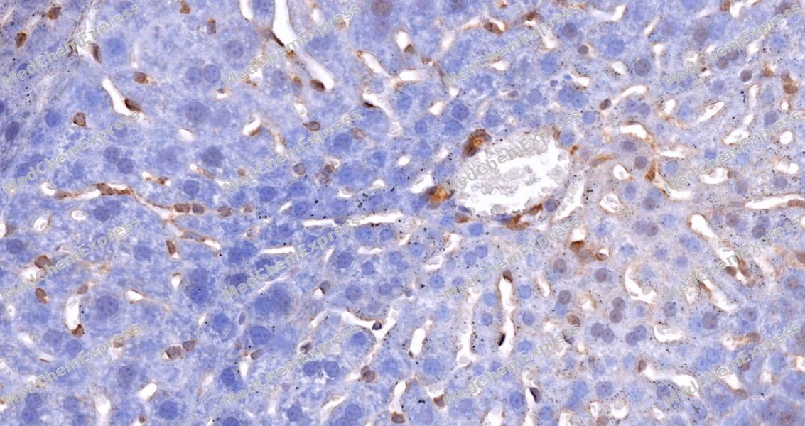
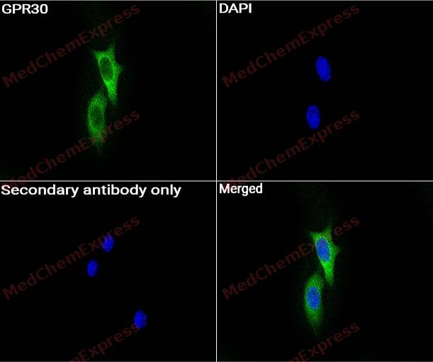
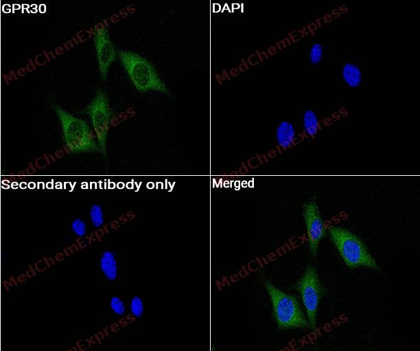
- Protéine GPCR/G
- Immunologie/Inflammation
- Signalisation JAK/STAT
- Voie MAPK/ERK
- Transporteurs membranaires/Canaux ioniques
- Enzyme métabolique/Protéase
- Signalisation neuronale
- NF-κB
- PI3K/Akt/mTOR
- PROTAC
- Protéines Tyrosine Kinase/RTK
- Cellules souches/Wnt
- TGF-bêta/Smad
- Récepteurs nucléaires/lié a la vitamine D
- Autres
Anti-infection
Apoptose
Cycle cellulaire/dommages à l'ADN
Cytosquelette
Épigénétique
Protéine GPCR/G
- Récepteur 5-HT
- Adénylate cyclase
- Adhesion G Protein-coupled Receptors (AGPCRs)
- Récepteur adrénergique
- Récepteur de l'amyline
- Récepteur de l'angiotensine
- Récepteur de l'apeline (APJ)
- Arf Family GTPase
- Arrestine
- Récepteur de la bombésine
- Récepteur de la bradykinine
- Récepteur cannabinoïde
- CaSR
- CCR
- Récepteur CGRP
- Récepteur de la chémérine
- Récepteur de la cholécystokinine
- CRFR
- CXCR
- EBI2/GPR183
Immunologie/Inflammation
Transporteurs membranaires/Canaux ioniques
Enzyme métabolique/Protéase
- 11β-HSD
- 15-PGDH
- 17β-HSD
- 3β-HSD
- 5 alpha réductase
- Acétolactate synthase (ALS)
- Acétyl-CoA carboxylase
- Acetyl-CoA synthetase
- ACSL Family
- Acyltransférase
- ADAMTS
- Récepteur de l'adiponectine
- Aldéhyde déshydrogénase (ALDH)
- Aldehyde Oxidase (AO)
- Aldose réductase
- Amine N-methyltransferase
- Amino Acid Oxidase
- Aminoacyl-ARNt synthétase
- Aminopeptidase
- Aminotransferases (Transaminases)
Signalisation neuronale
- Récepteur 5-HT
- AAK1
- Récepteur adrénergique
- Amino Acid Decarboxylase
- Amyloïde-β
- Bêta-sécrétase
- Calcineurine
- Canal calcique
- CaMK
- Récepteur cannabinoïde
- Récepteur CGRP
- Récepteur de la cholécystokinine
- Choline Kinase
- Cholinestérase (ChE)
- COMT
- Dihydroceramide Desaturase 1 (DES1)
- Dimethylargininase (DDAH)
- FAAH
- Récepteur GABA
- Glucosylcéramide synthase (GCS)
PROTAC
- ATTECs
- AUTACs
- AUTOTACs
- ByeTAC
- DUBTACs
- Conjugués Ligand-Linker pour E3 Ligase
- Hsp-targeting Chimeras
- HyT
- Ligands pour la ligase E3
- Ligands pour la protéine cible pour PROTAC
- LYTACs
- Colles moléculaires
- PhosTACs
- PROTAC Linkers
- Conjugués PROTAC-Linker pour PAC
- PROTACs
- Proteasome Cap Targeting Chimeras
- RIBOTAC
- SNIPERs
- Conjugués Ligand-Linker pour protéines cibles
Protéines Tyrosine Kinase/RTK
Cellules souches/Wnt
Récepteurs nucléaires/lié a la vitamine D
- Récepteur d'androgène
- Récepteur constitutif d'androstane
- Récepteur d'œstrogène/ERR
- Récepteur de glucocorticoïdes
- LXR
- Récepteur de minéralocorticoïdes
- Récepteur d'hormone nucléaire 4A/NR4A
- Orphan Nuclear Receptor
- PPAR
- Pregnane X Receptor (PXR)
- Récepteur de progestérone
- RAR/RXR
- REV-ERB
- ROR
- Récepteur d'hormone thyroïdienne
- VD/VDR
- VKOR
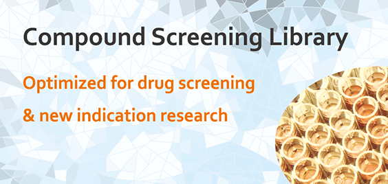
- Chimiothèques de criblage de composés
- Chimiothèques de criblage de composés bioactifs
- •Chimiothèques de composés bioactifs
- Séries de repositionnement de médicaments
- •Chimiothèques de médicaments approuvés par la FDA
- •Chimiothèques de composés pour repositionnement de médicaments
- Séries de produits naturels
- •Chimiothèques de produits naturels
- •Chimiothèques de composés similaires aux produits naturels
- Séries sur le métabolisme
- •Chimiothèques de composés métabolites endogènes humains
- Chimiothèques de composés liés aux maladies
- Séries sur les voies de signalisation
- Chimiothèques de fragments
- Chimiothèques de diversité de composés
- •Chimiothèques de diversité 50K
- •Chimiothèques "Scaffold" 5K
- •Chimiothèques de fragments divers en 3D
- Criblage virtuel
- •Chimiothèques de diversité virtuelle 50K
- •Chimiothèques de diversité virtuelle 10M
- Protéines recombinantes
- Cytokines et facteurs de croissance
- Protéines de points de contrôle immunitaire
- Protéines liées aux CAR-T
- Antigènes CD
- Récepteurs Fc
- Protéines réceptrices
- Enzymes et régulateurs
- Kinases
- Système du complément
- Protéines liées à l'ubiquitine
- Protéines virales
- Protéines biotinylées
- Protéines marquées par fluorescence
- Protéines de qualité GMP
- Protéines recombinantes sans origine animale
- Service d'expression de protéines
- Service de synthèse personnalisée
- Services personnalisés liés aux ADCs
- Services personnalisés liés aux PROTACs
- Chimiothèques de criblage de composés
- Séries de composés bioactifs
- •Chimiothèques de criblage de composés bioactifs
- •Séries de repositionnement de médicaments
- •Séries sur le métabolisme
- •Selon les caractéristiques du produit
- •Selon les structures
- •Selon les voies de signalisation ou les familles de protéines
- •Chimiothèques de composés liés aux maladies
- •Services de criblage des récepteurs nucléaires
- •Spectrométrie de masse d'affinité
- •Synthèse et criblage DEL
- •Criblage de médicaments piloté par IA
- •Service d'analyse d'interactions moléculaires
- •Service d'identification des cibles médicamenteuses
- Optimisation de chef de file
- Équipements et consommables
- Cytokines et facteurs de croissance
- Protéines de points de contrôle immunitaire
- Protéines liées aux CAR-T
- Antigènes CD
- Récepteurs Fc
- Protéines réceptrices
- Enzymes et régulateurs
- Kinases
- Système du complément
- Protéines liées à l'ubiquitine
- Protéines virales
- Protéines biotinylées
- Protéines marquées par fluorescence
- Protéines de qualité GMP
- Protéines recombinantes sans origine animale
- Autres
- Voir plus
Protéines liées aux CAR-T
- CD4
- CD19
- Ligand CD27/CD70
- CD123
- CD138/Syndecan-1
- Molécule d'adhésion aux cellules épithéliales (EpCAM)
- Récepteur de folate 1
- GPC-3
- GuanylateCyclase 2C
- ErbB2/HER2
- ErbB3/HER3
- c-Met/HGFR
- MSLN
- CA-125
- ROR1
- CEACAM-5
- Prostate Specific Membrane Antigen
- TROP-2
- Siglec-6
- Folate Receptor alpha (FR-alpha)
- CD314/NKG2D
- CD319/SLAMF7
- Siglec-3/CD33
- CD7
- MUC-1/CD227
Antigènes CD
- Protéines CD des cellules T
- Protéines CD des cellules B
- Protéines CD des cellules NK
- Protéines CD des macrophages
- Protéines CD des monocytes
- Protéines CD des cellules souches
- Protéines CD des plaquettes
- Protéines CD des érythrocytes
- Protéines CD des cellules dendritiques
- Protéines CD des cellules épithéliales
- Protéines CD des cellules endothéliales
- Protéines CD liées à la transduction du signal
- Protéines CD liées à l'adhésion cellulaire
Protéines réceptrices
- Récepteurs tyrosine kinases
- Récepteurs Sérine/Thréonine Kinases
- Récepteurs Tyrosine Phosphatase
- Récepteurs Famille Guanylyl Cyclase
- Molécules d'adhésion cellulaire (CAM)
- Récepteurs couplés aux protéines G (GPCR)
- Superfamille des récepteurs nucléaires
- Récepteurs de reconnaissance de formes
- Famille Notch
- Siglec
- Récepteurs de type immunoglobine leucocytaire
- Récepteurs de type immunoglobuline des cellules tueuses
- Récepteurs de cytokines
Enzymes et régulateurs
- Oxydoréductases (EC 1)
- Transférases (EC 2)
- Hydrolases (EC 3)
- Lyases (EC 4)
- Isomérases (EC 5)
- Ligases (EC 6)
- Translocases (EC 7)
- Métalloprotéases matricielles
- ADAM/ADAMTS
- Cathepsine
- Carboxypeptidase
- Enzymes de conversion de l'angiotensine
- Caspase
- Carbonic Anhydrase
- Serine/Threonine Kinase Proteins
- Protein Tyrosine Kinases
- Phosphatase
- Topoisomerase
- Protease Inhibitors
- Protein Kinase Inhibitor Peptide (PKI)
- Cyclin-Dependent Kinase Inhibitor Proteins
- Cystatin Family
Système du complément
- Composante complémentaire 1
- Composante complémentaire 2
- Composante complémentaire 3
- Composante complémentaire 4
- Composante complémentaire 5
- Composante complémentaire 6
- Composante complémentaire 7
- Composante complémentaire 8
- Protéine de liaison au mannose
- MASP-1
- MASP-2
- Protéines régulatrices du complément
- Récepteur du complément
- Complement Component 9
- Biologie moléculaire
- •Électrophorèse d'acides nucléiques sur gel
- •Construction de vecteur
- •Endonucléases de restriction
- •Matériels
- •PCR et qPCR
- •RT-PCR
- •Séquençage
- Biologie des protéines
- •Préparation d'échantillons de protéines
- •Purification des protéines
- •Électrophorèse des protéines et WB
- •Multiple Fluorescent Staining
- •Immunoassay
- •Immunoprecipitation Kit
- •Labeling Kit
- Biologie cellulaire
- •Culture cellulaire
- •Analyse des cellules
- •Culture cellulaire 3D
- •Isolement cellulaire
- Voir plus
Culture cellulaire
- Transfection cellulaire
- Élimination des mycoplasmes
- Solution antibiotique stérile
- Congélation des cellules
- Cocktail CEPT
- Milieux de culture cellulaire de base
- Solutions salines équilibrées
- Suppléments de culture cellulaire
- Réactifs de dissociation
- Cell Separation Media
- Specialized Cell Culture Medium
- Lentivirus
Analyse des cellules
- Prolifération cellulaire et cytotoxicité
- Test de gène rapporteur
- Conjugués de colorant phalloïdine
- Apoptose et nécrose cellulaires
- Milieu de montage anti-fade
- Isolement mitochondrial
- Isolement, purification et détection d'exosomes
- Recherche sur les organites
- Kit d'essai du métabolisme
- Mycoplasma Detection
- Biochemical Assay Kit
- Endotoxin Detection
- Staining Kit
- Système de management de la qualité
- Service de synthèse personnalisé
- Service de synthèse en vrac et personnalisé
- Services personnalisés liés aux ADC
- Services personnalisés liés aux PROTAC
- Produits standards de référence personnalisés
- Synthèse personnalisée de peptides
- Service d'expression protéique
- Service d'expression danticorps recombinants
- Élucidation de la structure cristalline des protéines
- Synthèse d'oligonucléotides
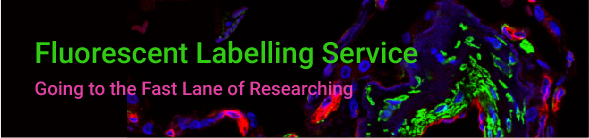
- Service de marquage fluorescent
- Synthèse personnalisée de composés marqués aux isotopes stables
- Service CDMO tout-en-un
- Plateforme de criblage de composés tout-en-un
- Criblage virtuel
- Simulation de dynamique moléculaire
- Criblage de composés sur cellules
- Criblage de canaux ioniques
- Service de criblage de kinases
- Service d'analyse SPR
- Services de criblage bioassay GPCR
- Services de criblage des récepteurs nucléaires
- Spectrométrie de masse d'affinité
- Synthèse et criblage DEL
- Service d'analyse d'interactions moléculaires
- Service d'identification des cibles médicamenteuses
- Criblage de médicaments piloté par IA
- Calculateur de molarité
- Calculateur de dilution


 Powered by Bioz


