



Cathepsin D Protein, Human (HEK293, His, solution)
Based on 1 publication(s) in Google Scholar
Cathepsin D Protein, Human (HEK293, His, solution) is an approximately 50.0 kDa cathepsin D protein with a His-flag. Cathepsin D is a representative lysosomal aspartic proteinases and belongs to the peptidase C1 family.
- Species: Human
- Source: HEK293
-
Storage:Stored at -80°C for 1 year from date of receipt. It is stable at -20°C for 3 months after opening. It is recommended to freeze aliquots at -80°C for extended storage. Avoid repeated freeze-thaw cycles.


To place orders, for customer services and technical support, please contact: MedChemExpress USA
Tel: 609-228-6898 E-mail: [email protected] [email protected]


-
 Biological Activity
-
Technical Parameters
-
Product Properties
- Documentation
- References
- Help & FAQs




Biological Activity
Description
Cathepsin D Protein, Human (HEK293, His, solution) is an approximately 50.0 kDa cathepsin D protein with a His-flag. Cathepsin D is a representative lysosomal aspartic proteinases and belongs to the peptidase C1 family[1].
Background
Cathepsin D (CTSD), a well-known lysosomal aspartyl protease and belongs to the peptidase C1 family, which is a normal and major component of lysosomes, and is found in almost all cells and tissues of mammals[1].Human cathepsin D is synthesized as a precursor protein, consisting of a signal peptide (residues 1-18), a propeptide (residues 19 64), and a mature chain (residues (65 412))[2].
Verified Bioactivity
Measured by its ability to cleave the fluorogenic peptide substrate, Mca-PLGL-Dpa-AR-NH2. The specific activity is >600 pmol/min/µg, as measured under the described conditions.
MCE Validation Data
-
Purity - SDS-PAGE
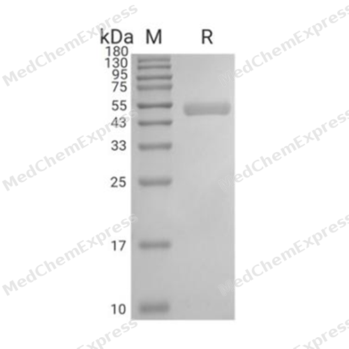
Purity - SDS-PAGE

Assay Procedure
Materials
Detection buffer: 0.1 M NaOAc, 0.2 M NaCl, pH 3.5
Cathepsin D Protein, Human (HEK293, His, solution) (HY-P7748)
Substrate: MOCAc-PLGL(Dpa)AR (HY-131498), 2 mM dissolved in DMSO
Standard: MCA-Pro-Leu-OH
Procedure
1. Standard Curve Preparation: Dilute the standard with the detection buffer to concentrations of 100, 50, 25, 12.5, 6.25, 3.125, 1.5625, and 0 μmol/L. Add 100 μL of each standard concentration to a 96-well plate. Read the plate in kinetic mode for 5 minutes with excitation and emission wavelengths set at 320 nm and 405 nm (top read), respectively. Plot the standard concentrations on the x-axis and the measured RFU (Relative Fluorescence Units) on the y-axis to generate the standard curve and obtain the standard curve equation.
2. Dilute the protein sample to 20 μg/mL with the detection buffer.
3. Incubate at 37℃ for 30 minutes.
4. Dilute the incubated protein sample to 1 μg/mL with the detection buffer.
5. Dilute the substrate to 60 μM with the detection buffer.
6. Add 50 μL of the diluted protein sample to the 96-well plate and initiate the reaction by adding 50 μL of 60 μM substrate. For the blank wells, add 50 μL of detection buffer and 50 μL of 60 μM substrate.
7. Read the plate in kinetic mode for 5 minutes with excitation and emission wavelengths set at 320 nm and 405 nm, respectively.
8. Calculate the specific activity:
Specific Activity (pmol/min/μg) = | Adjusted Vmax* (RFU/min) x Conversion Factor ** (pmol/RFU) |
| amount of enzyme (μg) |
*Adjusted for Control
**Derived using calibration standard
Per Well:
Human Cathepsin D: 0.05 μg
Substrate: 30 μM


Publications (1)
-
Journal Impact Factor
-
Most Recent
-
Adv Sci (Weinh)
Lysosomal Cathepsin S Escape Facilitates Near Infrared Light-Triggered Pyroptosis Via an Antibody-Indocyanine Green Conjugate. [Abstract]2025 Sep;12(34):e04851. PMID: 40539828


Technical Parameters
-
Species Human
-
Source HEK293
-
Tag C-6*His
-
Accession
P07339 (L21-L412)
-
Molecular Construction
-
N-term
-
Cathepsin D (L21-L412)
Accession # P07339 -
6*His
-
C-term
-
-
Protein Length
Full Length of Mature Protein (with Propeptide)
-
Synonyms
CTSD; Cathepsin D (Lysosomal Aspartyl Protease); Prev. CPSD; Lysosomal Aspartyl Protease; CLN10; Cathepsin D Isoform 2; Ceroid-Lipofuscinosis, Neuronal 10; HEL-S-130P; Epididymis Secretory Sperm Binding Protein Li 130P; Cathepsin D; Lysosomal Aspartyl Pep
-
AA Sequence
LVRIPLHKFTSIRRTMSEVGGSVEDLIAKGPVSKYSQAVPAVTEGPIPEVLKNYMDAQYYGEIGIGTPPQCFTVVFDTGSSNLWVPSIHCKLLDIACWIHHKYNSDKSSTYVKNGTSFDIHYGSGSLSGYLSQDTVSVPCQSASSASALGGVKVERQVFGEATKQPGITFIAAKFDGILGMAYPRISVNNVLPVFDNLMQQKLVDQNIFSFYLSRDPDAQPGGELMLGGTDSKYYKGSLSYLNVTRKAYWQVHLDQVEVASGLTLCKEGCEAIVDTGTSLMVGPVDEVRELQKAIGAVPLIQGEYMIPCEKVSTLPAITLKLGGKGYKLSPEDYTLKVSQAGKTLCLSGFMGMDIPPPSGPLWILGDVFIGRYYTVFDRDNNRVGFAEAARL
-
Predicted Molecular Mass
43.6 kDa
-
Molecular Weight
Approximately 50 kDa, based on SDS-PAGE under reducing conditions.
-
Glycosylation
Yes
-
Purity
≥ 95%, as determined by reducing SDS-PAGE.


Product Properties
Solution
Supplied as a 0.22 μm filter solution of 20 mM MES, 150 mM NaCl, pH 5.5.
<1 EU/μg, determined by LAL method.
Stored at -80°C for 1 year from date of receipt. It is stable at -20°C for 3 months after opening. It is recommended to freeze aliquots at -80°C for extended storage. Avoid repeated freeze-thaw cycles.
Shipping with dry ice.


Documentation
-
Data Sheet (263 KB)
-
SDS (252 KB)
- English - EN (252 KB)
- Français - FR (252 KB)
- Deutsch - DE (252 KB)
- Norwegian - NO (252 KB)
- Español - ES (252 KB)
- Swedish - SV (252 KB)
- Italian - IT (252 KB)
- Korean - KR (252 KB)
- Portuguese - PT (252 KB)
-
Handling Instructions (2659 KB)


References
[1]. T Tsukuba, et al. New functional aspects of cathepsin D and cathepsin E. Mol Cells. 2000 Dec 31;10(6):601-11. [Content Brief]
[2]. P L Faust, et al. Cloning and sequence analysis of cDNA for human cathepsin D. Proc Natl Acad Sci U S A. 1985 Aug;82(15):4910-4. [Content Brief]
[3]. B Redecker, et al. Molecular organization of the human cathepsin D gene. DNA Cell Biol [Content Brief]
[4]. T Tsukuba, et al. New functional aspects of cathepsin D and cathepsin E. Mol Cells [Content Brief]

Calculators
Concentration (start) × Volume (start) = Concentration (final) × Volume (final)


 Powered by Bioz

