



CD3D Protein, Mouse (HEK293, Fc)

Based on 1 Customer Validation
The CD3D protein is an important component of the TCR-CD3 complex on T lymphocytes and is critical for adaptive immune responses. After APC activates TCR, CD3D, together with CD3E, CD3G and CD3Z, transmits signals through ITAM and activates downstream pathways. CD3D Protein, Mouse (HEK293, Fc) is the recombinant mouse-derived CD3D protein, expressed by HEK293 , with C-hFc labeled tag.
- Species: Mouse
- Source: HEK293
-
Storage:Stored at -20°C for 2 years from date of receipt. After reconstitution, it is stable at 4°C for 1 week or -20°C for longer (with carrier protein). It is recommended to freeze aliquots at -20°C or -80°C for extended storage.


To place orders, for customer services and technical support, please contact: MedChemExpress USA
Tel: 609-228-6898 E-mail: [email protected] [email protected]


-
 Biological Activity
-
Technical Parameters
-
Product Properties
- Documentation
- Help & FAQs




Biological Activity
Description
The CD3D protein is an important component of the TCR-CD3 complex on T lymphocytes and is critical for adaptive immune responses. After APC activates TCR, CD3D, together with CD3E, CD3G and CD3Z, transmits signals through ITAM and activates downstream pathways. CD3D Protein, Mouse (HEK293, Fc) is the recombinant mouse-derived CD3D protein, expressed by HEK293 , with C-hFc labeled tag.
Background
The CD3D protein is a crucial component of the TCR-CD3 complex found on the surface of T-lymphocytes, playing a pivotal role in adaptive immune responses. Upon activation of the T-cell receptor (TCR) by antigen-presenting cells (APCs), CD3D, along with CD3E, CD3G, and CD3Z, transmits TCR-mediated signals across the cell membrane. These CD3 chains contain immunoreceptor tyrosine-based activation motifs (ITAMs) in their cytoplasmic domain, which, upon phosphorylation by LCK and FYN kinases, activate downstream signaling pathways. Beyond its role in signal transduction for T-cell activation, CD3D is indispensable in thymocyte differentiation, contributing to proper intracellular TCR-CD3 complex assembly and surface expression. Dysfunction in the TCR-CD3 complex leads to impaired thymocyte differentiation. CD3D further interacts with CD4 and CD8, establishing a functional link between the TCR and coreceptors CD4 and CD8, crucial for the activation and positive selection of CD4 or CD8 T-cells. The TCR-CD3 complex consists of CD3D/CD3E and CD3G/CD3E heterodimers, forming trimers that associate with TCRalpha and TCRbeta. Additionally, the hexamer interacts with CD3Z homodimer to complete the TCR-CD3 complex, wherein TCRalpha and TCRbeta can be replaced by TCRgamma and TCRdelta. This intricate interaction network highlights the multifaceted role of CD3D in orchestrating T-cell responses.
Verified Bioactivity
Immobilized Recombinant Human CD3 epsilon Protein at 10 µg/mL (100 µL/well) can bind Biotinylated Mouse CD3D protein. The ED50 for this effect is 4.477 μg/mL.
MCE Validation Data
-
Purity - SDS-PAGE
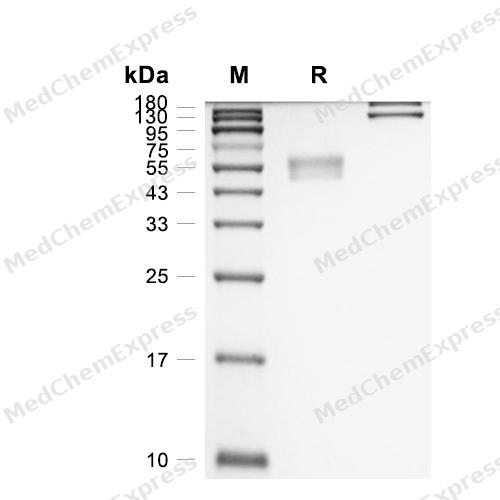
Purity - SDS-PAGE
-
Bioactivity - ELISA
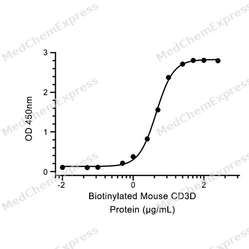
Bioactivity - ELISA


Technical Parameters
-
Species Mouse
-
Source HEK293
-
Tag C-hFc
-
Accession
P04235 (F22-A105)
-
Molecular Construction
-
N-term
-
CD3D (M1-A105)
Accession # P04235 -
hFc
-
C-term
-
-
Protein Length
Extracellular Domain
-
Synonyms
CD3D; CD3d Molecule, Delta (CD3-TCR Complex); Prev. T3D; CD3 Antigen, Delta Subunit; T-Cell Surface Glycoprotein CD3 Delta Chain; OKT3, Delta Chain; T-Cell Receptor T3 Delta Chain; CD3d Molecule; CD3-DELTA; CD3d Antigen; CD3DELTA; IMD19; CD3d Antigen, Del
-
AA Sequence
FKIQVTEYEDKVFVTCNTSVMHLDGTVEGWFAKNKTLNLGKGVLDPRGIYLCNGTEQLAKVVSSVQVHYRMCQNCVELDSGTMA
-
Molecular Weight
Approximately 44-48 kDa, based on SDS-PAGE under reducing conditions, due to the glycosylation.
-
Glycosylation
Yes
-
Purity
≥ 95%, as determined by reducing SDS-PAGE.


Product Properties
Lyophilized powder.
Lyophilized from a 0.22 μm filtered solution of PBS, pH 7.4.
<1 EU/μg, determined by LAL method.
It is not recommended to reconstitute to a concentration less than 100 μg/mL in ddH2O. For long term storage it is recommended to add a carrier protein (0.1% BSA, 5% HSA, 10% FBS or 5% Trehalose).
Stored at -20°C for 2 years from date of receipt. After reconstitution, it is stable at 4°C for 1 week or -20°C for longer (with carrier protein). It is recommended to freeze aliquots at -20°C or -80°C for extended storage.
Room temperature in continental US; may vary elsewhere.


Documentation
-
Data Sheet (236 KB)
-
SDS (252 KB)
- English - EN (252 KB)
- Français - FR (252 KB)
- Deutsch - DE (252 KB)
- Norwegian - NO (252 KB)
- Español - ES (252 KB)
- Swedish - SV (252 KB)
- Italian - IT (252 KB)
- Korean - KR (252 KB)
- Portuguese - PT (252 KB)
-
Handling Instructions (2659 KB)

Calculators
Concentration (start) × Volume (start) = Concentration (final) × Volume (final)


 Powered by Bioz

