



FSH Protein, Human (HEK293, Flag-His)
Based on 1 publication(s) in Google Scholar
Follicle-stimulating hormone (FSH) is a glycoprotein dimer polypeptide hormone produced by the anterior pituitary in response to gonadotropin-releasing hormone (GnRH) from the hypothalamus, which consists of Glycoprotein hormones α chain and Follitropin subunit β. FSH binds to FSHR, a G protein-coupled receptor, on target cells to activate downstream signaling pathways. FSH is involved in follicle development and spermatogenesis in reproductive organs. FSH Protein, Human (HEK293, Flag-His) is a recombinant protein with a Flag-His label that consisting of 116 amino acids of Glycoprotein hormones α chain and 129 amino acids of Follitropin subunit β, which is expressed in HEK293 cells.
- Species: Human
- Source: HEK293
-
Storage:Stored at -20°C for 2 years from date of receipt. After reconstitution, it is stable at 4°C for 1 week or -20°C for longer (with carrier protein). It is recommended to freeze aliquots at -20°C or -80°C for extended storage.


To place orders, for customer services and technical support, please contact: MedChemExpress USA
Tel: 609-228-6898 E-mail: [email protected] [email protected]


-
 Biological Activity
-
Technical Parameters
-
Product Properties
- Documentation
- References
- Help & FAQs


-

HY-P7109
EGF Protein, Human -
HY-P1777
IGF-I (24-41) -
HY-P70311
Activin A Protein, Human/Mouse/Rat (HEK293) -
HY-P7007
BMP-4 Protein, Human -
HY-P2293
Luteinizing hormone (human) -

HY-P1237
C-Type Natriuretic Peptide (CNP) (1-22), human -
HY-P73053
FGF-9 Protein, Human (HEK293, Fc, solution) -
HY-17394
Cisplatin -
HY-P7331
FGF basic/bFGF Protein, Human (154a.a) -
HY-P74133
FSH Protein, Human (HEK293, Fc) -
HY-P70578
Amphiregulin Protein, Human


Biological Activity
Description
Follicle-stimulating hormone (FSH) is a glycoprotein dimer polypeptide hormone produced by the anterior pituitary in response to gonadotropin-releasing hormone (GnRH) from the hypothalamus, which consists of Glycoprotein hormones α chain and Follitropin subunit β. FSH binds to FSHR, a G protein-coupled receptor, on target cells to activate downstream signaling pathways. FSH is involved in follicle development and spermatogenesis in reproductive organs. FSH Protein, Human (HEK293, Flag-His) is a recombinant protein with a Flag-His label that consisting of 116 amino acids of Glycoprotein hormones α chain and 129 amino acids of Follitropin subunit β, which is expressed in HEK293 cells[1][2][3][4][5][6][7][8][9].
Background
FSH Protein (Human) is a glycoprotein dimer with α and β subunits. The β subunit is unique to FSH, while the α subunit is shared as same as in thyroid stimulating hormone (TSH), choriogonadotropin (CG) and luteinizing hormone (LH)[1].
Follicle growth is directly dependent on FSH Protein (Human) stimulation. Granulosa cells from small follicles express FSH Protein (Human) receptors and respond to FSH Protein (Human) stimulation by synthesizing aromatase[1].
FSH Protein (Human) release is stimulated by gonadotropin-releasing hormone (GnRH): The hypothalamus produces GnRH, and it is released into the hypophyseal portal circulation to act on G-protein-coupled receptors at gonadotropic cells of the anterior pituitary. Gonadotropic cells produce FSH and luteinizing hormone (LH) and release them into the peripheral circulation[4].
FSH Protein (Human) regulates the development, growth, pubertal maturation and reproductive processes of the human body. FSH Protein (Human) is used commonly in infertility therapy, mainly for ovarian hyperstimulation. In some cases, it is used in ovulation induction for reversal of anovulation as well[5].
FSH Protein (Human) secretion is inhibited by negative feedback from estrogen levels in women and FSH Protein (Human) secretion is inhibited by levels of inhibin B (secreted by the Sertoli cells) via negative feedback in men[5,6].
FSH Protein (Human) Levels are associated with various diseases, such as Polycystic Ovarian Syndrome, male infertility, hypogonadotropic hypogonadism, Kallman Syndrome, Turner Syndrome, pituitary adenomas, and more[1,7,8,9].
In Vitro
The serum level of FSH Protein (Human) (13.7, 24.1 and 69 IU/L) increases as ovarian function declines, which acts a better predictor of in vitro fertilization (IVF) performance than age[3].
In Vivo
The plasma concentration of FSH Protein (Human) is profoundly reduced when a variety of α-adrenergic blocking agents interrupts the pulsatile discharges in ovariectomized rhesus monkeys, which suggests the production and secretion of FSH are critically dependent on hypothalamic GnRH[3].
Verified Bioactivity
1. Measured in a cell proliferation assay using SK-OV-3 cells. The ED50 for this effect is 0.048 ng/mL, corresponding to a specific activity is 2.083×10⁷ units/mg.
2. Human FSHR immobilized on CM5 Chip can bind FSH with an affinity constant of 102.9 nM as determined in a SPR assay.
3.Measured by its ability to induce cAMP accumulation in HEK293 human embryonic kidney cells transfected with human FSH R. The ED50 for this effect is <200 pg/mL.
MCE Validation Data
-
Purity - SDS-PAGE
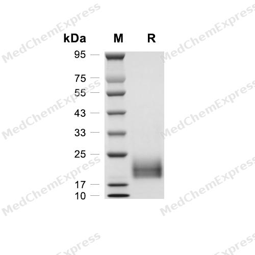
Purity - SDS-PAGE
-
Bioactivity - Cell-Based Assay
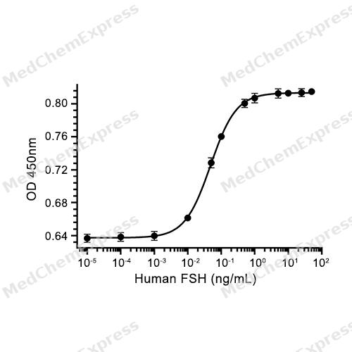
Bioactivity - Cell-Based Assay


Publications (1)
-
Journal Impact Factor
-
Most Recent
-
Adv Healthc Mater
Tuning the Testicular Microenvironment for Enhancing Human Sertoli Cells Maturation and Functionality In Vitro. [Abstract]2026 Jun;15(22):e05848. PMID: 42027040


Technical Parameters
-
Species Human
-
Source HEK293
-
Tag C-Flag;C-6*His
-
Synonyms
CGA; Anterior Pituitary Glycoprotein Hormones Common Subunit Alpha; Glycoprotein Hormones, Alpha Polypeptide; Follicle-Stimulating Hormone Alpha Subunit; Glycoprotein Hormones Alpha Chain; Chorionic Gonadotropin, Alpha Polypeptide; GPA1; Follicle-Stimulat
-
AA Sequence
APDVQDCPECTLQENPFFSQPGAPILQCMGCCFSRAYPTPLRSKKTMLVQKNVTSESTCCVAKSYNRVTVMGGFKVENHTACHCSTCYYHKS&NSCELTNITIAIEKEECRFCISINTTWCAGYCYTRDLVYKDPARPKIQKTCTFKELVYETVRVPGCAHHADSLYTYPVATQCHCGKCDSDSTDCTVRGLGPSYCSFGEMKE
-
Molecular Weight
Approximately 20-30 kDa, based on SDS-PAGE under reducing conditions.
-
Glycosylation
Yes
-
Purity
≥ 95%, as determined by reducing SDS-PAGE.


Product Properties
Lyophilized powder
1.Lyophilized from a 0.22 μm filtered solution of PBS, pH 7.4.
2.Lyophilized from a 0.22 μm filtered solution of PBS, pH 7.4, 8% trehalose.
Please refer to the lot-specific COA for specific buffer information.
<1 EU/μg, determined by LAL method.
It is not recommended to reconstitute to a concentration less than 100 μg/mL in ddH2O. For long term storage it is recommended to add a carrier protein (0.1% BSA, 5% HSA, 10% FBS or 5% Trehalose).
Stored at -20°C for 2 years from date of receipt. After reconstitution, it is stable at 4°C for 1 week or -20°C for longer (with carrier protein). It is recommended to freeze aliquots at -20°C or -80°C for extended storage.
Room temperature in continental US; may vary elsewhere.


Documentation
-
Data Sheet (266 KB)
-
SDS (254 KB)
- English - EN (254 KB)
- Français - FR (254 KB)
- Deutsch - DE (254 KB)
- Norwegian - NO (254 KB)
- Español - ES (254 KB)
- Swedish - SV (254 KB)
- Italian - IT (254 KB)
- Korean - KR (254 KB)
- Portuguese - PT (254 KB)
-
Handling Instructions (2659 KB)


References
[1]. Barbieri RL. The endocrinology of the menstrual cycle. Methods Mol Biol. 2014;1154:145-69. [Content Brief]
[2]. Toner JP, et al. Basal follicle-stimulating hormone level is a better predictor of in vitro fertilization performance than age. Fertil Steril. 1991 Apr;55(4):784-91. [Content Brief]
[3]. Knobil E. The neuroendocrine control of the menstrual cycle. Recent Prog Horm Res. 1980;36:53-88. [Content Brief]
[4]. Yonkers KA, et al. Premenstrual disorders. Am J Obstet Gynecol. 2018 Jan;218(1):68-74. [Content Brief]
[5]. Shaw ND, et al. Estrogen negative feedback on gonadotropin secretion: evidence for a direct pituitary effect in women. J Clin Endocrinol Metab. 2010 Apr;95(4):1955-61. [Content Brief]
[6]. Boepple PA, et al. Relative roles of inhibin B and sex steroids in the negative feedback regulation of follicle-stimulating hormone in men across the full spectrum of seminiferous epithelium function. J Clin Endocrinol Metab. 2008 May;93(5):1809-14. [Content Brief]
[7]. Nieschlag E, et al. Role of FSH in the regulation of spermatogenesis: clinical aspects. Clin Endocrinol (Oxf). 1999 Aug;51(2):139-46. [Content Brief]
[8]. Stamatiades GA, et al. Gonadotropin regulation by pulsatile GnRH: Signaling and gene expression. Mol Cell Endocrinol. 2018 Mar 5;463:131-141. [Content Brief]
[9]. Pouresmaeili F, et al. Premature ovarian failure: a critical condition in the reproductive potential with various genetic causes. Int J Fertil Steril. 2014 Apr;8(1):1-12. [Content Brief]

Calculators
Concentration (start) × Volume (start) = Concentration (final) × Volume (final)


 Powered by Bioz

