


3-TYP
Based on 105 publication(s) in Google Scholar
3-TYP is an inhibitor of SIRT3 (IC50of 38 μM) and an inhibitor of Methionine Adenosyltransferase (MAT) 2 and Indoleamine 2,3-Dioxygenase (IDO). There may be many off-target sites for 3-TYP that need to be examined, such as NAD-dependent enzymes, including dehydrogenases.
For research use only. We do not sell to patients.
- Purity: 99.90%
- CAS No.: 120241-79-4
- Formula: C7H6N4
- Molecular Weight:146.15
-
Storage:Powder -20°C, 3 years , 4°C, 2 years ; In solvent -80°C, 6 months , -20°C, 1 month

To place orders, for customer services and technical support, please contact: MedChemExpress USA
Tel: 609-228-6898 E-mail: [email protected] [email protected]


-
 Biological Activity
-
Chemical Information
-
Solvent & Solubility
- Protocol
- Purity & Documentation
- References
-
 Help & FAQs
-


Cell Cycle/DNA Damage Compound Library
HY-L004
-
Epigenetics Compound Library
HY-L005
-
Immunology/Inflammation Compound Library
HY-L007
-
Metabolism/Protease Compound Library
HY-L012
-
Histone Modification Research Compound Library
HY-L024
-
Anti-Cancer Compound Library
HY-L025
-
Small Molecule Immuno-Oncology Compound Library
HY-L031
-
Anti-Aging Compound Library
HY-L034
-
Antioxidant Compound Library
HY-L037
-
Reprogramming Compound Library
HY-L039
-
Oxygen Sensing Compound Library
HY-L045
-
Glutamine Metabolism Compound Library
HY-L064
-
Targeted Diversity Library
HY-L099
-
Heterocyclic Compound Library
HY-L138
-
Metabolic Enzyme Compound Library
HY-L146
-
Highly Selective Inhibitors Library
HY-L158
-
Multi-Target Compound Library
HY-L176
-
Radioprotector Library
HY-L178
-
Bioactive Compound Library Max
HY-L181
-
Anti-Aging Compound Library Mini
HY-L034M
-
Epigenetics Compound Library Mini
HY-L005M
-
MCE Bioactive Compound Library
HY-L001V
-
Bioactive Compound Library
HY-L001
-
Amino Acid Metabolism Compound Library
HY-L189
-
Non-Alcoholic Fatty Liver Disease (NAFLD) Compound Library
HY-L199
-
Cell Proliferation Compound Library
HY-L201
-
High-Throughput Bioactive Compound Library
HY-L205


Publications Citing Use of MedChemExpress (MCE) 3-TYP
More- Nat Immunol. 2023 Jan;24(1):162-173. [Abstract]
- Bone Res. 2025 Mar 14;13(1):36. [Abstract]
- J Adv Res. 2025 Nov 14:S2090-1232(25)00922-1. [Abstract]
- J Adv Res. 2025 Feb 1:S2090-1232(25)00062-1. [Abstract]
- Redox Biol. 2025 Sep:85:103751. [Abstract]
- Adv Sci (Weinh). 2025 Dec 16:e10368. [Abstract]
- Adv Sci (Weinh). 2025 Oct 14:e11734. [Abstract]
- Cell Death Dis. 2021 Sep 13;12(9):847. [Abstract]
- Cell Death Dis. 2021 May 18;12(6):501. [Abstract]
- Int J Biol Sci. 2026 Feb 4;22(5):2435-2451. [Abstract]
- Phytomedicine. 2026 Apr:153:157985. [Abstract]
- Phytomedicine. 2026 Jan 20:153:157860. [Abstract]
- Phytomedicine. 2025 May:140:156568. [Abstract]
- Phytomedicine. 2025 Jan:136:156260. [Abstract]
- Phytomedicine. 2024 Mar:125:155353. [Abstract]
- Phytomedicine. 2023 Jan:109:154556. [Abstract]
- Phytomedicine. 2020 Mar:68:153171. [Abstract]
- Acta Pharmacol Sin. 2022 Feb;43(2):457-469. [Abstract]
- Diabetes. 2023 May 1;72(5):611-626. [Abstract]
- Apoptosis. 2025 Apr;30(3-4):768-783. [Abstract]
- Int J Biol Macromol. 2025 Aug 5;322(Pt 1):146571. [Abstract]
- Int J Biol Macromol. 2025 Jul 5;320(Pt 1):145750. [Abstract]
- J Headache Pain. 2023 Jun 5;24(1):65. [Abstract]
- Int J Mol Med. 2026 Jun;57(6):162. [Abstract]
- Mol Med. 2025 Apr 30;31(1):161. [Abstract]
- Mol Med. 2024 Sep 19;30(1):154. [Abstract]
- Mol Med. 2024 Sep 12;30(1):148. [Abstract]
- Antioxidants (Basel). 2024 Aug 22;13(8):1023. [Abstract]
- Free Radic Biol Med. 2026 Aug 16:252:664-676. [Abstract]
- Free Radic Biol Med. 2026 Jun 8:254:31-45. [Abstract]
- Free Radic Biol Med. 2026 Jun:249:292-307. [Abstract]
- Free Radic Biol Med. 2024 Jan:210:378-389. [Abstract]
- Free Radic Biol Med. 2023 Oct:207:32-44. [Abstract]
- Free Radic Biol Med. 2023 Mar:198:59-67. [Abstract]
- Free Radic Biol Med. 2022 Jul:187:158-170. [Abstract]
- Cell Rep. 2025 Jan 28;44(1):115136. [Abstract]
- Br J Pharmacol. 2025 Mar 31. [Abstract]
- Br J Pharmacol. 2025 Apr 13. [Abstract]
- Chin Med. 2025 Dec 10;20(1):215. [Abstract]
- Redox Rep. 2026 Dec;31(1):2622255. [Abstract]
- J Agric Food Chem. 2023 Nov 1;71(43):16032-16042. [Abstract]
- Eur J Med Chem. 2020 Apr 15:192:112201. [Abstract]
- Cell Mol Life Sci. 2024 Dec 27;82(1):30. [Abstract]
- J Chem Inf Model. 2023 Aug 14;63(15):4780-4790. [Abstract]
- Food Funct. 2024 May 7;15(9):4954-4969. [Abstract]
- Food Funct. 2021 Sep 7;12(17):8026-8036. [Abstract]
- Food Funct. 2020 Mar 26;11(3):2535-2542. [Abstract]
- Food Biosci. 2025 Jan.
- Drug Des Devel Ther. 2026 Feb 25:20:578815. [Abstract]
- Nutrients. 2023 Jun 30;15(13):2973. [Abstract]
- Nutrients. 2023 Apr 23;15(9):2036. [Abstract]
- Nutrients. 2023 Jan 10;15(2):355. [Abstract]
- Cell Biol Toxicol. 2024 May 20;40(1):31. [Abstract]
- Eur J Pharmacol. 2021 Aug 15:905:174186. [Abstract]
- Int Immunopharmacol. 2026 Jan 15:169:116019. [Abstract]
- Int Immunopharmacol. 2025 Oct 30:164:115377. [Abstract]
- Biomolecules. 2024 May 18;14(5):598. [Abstract]
- Int Immunopharmacol. 2024 Mar 10:129:111636. [Abstract]
- Int Immunopharmacol. 2022 May:106:108600. [Abstract]
- Inflammation. 2021 Oct;44(5):1782-1792. [Abstract]
- Front Cell Dev Biol. 2021 Nov 18:9:766512. [Abstract]
- Chem Biol Interact. 2026 Jul 1:434:112089. [Abstract]
- J Integr Med. 2026 Jan 28:S2095-4964(26)00008-7. [Abstract]
- Chem Biol Interact. 2025 Nov 1:421:111756. [Abstract]
- Biochim Biophys Acta Mol Basis Dis. 2024 Oct;1870(7):167433. [Abstract]
- Sci Rep. 2025 Sep 29;15(1):33699. [Abstract]
- Cell Signal. 2025 Jul:131:111765. [Abstract]
- J Inflamm Res. 2024 Oct 24:17:7675-7685. [Abstract]
- BMC Complement Med Ther. 2024 Jan 9;24(1):29. [Abstract]
- FEBS J. 2023 Jul;290(14):3629-3645. [Abstract]
- Fish Shellfish Immunol. 2022 Oct:129:85-95. [Abstract]
- Microbiol Spectr. 2024 Aug 6;12(8):e0074924. [Abstract]
- ACS Pharmacol Transl Sci. 2026 Apr 3;9(5):1134-1152. [Abstract]
- J Funct Foods. August 2022, 105178.
- Nutr Metab. 2021 Sep 9;18(1):83. [Abstract]
- Toxicol Appl Pharmacol. 2024 Oct:491:117066. [Abstract]
- Photochem Photobiol Sci. 2025 Feb;24(2):293-306. [Abstract]
- Neural Plast. 2022 Jan 19:2022:5567174. [Abstract]
- Exp Eye Res. 2026 Jun:267:110960. [Abstract]
- Exp Eye Res. 2025 Jun:255:110345. [Abstract]
- Exp Eye Res. 2025 May:254:110328. [Abstract]
- Am J Cancer Res. 2022 May 15;12(5):2310-2322. [Abstract]
- J Membr Biol. 2025 Apr;258(2):123-133. [Abstract]
- Biochem Biophys Res Commun. 2025 Jul 12:770:151957. [Abstract]
- Cell Physiol Biochem. 2017;44(6):2212-2227. [Abstract]
- J Food Biochem. 2022 Sep 20;e14428. [Abstract]
- J Food Biochem. 2022 Mar;46(3):e13820. [Abstract]
- Neuroreport. 2025 Jul 2;36(10):514-523. [Abstract]
- Exp Anim. 2023 Aug 7;72(3):346-355. [Abstract]
- Curr Med Sci. 2022 Oct;42(5):981-990. [Abstract]
- Biomed Pharmacother. 2025 May:186:118043. [Abstract]
- Heliyon. 2024 Oct 5;10(21):e39031. [Abstract]
- Res Sq. 2024 Jun 09.
- Biomed Pharmacother. 2023 Nov:167:115618. [Abstract]
- Oxid Med Cell Longev. 2023 Feb 17:2023:1708251. [Abstract]
- Ann Transl Med. 2023 Jan 31;11(2):72. [Abstract]
- Oxid Med Cell Longev. 2023 Jan 14:2023:9966355. [Abstract]
- Oxid Med Cell Longev. 2022 Sep 20;2022:2213503. [Abstract]
- Research Square Print. 2022 Aug.
- Research Square Preprint. 2022 Jul.
- Oxid Med Cell Longev. 2021 Nov 9;2021:5577019. [Abstract]
- Research Square Preprint. 2021 Aug.
- Aging (Albany NY). 2021 Jun 11;13(12):16105-16123. [Abstract]
- Research Square Preprint. 2021 Mar.
- Aging (Albany NY). 2020 Apr 14;12(7):6415-6435. [Abstract]
 Customer Validation & Images

-
WB
-
IHC

-
WB
-
WB


Biological Activity
|
SIRT3 38 μM (IC50) |
Methionine Aminopeptidase 2 |
|
Cell Line
|
Type | Value | Description | References |
|---|---|---|---|---|
| HeLa | IC50 |
4.7 μM
Compound: 2
|
Cytotoxicity against human HeLa cells assessed as cell death after 1 to 6 hrs
Cytotoxicity against human HeLa cells assessed as cell death after 1 to 6 hrs
|
[PMID: 22835719] |
| HL-60 | IC50 |
>50 μM
Compound: 3-TYP
|
Antiproliferative activity against human HL-60 cells assessed as inhibition of cell growth incubated for 24 hrs by MTT assay
Antiproliferative activity against human HL-60 cells assessed as inhibition of cell growth incubated for 24 hrs by MTT assay
|
[PMID: 39053191] |
| MOLM-13 | IC50 |
12.55 μM
Compound: 3-TYP
|
Antiproliferative activity against human MOLM-13 cells assessed as inhibition of cell growth incubated for 24 hrs by MTT assay
Antiproliferative activity against human MOLM-13 cells assessed as inhibition of cell growth incubated for 24 hrs by MTT assay
|
[PMID: 39053191] |
| MV4-11 | IC50 |
27.76 μM
Compound: 3-TYP
|
Antiproliferative activity against human MV4-11 cells assessed as inhibition of cell growth incubated for 24 hrs by MTT assay
Antiproliferative activity against human MV4-11 cells assessed as inhibition of cell growth incubated for 24 hrs by MTT assay
|
[PMID: 39053191] |
3-TYP inhibits melatonin-enhanced SIRT3 activity but does not affect SIRT3 protein expression. 3-TYP pretreatment reverses the protective effects of melatonin on cadmium (Cd)-induced mitochondrial-derived O2- production and autophagic cell death. 3-TYP significantly attenuates melatonin-induced increases in deacetylated-SOD2 expression and SOD2 activity in HepG2 cells exposed to Cd[1].
MedChemExpress (MCE) has not independently confirmed the accuracy of these methods. They are for reference only.
MedChemExpress (MCE) has not independently confirmed the accuracy of these methods. They are for reference only.


Chemical Information
-
CAS No. 120241-79-4
-
Appearance Solid
-
Molecular Weight 146.15
-
Formula C7H6N4
-
Color White to off-white
-
SMILES
C1(C2=CN=NN2)=CC=CN=C1
-
Shipping
Room temperature in continental US; may vary elsewhere.
-
Storage
Powder -20°C 3 years 4°C 2 years In solvent -80°C 6 months -20°C 1 month


Publications (105)
-
Journal Impact Factor
-
Most Recent
-
Nat Immunol
Ammonia detoxification promotes CD8+ T cell memory development by urea and citrulline cycles. [Abstract]2023 Jan;24(1):162-173. PMID: 36471170
3-TYP purchased from MedChemExpress. Usage Cited in: Nat Immunol. 2023 Jan;24(1):162-173. [Abstract]
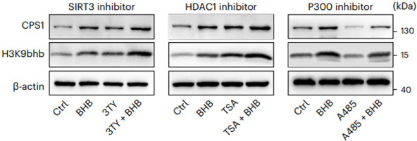
A485 (10 μM; 24 h) leads to the downregulation of H3K9bhb and CPS1 in BHB-treated CD8+ TM cells; however, the use of 3-TYP (10 μM; 24 h) or Trichostatin A (TSA; 5 nM; 24 h) result in the increase of H3K9bhb and CPS1.
-
Bone Res
SIRT3-PINK1-PKM2 axis prevents osteoarthritis via mitochondrial renewal and metabolic switch. [Abstract]2025 Mar 14;13(1):36. PMID: 40087281 -
J Adv Res
SIRT3-activating, biodegradable poly-honokiol with high drug loading for thoracic aortic dissection therapy. [Abstract]2025 Nov 14:S2090-1232(25)00922-1. PMID: 41242496 -
J Adv Res
Period3 modulates the NAD+-SIRT3 axis to alleviate depression-like behaviour by enhancing NAMPT activity in mice. [Abstract]2025 Feb 1:S2090-1232(25)00062-1. PMID: 39894345 -
Redox Biol
2025 Sep:85:103751. PMID: 40628122 -
Adv Sci (Weinh)
Skeletal Muscle HSF1 Alleviates Age-Associated Sarcopenia and Mitochondrial Function Decline via SIRT3-PGC1α Axis. [Abstract]2025 Dec 16:e10368. PMID: 41400028 -
Adv Sci (Weinh)
Punicalagin Enhances Autophagy Through Sirtuin 1/FoxO3a Axis to Inhibit Intracellular Mycobacterium Abscessus Infection. [Abstract]2025 Oct 14:e11734. PMID: 41085014 -
Cell Death Dis
Sirtuin 3 regulates mitochondrial protein acetylation and metabolism in tubular epithelial cells during renal fibrosis. [Abstract]2021 Sep 13;12(9):847. PMID: 34518519 -
Cell Death Dis
IL-4 inhibits regulatory T cells differentiation by HDAC9-mediated epigenetic regulation. [Abstract]2021 May 18;12(6):501. PMID: 34006836 -
Int J Biol Sci
DNA Polymerase Gamma Acetylation Governs Mitochondrial Homeostasis and Vascular Cell Senescence. [Abstract]2026 Feb 4;22(5):2435-2451. PMID: 41800262 -
Phytomedicine
Kakkalide promotes spinal cord injury repair by regulating microglial M2 polarization via mitophagy. [Abstract]2026 Apr:153:157985. PMID: 41720005 -
Phytomedicine
Anti-tumor effects of Guggulsterone in osteosarcoma: Role of SIRT3-mediated PINK1-Parkin mitophagy activation. [Abstract]2026 Jan 20:153:157860. PMID: 41650525 -
Phytomedicine
Effective compound combination of Bufei Yishen formula ameliorates PM2.5-induced COPD by inhibiting mitochondrial oxidative stress through SIRT3-mediated FOXO3 deacetylation. [Abstract]2025 May:140:156568. PMID: 40056638 -
Phytomedicine
Salvianolic acid B alleviated myocardial ischemia-reperfusion injury via modulating SIRT3-mediated crosstalk between mitochondrial ROS and NLRP3. [Abstract]2025 Jan:136:156260. PMID: 39579610 -
Phytomedicine
Isolation of Calenduloside E from Achyranthes bidentata Blume and its effects on LPS/D-GalN-induced acute liver injury in mice by regulating the AMPK-SIRT3 signaling pathway. [Abstract]2024 Mar:125:155353. PMID: 38241918 -
Phytomedicine
Chinese herbal medicine alleviates the pathogenesis of polycystic ovary syndrome by improving oxidative stress and glucose metabolism via mitochondrial Sirtuin 3 signaling. [Abstract]2023 Jan:109:154556. PMID: 36610149 -
Phytomedicine
High content screening identifies licoisoflavone A as a bioactive compound of Tongmaiyangxin Pills to restrain cardiomyocyte hypertrophy via activating Sirt3. [Abstract]2020 Mar:68:153171. PMID: 32018211 -
Acta Pharmacol Sin
2022 Feb;43(2):457-469. PMID: 33850273 -
Diabetes
Metrnl alleviates lipid accumulation by modulating mitochondrial homeostasis in diabetic nephropathy. [Abstract]2023 May 1;72(5):611-626. PMID: 36812572 -
Apoptosis
Brazilin alleviates acute lung injury via inhibition of ferroptosis through the SIRT3/GPX4 pathway. [Abstract]2025 Apr;30(3-4):768-783. PMID: 39720978 -
Int J Biol Macromol
KAT8-mediated MDH2 lactylation promotes renal cancer progression by enhancing mitochondrial function and stress resistance. [Abstract]2025 Aug 5;322(Pt 1):146571. PMID: 40769364 -
Int J Biol Macromol
Hesperidin mitigates copper nanoparticle exposure-induced mitochondrial unfolded protein response through the SIRT3-FOXO3A signaling pathway. [Abstract]2025 Jul 5;320(Pt 1):145750. PMID: 40617440 -
J Headache Pain
SS-31 alleviated nociceptive responses and restored mitochondrial function in a headache mouse model via Sirt3/Pgc-1α positive feedback loop. [Abstract]2023 Jun 5;24(1):65. PMID: 37271805 -
Int J Mol Med
IDH2 lactylation regulates mitochondrial dysfunction injury induced by myocardial ischemia‑reperfusion via the AMPK signaling pathway. [Abstract]2026 Jun;57(6):162. PMID: 41992974 -
Mol Med
Surfactant protein D alleviates chondrocytes senescence by upregulating SIRT3/SOD2 pathway in osteoarthritis. [Abstract]2025 Apr 30;31(1):161. PMID: 40307686 -
Mol Med
ACE2 deficiency inhibits thoracic aortic dissection by enhancing SIRT3 mediated inhibition of inflammation and VSCMs phenotypic switch. [Abstract]2024 Sep 19;30(1):154. PMID: 39300372 -
Mol Med
Capsaicin mitigates ventilator-induced lung injury by suppressing ferroptosis and maintaining mitochondrial redox homeostasis through SIRT3-dependent mechanisms. [Abstract]2024 Sep 12;30(1):148. PMID: 39266965 -
Antioxidants (Basel)
A Multi-Target Pharmacological Correction of a Lipoyltransferase LIPT1 Gene Mutation in Patient-Derived Cellular Models. [Abstract]2024 Aug 22;13(8):1023. PMID: 39199267 -
Free Radic Biol Med
Vitamin D receptor/sirtuin 3 pathway mediates the cardioprotective effects of aerobic exercise in diabetic mice. [Abstract]2026 Aug 16:252:664-676. PMID: 42061479 -
Free Radic Biol Med
Thiomyristoyl promotes type 2 diabetic wound healing and inhibits scarring via the PPARγ/Sirt3/SOD2 axis. [Abstract]2026 Jun 8:254:31-45. PMID: 42264197 -
Free Radic Biol Med
Cornuside alleviates microglia-mediated neuroinflammation to ameliorate chronic neuropathic pain-induced cognitive impairment via the Nrf2/Sirt3 signaling pathway. [Abstract]2026 Jun:249:292-307. PMID: 41831801 -
Free Radic Biol Med
Sentrin-specific protease 1 maintains mitochondrial homeostasis through targeting the deSUMOylation of sirtuin-3 to alleviate oxidative damage induced by hepatic ischemia/reperfusion. [Abstract]2024 Jan:210:378-389. PMID: 38052275 -
Free Radic Biol Med
DMT1 differentially regulates mitochondrial complex activities to reduce glutathione loss and mitigate ferroptosis. [Abstract]2023 Oct:207:32-44. PMID: 37419216 -
Free Radic Biol Med
Sirt3-dependent regulation of mitochondrial oxidative stress and apoptosis contributes to the dysfunction of pancreatic islets after severe burns. [Abstract]2023 Mar:198:59-67. PMID: 36738799 -
Free Radic Biol Med
NAC alleviative ferroptosis in diabetic nephropathy via maintaining mitochondrial redox homeostasis through activating SIRT3-SOD2/Gpx4 pathway. [Abstract]2022 Jul:187:158-170. PMID: 35660452 -
Cell Rep
2025 Jan 28;44(1):115136. PMID: 39932192 -
Br J Pharmacol
Rhynchophylline as an agonist of sirtuin 3 ameliorates endothelial dysfunction via antagonizing mitochondrial damage of endothelial progenitor cells. [Abstract]2025 Mar 31. PMID: 40164963 -
Br J Pharmacol
Berberine partially ameliorates cardiolipotoxicity in diabetic cardiomyopathy by modulating SIRT3-mediated lipophagy to remodel lipid droplets homeostasis. [Abstract]2025 Apr 13. PMID: 40222752 -
Chin Med
Fat-targeted small molecule alleviates abnormal adipose tissue remodeling in obesity via SIRT3-driven mitophagy and inflammasome inhibition. [Abstract]2025 Dec 10;20(1):215. PMID: 41372934 -
Redox Rep
Urolithin A alleviates vascular remodeling through mitochondrial SIRT3-mediated SOD2 deacetylation and antioxidation in hypertensive rats. [Abstract]2026 Dec;31(1):2622255. PMID: 41645805 -
J Agric Food Chem
5-Heptadecylresorcinol Ameliorates Obesity-Associated Skeletal Muscle Mitochondrial Dysfunction through SIRT3-Mediated Mitophagy. [Abstract]2023 Nov 1;71(43):16032-16042. PMID: 37862266 -
Eur J Med Chem
2020 Apr 15:192:112201. PMID: 32163813 -
Cell Mol Life Sci
Endothelial CD38-induced endothelial-to-mesenchymal transition is a pivotal driver in pulmonary fibrosis. [Abstract]2024 Dec 27;82(1):30. PMID: 39725783 -
J Chem Inf Model
Discovery of Novel SIRT1/2 Inhibitors with Effective Cytotoxicity against Human Leukemia Cells. [Abstract]2023 Aug 14;63(15):4780-4790. PMID: 37486605 -
Food Funct
Cynarin alleviates acetaminophen-induced acute liver injury through the activation of Keap1/Nrf2-mediated lipid peroxidation defense via the AMPK/SIRT3 signaling pathway. [Abstract]2024 May 7;15(9):4954-4969. PMID: 38602356 -
Food Funct
Pine nut antioxidant peptides ameliorate the memory impairment in a scopolamine-induced mouse model via SIRT3-induced synaptic plasticity. [Abstract]2021 Sep 7;12(17):8026-8036. PMID: 34269783 -
Food Funct
5-Heptadecylresorcinol attenuates oxidative damage and mitochondria-mediated apoptosis through activation of the SIRT3/FOXO3a signaling pathway in neurocytes. [Abstract]2020 Mar 26;11(3):2535-2542. PMID: 32141452 -
-
Drug Des Devel Ther
(+)-JQ1 Upregulates SIRT3 to Suppress cGAS/STING Pathway-Mediated Neuronal Inflammation and Ferroptosis After Hypoxic-Ischemic Encephalopathy. [Abstract]2026 Feb 25:20:578815. PMID: 41778146 -
Nutrients
Piceatannol Protects PC-12 Cells against Oxidative Damage and Mitochondrial Dysfunction by Inhibiting Autophagy via SIRT3 Pathway. [Abstract]2023 Jun 30;15(13):2973. PMID: 37447299 -
Nutrients
Pterostilbene Attenuates High-Intensity Swimming Exercise-Induced Glucose Absorption Dysfunction Associated with the Inhibition of NLRP3 Inflammasome-Induced IECs Pyroptosis. [Abstract]2023 Apr 23;15(9):2036. PMID: 37432144 -
Nutrients
Dihydromyricetin Protects Intestinal Barrier Integrity by Promoting IL-22 Expression in ILC3s through the AMPK/SIRT3/STAT3 Signaling Pathway. [Abstract]2023 Jan 10;15(2):355. PMID: 36678226 -
Cell Biol Toxicol
SIRT1 restores mitochondrial structure and function in rats by activating SIRT3 after cerebral ischemia/reperfusion injury. [Abstract]2024 May 20;40(1):31. PMID: 38767771 -
Eur J Pharmacol
2021 Aug 15:905:174186. PMID: 34033817 -
Int Immunopharmacol
Melatonin alleviates atherosclerosis by inhibiting pro-inflammatory differentiation of macrophages via regulating Sirt3-Drp1 mediated mitochondrial fission. [Abstract]2026 Jan 15:169:116019. PMID: 41389666 -
Int Immunopharmacol
SIRT3 attenuates sepsis-induced EndMT and cardiac remodeling by facilitating mitophagy process via PINK1/Parkin signaling. [Abstract]2025 Oct 30:164:115377. PMID: 40840139 -
Biomolecules
Polydatin and Nicotinamide Rescue the Cellular Phenotype of Mitochondrial Diseases by Mitochondrial Unfolded Protein Response (mtUPR) Activation. [Abstract]2024 May 18;14(5):598. PMID: 38786005 -
Int Immunopharmacol
Oroxylin A suppress LL-37 generated rosacea-like skin inflammation through the modulation of SIRT3-SOD2-NF-κB signaling pathway. [Abstract]2024 Mar 10:129:111636. PMID: 38364746 -
Int Immunopharmacol
SIRT3-AMPK signaling pathway as a protective target in endothelial dysfunction of early sepsis. [Abstract]2022 May:106:108600. PMID: 35217431 -
Inflammation
Protective Effect of Sirtuin 3 on CLP-Induced Endothelial Dysfunction of Early Sepsis by Inhibiting NF-κB and NLRP3 Signaling Pathways. [Abstract]2021 Oct;44(5):1782-1792. PMID: 33770326 -
Front Cell Dev Biol
Sirtuin-3 Protects Cochlear Hair Cells Against Noise-Induced Damage via the Superoxide Dismutase 2/Reactive Oxygen Species Signaling Pathway. [Abstract]2021 Nov 18:9:766512. PMID: 34869361 -
Chem Biol Interact
Exogenous pyruvate restores mitochondrial bioenergetics by synergizing with the AMPK-mTOR-SIRT3 pathway to alleviate sepsis-associated acute kidney injury. [Abstract]2026 Jul 1:434:112089. PMID: 42031090 -
J Integr Med
Honokiol protects against acute pancreatitis by activating SIRT3 to restore mitochondrial oxidative phosphorylation and alleviate hyperacetylation. [Abstract]2026 Jan 28:S2095-4964(26)00008-7. PMID: 41656152 -
Chem Biol Interact
Sirtuin 1/3 regulates p53 deacetylation to inhibit iron poisoning-induced alveolar epithelial cell death and contributes to Rapamycin-mediated protection against limb ischemia/reperfusion-induced lung injury. [Abstract]2025 Nov 1:421:111756. PMID: 40992491 -
Biochim Biophys Acta Mol Basis Dis
Dapagliflozin attenuates AKI to CKD transition in diabetes by activating SIRT3/PGC1-α signaling and alleviating aberrant metabolic reprogramming. [Abstract]2024 Oct;1870(7):167433. PMID: 39067538 -
Sci Rep
Dexmedetomidine regulates the SIRT3-mediated JAK2/STAT3 signaling pathway to protect against sepsis-induced intestinal injury. [Abstract]2025 Sep 29;15(1):33699. PMID: 41023284 -
Cell Signal
Inhibition of DJ-1 induces TFAM secretion from cancer cells to suppress tumor growth via promoting M1 macrophage polarization. [Abstract]2025 Jul:131:111765. PMID: 40147549 -
J Inflamm Res
Menaquinone-4 Alleviates Sepsis-Associated Acute Lung Injury via Activating SIRT3-p53/SLC7A11 Pathway. [Abstract]2024 Oct 24:17:7675-7685. PMID: 39469061 -
BMC Complement Med Ther
Diosmin ameliorates renal fibrosis through inhibition of inflammation by regulating SIRT3-mediated NF-κB p65 nuclear translocation. [Abstract]2024 Jan 9;24(1):29. PMID: 38195573 -
FEBS J
Radiation resistance of cancer cells caused by mitochondrial dysfunction depends on SIRT3-mediated mitophagy. [Abstract]2023 Jul;290(14):3629-3645. PMID: 36871142 -
Fish Shellfish Immunol
Triclocarban evoked neutrophil extracellular trap formation in common carp (Cyprinus carpio L.) by modulating SIRT3-mediated ROS crosstalk with ERK1/2/p38 signaling. [Abstract]2022 Oct:129:85-95. PMID: 36057428 -
Microbiol Spectr
Quantitative analysis of the lysine acetylome reveals the role of SIRT3-mediated HSP60 deacetylation in suppressing intracellular Mycobacterium tuberculosis survival. [Abstract]2024 Aug 6;12(8):e0074924. PMID: 38916288 -
ACS Pharmacol Transl Sci
Dapagliflozin Protects Cardiomyocytes against Doxorubicin-Induced Toxicity by Modulating Sirtuin 1/Sirtuin 3 and Ferroptosis Pathway. [Abstract]2026 Apr 3;9(5):1134-1152. PMID: 42130718 -
-
Nutr Metab
Dihydromyricetin attenuates palmitic acid-induced oxidative stress by promoting autophagy via SIRT3-ATG4B signaling in hepatocytes. [Abstract]2021 Sep 9;18(1):83. PMID: 34503544 -
Toxicol Appl Pharmacol
Nicotinamide riboside activates SIRT3 to prevent paclitaxel-induced peripheral neuropathy. [Abstract]2024 Oct:491:117066. PMID: 39128506
3-TYP purchased from MedChemExpress. Usage Cited in: Toxicol Appl Pharmacol. 2024 Oct:491:117066. [Abstract]
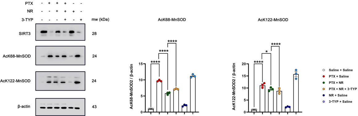
WB images of SIRT3, AcK68-MnSOD and AcK122-MnSOD expressions in DRG tissues. Quantification of SIRT3, AcK68-MnSOD and AcK122-MnSOD expressions normalized with β-actin expressions and the Saline+Saline treated samples was set to 1 (NR (Nicotinamide riboside chloride); PTX (4 mg/kg, i. p.) Group, PTX (4 mg/kg, i.p.) + NR (400 mg/kg, i.g.) Group, PTX (4 mg/kg, I.P.) + NR (400 mg/kg, i.g.) +3-TYP (50 mg/kg, i.p.) group, NR (400 mg/kg, i.p.) group, 3-TYP (50 mg/kg, i.p.) group in SIRT3 WT mice)
3-TYP purchased from MedChemExpress. Usage Cited in: Toxicol Appl Pharmacol. 2024 Oct:491:117066. [Abstract]
The representative images depicting the expression of AcK68-MnSOD in each group were utilized for immunohistochemical detection (NR (Nicotinamide riboside chloride); PTX (4 mg/kg, i. p.) Group, PTX (4 mg/kg, i.p.) + NR (400 mg/kg, i.g.) Group, PTX (4 mg/kg, I.P.) + NR (400 mg/kg, i.g.) +3-TYP (50 mg/kg, i.p.) group, NR(400 mg/kg, i.p.) group, 3-TYP (50 mg/kg, i.p.) group in SIRT3 WT mice)
-
Photochem Photobiol Sci
β-Nicotinamide mononucleotide blocks UVB-induced collagen reduction via regulation of ROS/MAPK/AP-1 and stimulation of mitochondrial proline biosynthesis. [Abstract]2025 Feb;24(2):293-306. PMID: 40025354 -
Neural Plast
ROS-Induced Oxidative Damage and Mitochondrial Dysfunction Mediated by Inhibition of SIRT3 in Cultured Cochlear Cells. [Abstract]2022 Jan 19:2022:5567174. PMID: 35096052 -
Exp Eye Res
SIRT3 mediates mitochondrial protection and attenuates mtROS-TXNIP-NLRP3 signaling activation in dry eye disease. [Abstract]2026 Jun:267:110960. PMID: 41791477 -
Exp Eye Res
Oroxylin A alleviates pyroptosis and apoptosis in human corneal epithelial cells under hyperosmotic stress by activating the SIRT3-SOD2/HIF-1α pathway. [Abstract]2025 Jun:255:110345. PMID: 40096905 -
Exp Eye Res
SIRT3 mitigates dry eye disease through the activation of autophagy by deacetylation of FOXO1. [Abstract]2025 May:254:110328. PMID: 40064414 -
Am J Cancer Res
2022 May 15;12(5):2310-2322. PMID: 35693089 -
J Membr Biol
Schisandrin B Improves Mitochondrial Function and Inhibits HT22 Cell Apoptosis by Regulating Sirt3 Protein. [Abstract]2025 Apr;258(2):123-133. PMID: 39939534 -
Biochem Biophys Res Commun
Alleviation of fibrosis and oxidative stress in pressure overload-induced cardiac remodeling and heart failure via SIRT3 activation by colchicine. [Abstract]2025 Jul 12:770:151957. PMID: 40373382 -
Cell Physiol Biochem
2017;44(6):2212-2227. PMID: 29248930
3-TYP purchased from MedChemExpress. Usage Cited in: Cell Physiol Biochem. 2017;44(6):2212-2227. [Abstract]
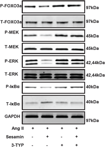
MEK, ERK1/2 and IκBα phosphorylation and their total protein expression are detected by Western blot. Cardiac myocytes are stimulated by Ang II for 48h after treatment with Sesamin (10 mM) or 3-TYP (50 μM).
-
J Food Biochem
Biochanin A ameliorated oleate-induced steatosis in HepG2 cells by activating the SIRT3/AMPK/ULK-1 signaling pathway. [Abstract]2022 Sep 20;e14428. PMID: 36125796 -
J Food Biochem
Piceatannol alleviate ROS-mediated PC-12 cells damage and mitochondrial dysfunction through SIRT3/FOXO3a signaling pathway. [Abstract]2022 Mar;46(3):e13820. PMID: 34132394 -
Neuroreport
Tetrandrine mediates autophagy via sirtuin 3/adenosine 5-monophosphate-activated protein kinase/mammalian target of rapamycin signal pathway to attenuate early brain injury after subarachnoid hemorrhage. [Abstract]2025 Jul 2;36(10):514-523. PMID: 40377960 -
Exp Anim
SIRT3 confers protection against acute pulmonary embolism by anti-inflammation, anti-oxidative stress, anti-apoptosis: participation of AMPK/mTOR pathway. [Abstract]2023 Aug 7;72(3):346-355. PMID: 36858596 -
Curr Med Sci
Green Tea Polyphenols Prevent Early Vascular Aging Induced by High-Fat Diet via Promoting Autophagy in Young Adult Rats. [Abstract]2022 Oct;42(5):981-990. PMID: 35896932 -
Biomed Pharmacother
Crocin-I mitigates diquat-induced pulmonary fibrosis via activation of the SIRT3/FOXO3a pathway. [Abstract]2025 May:186:118043. PMID: 40194334 -
Heliyon
Quercetin inhibits cardiomyocyte apoptosis via Sirt3/SOD2/mitochondrial reactive oxygen species during myocardial ischemia-reperfusion injury. [Abstract]2024 Oct 5;10(21):e39031. PMID: 39568838 -
-
Biomed Pharmacother
Cyclovirobuxine D alleviates aldosterone-induced myocardial hypertrophy by protecting mitochondrial function depending on the mutual regulation of Nrf2-SIRT3. [Abstract]2023 Nov:167:115618. PMID: 37793277 -
Oxid Med Cell Longev
Curcumin Ameliorates Age-Induced Tight Junction Impaired in Porcine Sertoli Cells by Inactivating the NLRP3 Inflammasome through the AMPK/SIRT3/SOD2/mtROS Signaling Pathway. [Abstract]2023 Feb 17:2023:1708251. PMID: 36846717 -
Ann Transl Med
Lycium barbarum polysaccharide protects cardiomyocytes from hypoxia/reoxygenation injury via activation of SIRT3/CypD signaling. [Abstract]2023 Jan 31;11(2):72. PMID: 36819526 -
Oxid Med Cell Longev
2023 Jan 14:2023:9966355. PMID: 36691640 -
Oxid Med Cell Longev
Mitochondria-Targeted Antioxidant Mitoquinone Maintains Mitochondrial Homeostasis through the Sirt3-Dependent Pathway to Mitigate Oxidative Damage Caused by Renal Ischemia/Reperfusion. [Abstract]2022 Sep 20;2022:2213503. PMID: 36193071 -
-
-
Oxid Med Cell Longev
Tubeimoside I Ameliorates Myocardial Ischemia-Reperfusion Injury through SIRT3-Dependent Regulation of Oxidative Stress and Apoptosis. [Abstract]2021 Nov 9;2021:5577019. PMID: 34795840 -
-
Aging (Albany NY)
Melatonin protects against focal cerebral ischemia-reperfusion injury in diabetic mice by ameliorating mitochondrial impairments: involvement of the Akt-SIRT3-SOD2 signaling pathway. [Abstract]2021 Jun 11;13(12):16105-16123. PMID: 34118791 -
-
Aging (Albany NY)
Electrical stimulation inhibits Val-boroPro-induced pyroptosis in THP-1 macrophages via sirtuin3 activation to promote autophagy and inhibit ROS generation. [Abstract]2020 Apr 14;12(7):6415-6435. PMID: 32289749


Solvent & Solubility
DMSO : 125 mg/mL (855.29 mM; Need ultrasonic; Hygroscopic DMSO has a significant impact on the solubility of product, please use newly opened DMSO)
Ethanol : 16.67 mg/mL (114.06 mM; Need ultrasonic)
H2O : 1 mg/mL (6.84 mM; ultrasonic and warming and heat to 60°C)
Please refer to the solubility information to select the appropriate solvent. Once prepared, please aliquot and store the solution to prevent product inactivation from repeated freeze-thaw cycles.
Storage method and period of stock solution: -80°C, 6 months; -20°C, 1 month. When stored at -80°C, please use it within 6 months. When stored at -20°C, please use it within 1 month.
* Note: If you choose water as the stock solution, please dilute it to the working solution, then filter and sterilize it with a 0.22 μm filter before use.
Concentration (start) × Volume (start) = Concentration (final) × Volume (final)
Select the appropriate dissolution method based on your experimental animal and administration route.
- For the following dissolution methods, please ensure to first prepare a clear stock solution using an In Vitro approach and then sequentially add co-solvents:
- To ensure reliable experimental results, the clarified stock solution can be appropriately stored based on storage conditions. As for the working solution for In Vivo experiments, it is recommended to prepare freshly and use it on the same day.
- The percentages shown for the solvents indicate their volumetric ratio in the final prepared solution. If precipitation or phase separation occurs during preparation, heat and/or sonication can be used to aid dissolution.
Add each solvent one by one: 10% DMSO 40% PEG300 5% Tween-80 45% Saline
Solubility: ≥ 2.08 mg/mL (14.23 mM); Clear solution
This protocol yields a clear solution of ≥ 2.08 mg/mL (saturation unknown).
Taking 1 mL working solution as an example, add 100 μL DMSO stock solution (20.8 mg/mL) to 400 μL PEG300, and mix evenly; then add 50 μL Tween-80 and mix evenly; then add 450 μL Saline to adjust the volume to 1 mL.
Preparation of Saline: Dissolve 0.9 g sodium chloride in ddH₂O and dilute to 100 mL to obtain a clear Saline solution.
Add each solvent one by one: 10% DMSO 90% (20% SBE-β-CD in Saline)
Solubility: ≥ 2.08 mg/mL (14.23 mM); Clear solution
This protocol yields a clear solution of ≥ 2.08 mg/mL (saturation unknown).
Taking 1 mL working solution as an example, add 100 μL DMSO stock solution (20.8 mg/mL) to 900 μL 20% SBE-β-CD in Saline, and mix evenly.
Preparation of 20% SBE-β-CD in Saline (4°C, storage for one week): 2 g SBE-β-CD powder is dissolved in 10 mL Saline, completely dissolve until clear.
For the following dissolution methods, please prepare the working solution directly:
It is recommended to prepare fresh solutions and use them promptly within a short period of time.
The percentages shown for the solvents indicate their volumetric ratio in the final prepared solution. If precipitation or phase separation occurs during preparation, heat and/or sonication can be used to aid dissolution.
Add each solvent one by one: 50% PEG300 50% Saline
Solubility: 25 mg/mL (171.06 mM); Clear solution; Need ultrasonic
Please enter the basic information of animal experiments:
-
-
-
-
Recommended: Prepare an additional quantity of animals to account for potential losses during experiments.
Please enter your animal formula composition:
-
%DMSO +
Recommended: Keep the proportion of DMSO in working solution below 2% if your animal is weak.
-
%+
-
+%Tween-80 + +
-
%Saline +
The co-solvents required include: DMSO, . All of co-solvents are available by MedChemExpress (MCE). , Tween 80. All of co-solvents are available by MedChemExpress (MCE).
Working solution concentration: 0.22 mg/mL
Method for preparing stock solution: mg drug dissolved in μL DMSO. Stock solution concentration: mg/mL.
1. Take μL DMSO stock solution;
2. Add μL .
μL , mix evenly;
3. Then add μL Tween 80, mix evenly;
4. Then add μL
Please ensure that the stock solution in the first step is dissolved to a clear state, and add co-solvents in sequence. You can use ultrasonic heating (ultrasonic cleaner, recommended frequency 20-40 kHz), vortexing, etc. to assist dissolution.

Protocol
Cell viability is analyzed using Cell Counting Kit-8. Briefly, 1×10⁴ cells are inoculated into 96-well plates. After being treated, 90 μL of medium and 10 μL of CCK-8 solution are added to each well. The cells are then incubated at 37°C for 2 h. After incubation, the absorption at 450 nm is measured using an Infinite™ M200 Microplate Reader. The results are expressed as a percentage of the control. The cell death is also evaluated using the trypan blue assay. HepG2 cells are plated in the 6-well plates (5×10⁵ cells per well) and incubated for 24 h. After being treated with Cd or melatonin, the cells are detached with 300 μL trypsin-EDTA solution. The mixture of detached cells is centrifugated at 300 g for 5 min. Then, the residue is combined with 800 μL trypan blue solution and dispersed. After 3 min staining, cells are counted using an automated cell counter. The dead cells are stained with the blue color. Cell mortality (%) is expressed as percentage of the dead cell number/the total cell number.
MedChemExpress (MCE) has not independently confirmed the accuracy of these methods. They are for reference only.
In brief, male C57BL/6 mice are anesthetized with 2% isoflurane, and myocardial ischemia is produced by temporarily exteriorizing the heart via a left thoracic incision and placing a 6-0 silk suture slipknot around the left anterior descending coronary artery. After 30 minutes of myocardial ischemia, the slipknot is released, and the myocardium is reperfused for 3 hour (for western blot analysis and oxidative stress measurement) or 24 hour (for cardiac function, apoptotic index and infarct size determination). Sham-operated mice undergo the same surgical procedures except the suture placed under the left coronary artery is not tied. Ten minutes before reperfusion, mice are randomized to receive either vehicle (1% ethanol) or melatonin (20 mg/kg) by intraperitoneal injection. C57BL/6 mice are randomly divided into the following groups: (i) Sham group: mice underwent the sham operation and are treated with vehicle (1% ethanol); (ii) Mel group: mice are treated with melatonin (20 mg/kg via intraperitoneal injection); (iii) IR+V group: mice underwent the MI/R operation and are treated with vehicle (1% ethanol); (iv) IR+Mel group: mice underwent the MI/R operation and are treated with melatonin (20 mg/kg via intraperitoneal injection 10 minutes before reperfusion); (v) IR+Mel+3-TYP group: mice are pretreated with 3-TYP (3-TYP is intraperitoneally injected at a dose of 50 mg/kg every 2 days for a total of three doses prior to the MI/R surgery), subjected to the MI/R operation, and treated with melatonin (20 mg/kg via intraperitoneal injection 10 minutes before reperfusion); and (vi) IR+3-TYP group: mice are pretreated with 3-TYP and then subjected to the MI/R operation.
MedChemExpress (MCE) has not independently confirmed the accuracy of these methods. They are for reference only.


Purity & Documentation
-
Data Sheet (283 KB)
-
SDS (396 KB)
- English - EN (396 KB)
- Français - FR (396 KB)
- Deutsch - DE (396 KB)
- Norwegian - NO (396 KB)
- Español - ES (396 KB)
- Swedish - SV (396 KB)
- Italian - IT (396 KB)
- Korean - KR (396 KB)
- Portuguese - PT (396 KB)
-
Handling Instructions (2659 KB)
References
[1]. Pi H, et al. SIRT3-SOD2-mROS-dependent autophagy in cadmium-induced hepatotoxicity and salvage by melatonin. Autophagy. 2015;11(7):1037-51. [Content Brief]
[2]. Zhai M, et al. Melatonin ameliorates myocardial ischemia reperfusion injury through SIRT3-dependent regulation of oxidative stress and apoptosis. J Pineal Res. 2017 Sep;63(2). [Content Brief]
[3]. Galli U, et al. Identification of a sirtuin 3 inhibitor that displays selectivity over sirtuin 1 and 2. Eur J Med Chem. 2012 Sep;55:58-66. [Content Brief]


Complete Stock Solution Preparation Table
Please refer to the solubility information to select the appropriate solvent. Once prepared, please aliquot and store the solution to prevent product inactivation from repeated freeze-thaw cycles.
Storage method and period of stock solution: -80°C, 6 months; -20°C, 1 month. When stored at -80°C, please use it within 6 months. When stored at -20°C, please use it within 1 month.
| Optional Solvent | Concentration Solvent Mass | 1 mg | 5 mg | 10 mg | 25 mg |
|---|---|---|---|---|---|
| H2O / Ethanol / DMSO | 1 mM | 6.8423 mL | 34.2114 mL | 68.4229 mL | 171.0571 mL |
| 5 mM | 1.3685 mL | 6.8423 mL | 13.6846 mL | 34.2114 mL | |
| Ethanol / DMSO | 10 mM | 0.6842 mL | 3.4211 mL | 6.8423 mL | 17.1057 mL |
| 15 mM | 0.4562 mL | 2.2808 mL | 4.5615 mL | 11.4038 mL | |
| 20 mM | 0.3421 mL | 1.7106 mL | 3.4211 mL | 8.5529 mL | |
| 25 mM | 0.2737 mL | 1.3685 mL | 2.7369 mL | 6.8423 mL | |
| 30 mM | 0.2281 mL | 1.1404 mL | 2.2808 mL | 5.7019 mL | |
| 40 mM | 0.1711 mL | 0.8553 mL | 1.7106 mL | 4.2764 mL | |
| 50 mM | 0.1368 mL | 0.6842 mL | 1.3685 mL | 3.4211 mL | |
| 60 mM | 0.1140 mL | 0.5702 mL | 1.1404 mL | 2.8510 mL | |
| 80 mM | 0.0855 mL | 0.4276 mL | 0.8553 mL | 2.1382 mL | |
| 100 mM | 0.0684 mL | 0.3421 mL | 0.6842 mL | 1.7106 mL |
* Note: If you choose water as the stock solution, please dilute it to the working solution, then filter and sterilize it with a 0.22 μm filter before use.


 Powered by Bioz