


Mdivi-1
Based on 346 publication(s) in Google Scholar
Mdivi-1 is a selective dynamin-related protein 1 (Drp1) inhibitor. Mdivi-1 is a mitochondrial division/mitophagy inhibitor.
For research use only. We do not sell to patients.
- Purity: 99.96%
- CAS No.: 338967-87-6
- Formula: C15H10Cl2N2O2S
- Molecular Weight:353.22
-
Storage:Powder -20°C, 3 years , 4°C, 2 years ; In solvent -80°C, 6 months , -20°C, 1 month

To place orders, for customer services and technical support, please contact: MedChemExpress USA
Tel: 609-228-6898 E-mail: [email protected] [email protected]


-
 Biological Activity
-
Chemical Information
-
Solvent & Solubility
- Protocol
- Purity & Documentation
- References
-
 Help & FAQs
-


Apoptosis Compound Library
HY-L003
-
Immunology/Inflammation Compound Library
HY-L007
-
Anti-Cancer Compound Library
HY-L025
-
Autophagy Compound Library
HY-L029
-
Anti-Aging Compound Library
HY-L034
-
Cytoskeleton Compound Library
HY-L060
-
Mitochondria-Targeted Compound Library
HY-L089
-
Targeted Diversity Library
HY-L099
-
Human Metabolite Library
HY-L123
-
Mitochondrial Protection Compound Library
HY-L144
-
Cysteine Targeted Covalent Library
HY-L153
-
Mitochondrial Toxicity Compound Library
HY-L155
-
Highly Selective Inhibitors Library
HY-L158
-
Highly Selective Activators Library
HY-L159
-
Cell Death Library
HY-L162
-
Mitophagy Compound Library
HY-L180
-
Bioactive Compound Library Max
HY-L181
-
MCE Bioactive Compound Library
HY-L001V
-
Bioactive Compound Library
HY-L001
-
Non-Alcoholic Fatty Liver Disease (NAFLD) Compound Library
HY-L199
-
High-Throughput Bioactive Compound Library
HY-L205
-
Mass Spectrometry Human Metabolite Library
HY-L215


Publications Citing Use of MedChemExpress (MCE) Mdivi-1
More- Signal Transduct Target Ther. 2025 Dec 17;10(1):413. [Abstract]
- Nature. 2026 May;653(8114):548-557. [Abstract]
- Nature. 2026 Jan;649(8098):1022-1031. [Abstract]
- Cell Metab. 2025 Oct 24:S1550-4131(25)00395-X. [Abstract]
- Mil Med Res. 2026 Dec;13(1):100004. [Abstract]
- Cell Mol Immunol. 2022 Jan;19(1):67-78. [Abstract]
- Cell Stem Cell. 2025 Mar 20:S1934-5909(25)00084-0. [Abstract]
- Bone Res. 2025 Mar 3;13(1):30. [Abstract]
- Bone Res. 2025 Jan 26;13(1):18. [Abstract]
- Autophagy. 2026 Jun 16:1-20. [Abstract]
- Autophagy. 2026 Mar;22(3):445-467. [Abstract]
- Autophagy. 2026 Jan 1:1-19. [Abstract]
- Autophagy. 2026 Jan 1:1-17. [Abstract]
- Autophagy. 2025 Jun;21(6):1228-1244. [Abstract]
- Autophagy. 2025 Jan;21(1):102-119. [Abstract]
- Autophagy. 2021 Nov;17(11):3592-3606. [Abstract]
- Autophagy. 2020 Aug;16(8):1413-1435. [Abstract]
- Nat Commun. 2026 Feb 9;17(1):2544. [Abstract]
- Nat Commun. 2025 Aug 14;16(1):7564. [Abstract]
- Nat Commun. 2024 Oct 16;15(1):8927. [Abstract]
- Nat Commun. 2022 Jul 6;13(1):3882. [Abstract]
- ACS Nano. 2025 Apr 23. [Abstract]
- J Adv Res. 2025 Dec 19:S2090-1232(25)01014-8. [Abstract]
- J Adv Res. 2025 Nov 20:S2090-1232(25)00927-0. [Abstract]
- J Adv Res. 2025 Jul:73:199-217. [Abstract]
- Metabolism. 2026 May:178:156567. [Abstract]
- Redox Biol. 2025 Mar:80:103511. [Abstract]
- Redox Biol. 2025 Feb:79:103471. [Abstract]
- Redox Biol. 2023 Jun:62:102663. [Abstract]
- Redox Biol. 2022 Feb;49:102216. [Abstract]
- Redox Biol. 2020 Jul:34:101523. [Abstract]
- Mol Cell. 2026 Feb 24:S1097-2765(26)00071-7. [Abstract]
- Gut Microbes. Jan-Dec 2022;14(1):2096989. [Abstract]
- J Nanobiotechnology. 2025 Jul 16;23(1):521. [Abstract]
- J Nanobiotechnology. 2025 Feb 17;23(1):114. [Abstract]
- Theranostics. 2021 Jan 1;11(2):522-539. [Abstract]
- Theranostics. 2019 Feb 28;9(6):1698-1713. [Abstract]
- Genes Dis. 2026 Jun 15.
- Genes Dis. 2022 Sep 10;10(5):2151-2166. [Abstract]
- Adv Sci (Weinh). 2026 May 19:e75784. [Abstract]
- Adv Sci (Weinh). 2026 Apr;13(21):e17108. [Abstract]
- Adv Sci (Weinh). 2025 Aug 11:e01041. [Abstract]
- Cell Rep Med. 2025 Apr 15;6(4):102039. [Abstract]
- Biomaterials. 2026 May 15:334:124316. [Abstract]
- Cell Death Differ. 2022 Dec;29(12):2417-2428. [Abstract]
- Carbohydr Polym. 2022 Nov 1:295:119841. [Abstract]
- Cell Death Dis. 2026 Apr 23;17(1):537. [Abstract]
- Pharmacol Res. 2025 Dec:222:108028. [Abstract]
- Environ Sci Technol. 2025 Oct 28;59(42):22423-22438. [Abstract]
- Environ Sci Technol. 2025 Aug 5;59(30):15705-15719. [Abstract]
- Pharmacol Res. 2024 Feb:200:107051. [Abstract]
- Pharmacol Res. 2021 Aug:170:105712. [Abstract]
- Cell Death Dis. 2019 Mar 8;10(3):232. [Abstract]
- Pharmacol Res. 2019 Jan:139:273-285. [Abstract]
- Cell Death Dis. 2018 Sep 5;9(9):907. [Abstract]
- Small. 2025 Jul;21(26):e2502939. [Abstract]
- Cancer Lett. 2024 Oct 10:602:217192. [Abstract]
- Small. 2023 Aug;19(33):e2301497. [Abstract]
- Int J Biol Sci. 2024 Aug 12;20(11):4382-4406. [Abstract]
- Int J Biol Sci. 2024 Apr 29;20(7):2658-2685. [Abstract]
- J Neuroinflammation. 2025 Apr 17;22(1):108. [Abstract]
- J Neuroinflammation. 2022 Apr 12;19(1):87. [Abstract]
- Phytomedicine. 2026 Apr:153:157985. [Abstract]
- Phytomedicine. 2026 Jan 20:153:157860. [Abstract]
- Phytomedicine. 2025 Nov 25:148:157486. [Abstract]
- Phytomedicine. 2025 Feb 21:139:156500. [Abstract]
- Phytomedicine. 2023 Jul 25:116:154901. [Abstract]
- Mater Today Bio. 2026 Jun 23;39:103393. [Abstract]
- Adv Healthc Mater. 2026 Jan 19:e05255. [Abstract]
- Arch Toxicol. 2025 Aug 20. [Abstract]
- Environ Health Perspect. 2022 May;130(5):57004. [Abstract]
- J Hazard Mater. 2025 Dec 5:500:140498. [Abstract]
- J Hazard Mater. 2022 Feb 15;424(Pt A):127268. [Abstract]
- Cell Death Discov. 2026 Feb 26. [Abstract]
- Cell Death Discov. 2026 Jan 6;12(1):81. [Abstract]
- Mol Psychiatry. 2025 Sep 16. [Abstract]
- Cell Death Discov. 2022 Feb 17;8(1):69. [Abstract]
- Environ Int. 2021 Feb:147:106319. [Abstract]
- J Orthop Translat. 2026 Jan 6.
- J Transl Med. 2025 Apr 5;23(1):402. [Abstract]
- J Transl Med. 2025 Feb 4;23(1):156. [Abstract]
- J Transl Med. 2023 Oct 10;21(1):711. [Abstract]
- J Transl Med. 2023 Mar 25;21(1):218. [Abstract]
- J Colloid Interface Sci. 2025 Sep 25;703(Pt 1):139108. [Abstract]
- Basic Res Cardiol. 2021 Dec 16;116(1):65. [Abstract]
- Chin Med J (Engl). 2026 Mar 5;139(5):699-709. [Abstract]
- Oncogene. 2025 Aug;44(32):2893-2906. [Abstract]
- Int J Surg. 2025 Jul 2. [Abstract]
- Int J Biol Macromol. 2026 Apr:353:151239. [Abstract]
- Int J Biol Macromol. 2025 Sep;322(Pt 3):146900. [Abstract]
- BMC Med. 2025 Mar 31;23(1):189. [Abstract]
- Int J Nanomedicine. 2021 Sep 29;16:6661-6679. [Abstract]
- Neural Regen Res. 2025 March 25.
- Mol Med. 2025 May 29;31(1):211. [Abstract]
- Antioxidants (Basel). 2023 Oct 31;12(11):1939. [Abstract]
- Antioxidants (Basel). 2023 Mar 26;12(4):806. [Abstract]
- Phytother Res. 2026 Jul;40(7):3990-4004. [Abstract]
- Phytother Res. 2023 Dec;37(12):5916-5931. [Abstract]
- Free Radic Biol Med. 2026 Jun 7:254:307-322. [Abstract]
- Free Radic Biol Med. 2026 Jun 27:254:391-404. [Abstract]
- Free Radic Biol Med. 2026 May:248:162-176. [Abstract]
- Free Radic Biol Med. 2026 May:248:255-271. [Abstract]
- Free Radic Biol Med. 2026 Mar 25:250:153-167. [Abstract]
- Free Radic Biol Med. 2026 Mar 16:246:93-106. [Abstract]
- Free Radic Biol Med. 2025 Dec 30:S0891-5849(25)01474-1. [Abstract]
- Free Radic Biol Med. 2025 Dec 16:244:380-394. [Abstract]
- Free Radic Biol Med. 2025 Nov 23:243:398-413. [Abstract]
- Free Radic Biol Med. 2025 Aug 13:240:59-70. [Abstract]
- Free Radic Biol Med. 2024 Nov 20:225:699-710. [Abstract]
- Free Radic Biol Med. 2024 Nov 1:224:521-539. [Abstract]
- Free Radic Biol Med. 2024 Nov 20:225:665-676. [Abstract]
- Free Radic Biol Med. 2024 Jun:218:132-148. [Abstract]
- Free Radic Biol Med. 2024 Mar:213:150-163. [Abstract]
- Free Radic Biol Med. 2021 Aug 20:172:19-32. [Abstract]
- Free Radic Biol Med. 2017 Nov:112:336-349. [Abstract]
- Clin Transl Med. 2024 Apr;14(4):e1653. [Abstract]
- J Anim Sci Biotechnol. 2020 Aug 5:11:77. [Abstract]
- Stem Cell Res Ther. 2025 Oct 28;16(1):584. [Abstract]
- Stem Cell Res Ther. 2025 Mar 5;16(1):116. [Abstract]
- Cell Rep. 2026 May 26;45(5):117321. [Abstract]
- Aging Cell. 2024 Nov;23(11):e14294. [Abstract]
- Cell Rep. 2022 Apr 12;39(2):110635. [Abstract]
- World J Gastroenterol. 2020 Apr 21;26(15):1758-1774. [Abstract]
- Cell Prolif. 2026 Apr 27:e70216. [Abstract]
- Cell Prolif. 2025 Jul 4:e70085. [Abstract]
- Cell Prolif. 2025 May;58(5):e13799. [Abstract]
- Cell Prolif. 2021 Jun;54(6):e13048. [Abstract]
- J Pineal Res. 2018 Sep;65(2):e12491. [Abstract]
- J Pineal Res. 2018 Jan;64(1). [Abstract]
- Clin Sci. 2025 Oct 22;139(20):1163-1185. [Abstract]
- Environ Pollut. 2024 Dec 1:362:124935. [Abstract]
- Environ Pollut. 2024 Apr 15:347:123740. [Abstract]
- Lebensm Wiss Technol. 2025 Nov 25,238:118816.
- Atherosclerosis. 2025 Jun:405:119216. [Abstract]
- Atherosclerosis. 2022 Apr:346:36-45. [Abstract]
- Cancer Cell Int. 2019 Dec 10:19:332. [Abstract]
- Redox Rep. 2024 Dec;29(1):2377870. [Abstract]
- Lipids Health Dis. 2024 Sep 3;23(1):279. [Abstract]
- J Ethnopharmacol. 2026 May 23:363:121445. [Abstract]
- J Ethnopharmacol. 2026 May 25:369:121896. [Abstract]
- J Ethnopharmacol. 2026 Feb 28:357:120899. [Abstract]
- Antioxid Redox Signal. 2025 Dec 26. [Abstract]
- Antioxid Redox Signal. 2025 Feb;42(4-6):249-264. [Abstract]
- J Agric Food Chem. 2024 Jun 5;72(22):12775-12787. [Abstract]
- J Agric Food Chem. 2024 Jan 31;72(4):2120-2134. [Abstract]
- Ecotoxicol Environ Saf. 2025 Oct 1:304:119133. [Abstract]
- Ecotoxicol Environ Saf. 2025 Oct 15:305:119206. [Abstract]
- Ecotoxicol Environ Saf. 2024 Jun 1:277:116392. [Abstract]
- CNS Neurosci Ther. 2024 Jun;30(6):e14800. [Abstract]
- Biochem Pharmacol. 2026 Jul:249:117891. [Abstract]
- J Environ Sci. 2026 May 14.
- Cell Biosci. 2021 Jan 7;11(1):9. [Abstract]
- Precis Clin Med. 2026 Mar 14;9(2):pbag009. [Abstract]
- Cancer Gene Ther. 2024 Jan;31(1):43-57. [Abstract]
- Life Sci. 2020 Sep 15:257:118116. [Abstract]
- Food Funct. 2026 Jan 26;17(2):1045-1060. [Abstract]
- Prog Orthod. 2026 Jan 13;27(1):1. [Abstract]
- Food Funct. 2022 Jul 18;13(14):7666-7683. [Abstract]
- Food Biosci. 2025 Jun.
- Food Biosci. 2025 Apr 21.
- Transl Res. 2018 Apr:194:68-78. [Abstract]
- Pharm Biol. 2025 Dec;63(1):188-200. [Abstract]
- Commun Biol. 2025 Feb 28;8(1):338. [Abstract]
- Radiother Oncol. 2024 Jan:190:110028. [Abstract]
- Commun Biol. 2022 Jun 22;5(1):616. [Abstract]
- Eur J Pharmacol. 2025 Jun 15:997:177468. [Abstract]
- Cell Biol Toxicol. 2024 Oct 30;40(1):93. [Abstract]
- Eur J Pharmacol. 2024 Aug 15:977:176736. [Abstract]
- Respir Res. 2023 Sep 6;24(1):216. [Abstract]
- Eur J Pharmacol. 2023 Mar 5:942:175531. [Abstract]
- Cell Biol Toxicol. 2022 Jun;38(3):487-504. [Abstract]
- Biomolecules. 2026 Apr 30;16(5):664. [Abstract]
- Biomolecules. 2026 Mar 13;16(3):429. [Abstract]
- Int Immunopharmacol. 2026 Feb 1:170:116049. [Abstract]
- Int J Mol Sci. 2026 Jan 30;27(3):1390. [Abstract]
- Int Immunopharmacol. 2025 Nov 14:165:115443. [Abstract]
- Int Immunopharmacol. 2025 Sep 10:165:115526. [Abstract]
- Int Immunopharmacol. 2025 Jun 17:158:114863. [Abstract]
- Int Immunopharmacol. 2025 Jun 26:159:114911. [Abstract]
- Int Immunopharmacol. 2025 Jun 5:157:114741. [Abstract]
- Int J Mol Sci. 2025 Apr 27;26(9):4162. [Abstract]
- Biomolecules. 2025 Apr 9;15(4):556. [Abstract]
- Int Immunopharmacol. 2024 Nov 15:141:113011. [Abstract]
- Int Immunopharmacol. 2024 Oct 25:140:112831. [Abstract]
- Int Immunopharmacol. 2024 Sep 10:138:112652. [Abstract]
- Int Immunopharmacol. 2024 Sep 23;142(Pt B):113092. [Abstract]
- Int J Mol Sci. 2024 Apr 29;25(9):4853. [Abstract]
- Int J Mol Sci. 2023 May 11;24(10):8609. [Abstract]
- Int J Mol Sci. 2023 Mar 20;24(6):5896. [Abstract]
- Int J Mol Sci. 2022 Apr 28;23(9):4907. [Abstract]
- Mol Neurobiol. 2025 Nov 26;63(1):177. [Abstract]
- Mol Neurobiol. 2025 Nov 19;63(1):76. [Abstract]
- Mol Neurobiol. 2025 Mar;62(3):3125-3142. [Abstract]
- Mol Neurobiol. 2022 Jun;59(6):3933-3946. [Abstract]
- mBio. 2025 Sep 10;16(9):e0143625. [Abstract]
- Am J Physiol Cell Physiol. 2025 Aug 20. [Abstract]
- Am J Physiol Cell Physiol. 2024 Dec 31. [Abstract]
- Inflammation. 2024 Dec 30. [Abstract]
- mBio. 2023 Dec 19;14(6):e0148023. [Abstract]
- Front Pharmacol. 2021 Mar 9;12:616803. [Abstract]
- Front Pharmacol. 2019 Sep 3:10:968. [Abstract]
- Int Dent J. 2025 Jun 14;75(4):100853. [Abstract]
- Chem Biol Interact. 2024 Apr 1:392:110904. [Abstract]
- Chem Biol Interact. 2023 Jul 1:379:110523. [Abstract]
- Toxicology. 2022 Nov:481:153348. [Abstract]
- Toxicology. 2022 Jan 30:466:153082. [Abstract]
- Exp Gerontol. 2026 Jun 15:219:113157. [Abstract]
- J Physiol Biochem. 2024 Nov;80(4):935-948. [Abstract]
- Eur J Pharm Sci. 2018 Aug 30:121:243-250. [Abstract]
- Biochim Biophys Acta Mol Basis Dis. 2025 Nov 19;1872(3):168106. [Abstract]
- Biochim Biophys Acta Mol Basis Dis. 2025 Oct 15;1872(2):168074. [Abstract]
- Hypertens Res. 2024 May;47(5):1338-1349. [Abstract]
- J Mol Med (Berl). 2021 Mar;99(3):359-371. [Abstract]
- Sci Rep. 2026 Jun 11. [Abstract]
- Sci Rep. 2026 Apr 8;16(1):16667. [Abstract]
- PLoS Pathog. 2026 Feb 12;22(2):e1013975. [Abstract]
- Mediators Inflamm. 2026 Jan 8:2026:7600668. [Abstract]
- Sci Rep. 2025 May 4;15(1):15571. [Abstract]
- Sci Rep. 2024 Dec 28;14(1):31291. [Abstract]
- Sci Rep. 2024 Jun 6;14(1):13063. [Abstract]
- PLoS Pathog. 2016 Aug 24;12(8):e1005823. [Abstract]
- Cancers (Basel). 2025 Dec 27;18(1):92. [Abstract]
- Exp Neurol. 2017 Sep:295:116-124. [Abstract]
- Cell Signal. 2026 Oct:146:112679. [Abstract]
- Mol Cell Biochem. 2026 Jun 2. [Abstract]
- Cell Signal. 2025 Sep:133:111868. [Abstract]
- J Cell Mol Med. 2024 Jul;28(14):e18375. [Abstract]
- J Cell Mol Med. 2024 May;28(9):e18353. [Abstract]
- Virol Sin. 2023 Aug;38(4):520-530. [Abstract]
- J Cell Mol Med. 2023 Feb;27(3):412-421. [Abstract]
- Cell Signal. 2023 Jan:101:110500. [Abstract]
- J Cell Mol Med. 2022 Feb;26(3):893-912. [Abstract]
- Oncol Rep. 2020 Mar;43(3):1010-1018. [Abstract]
- J Inflamm Res. 2026 Feb 2:19:540176. [Abstract]
- J Inflamm Res. 2025 Oct 8:18:13965-13984. [Abstract]
- J Neurochem. 2022 Dec;163(5):419-437. [Abstract]
- Brain Res Bull. 2026 Jun 10:243:111997. [Abstract]
- Brain Res Bull. 2025 Jul:227:111404. [Abstract]
- Poult Sci. 2025 Jun 26;104(9):105491. [Abstract]
- iScience. 2024 Jan 20;27(2):108982. [Abstract]
- iScience. 2023 Oct 20;26(11):108270. [Abstract]
- iScience. 2022 Jun 11;25(7):104582. [Abstract]
- Neurochem Res. 2021 Mar;46(3):573-583. [Abstract]
- Aquaculture. 2025 Feb 15.
- FASEB J. 2026 Jun 30;40(12):e72050. [Abstract]
- Biol Trace Elem Res. 2026 Mar 27. [Abstract]
- Phytopathol Res. 2025 Jul 15.
- Biochim Biophys Acta Mol Cell Res. 2025 Mar;1872(3):119911. [Abstract]
- FASEB J. 2024 Jun 30;38(12):e23723. [Abstract]
- Biochim Biophys Acta Mol Cell Res. 2023 Apr;1870(4):119450. [Abstract]
- Fish Shellfish Immunol. 2026 Jul 6:177:111583. [Abstract]
- Fish Shellfish Immunol. 2026 Jun:173:111248. [Abstract]
- FEBS J. 2023 Jul;290(14):3629-3645. [Abstract]
- Drug Dev Res. 2019 Jun;80(4):481-489. [Abstract]
- J Virol. 2025 Aug 19;99(8):e0049825. [Abstract]
- Cancer Res Commun. 2024 May 3;4(5):1189-1198. [Abstract]
- Environ Microbiol. 2021 Feb;23(2):774-790. [Abstract]
- J Funct Foods. 2026 Apr 11;140:107290.
- Biochim Biophys Acta Mol Cell Biol Lipids. 2024 Mar;1869(2):159428. [Abstract]
- Biochim Biophys Acta Mol Cell Biol Lipids. 2024 Jan;1869(1):159425. [Abstract]
- J Orthop Surg Res. 2023 Aug 24;18(1):620. [Abstract]
- ESC Heart Fail. 2023 Feb;10(1):366-376. [Abstract]
- Viruses. 2020 Mar 6;12(3):289. [Abstract]
- J Anim Sci. 2025 Jan 4:103:skaf384. [Abstract]
- Mol Immunol. 2025 Oct:186:48-62. [Abstract]
- J Med Virol. 2024 Nov;96(11):e70062. [Abstract]
- Toxicol Appl Pharmacol. 2026 May:510:117772. [Abstract]
- Toxicol Appl Pharmacol. 2020 Feb 1:388:114874. [Abstract]
- Appl Biochem Biotechnol. 2026 May 20. [Abstract]
- Int J Biochem Cell Biol. 2026 Mar:192:106910. [Abstract]
- Cardiovasc Drugs Ther. 2026 Jan 12. [Abstract]
- Arch Biochem Biophys. 2025 May:767:110354. [Abstract]
- Int J Biochem Cell Biol. 2024 Mar:168:106517. [Abstract]
- Arch Biochem Biophys. 2023 Apr:738:109558. [Abstract]
- Arch Biochem Biophys. 2022 Mar 30;718:109147. [Abstract]
- Int J Biochem Cell Biol. 2021 Jul:136:106003. [Abstract]
- Arch Biochem Biophys. 2020 May 30:685:108284. [Abstract]
- Arch Biochem Biophys. 2019 Aug 15:671:42-51. [Abstract]
- Reprod Toxicol. 2022 Oct:113:18-29. [Abstract]
- Cell Immunol. 2026 Jul:425:105102. [Abstract]
- Food Chem Toxicol. 2026 Jul:213:116103. [Abstract]
- Immunol Lett. 2026 Jun 6:281:107198. [Abstract]
- Food Chem Toxicol. 2023 Dec:182:114129. [Abstract]
- IUBMB Life. 2022 Jun;74(6):519-531. [Abstract]
- Brain Res. 2019 May 15:1711:68-76. [Abstract]
- Kaohsiung J Med Sci. 2026 Jun 9:e70242. [Abstract]
- Toxicol Lett. 2026 May 15:420:111896. [Abstract]
- 3 Biotech. 2025 Oct;15(10):341. [Abstract]
- Exp Eye Res. 2024 May:242:109860. [Abstract]
- Toxicol Lett. 2023 Sep 15:387:1-13. [Abstract]
- Exp Eye Res. 2022 Apr 28;220:109095. [Abstract]
- Expert Rev Anticancer Ther. 2025 Sep 3:1-10. [Abstract]
- Theriogenology. 2026 Jul 1:258:117890. [Abstract]
- Biochim Biophys Acta Gen Subj. 2025 Oct;1869(11):130851. [Abstract]
- Behav Brain Res. 2023 Feb 25:440:114264. [Abstract]
- Toxicol Res (Camb). 2022 May 23;11(3):486-497. [Abstract]
- Vet Microbiol. 2025 Sep 6:310:110721. [Abstract]
- Vet Microbiol. 2023 May:280:109697. [Abstract]
- Clin Rheumatol. 2025 Nov 20. [Abstract]
- Toxicol In Vitro. 2023 Apr:88:105552. [Abstract]
- Steroids. 2020 Nov:163:108699. [Abstract]
- Biochem Biophys Res Commun. 2026 May 7:812:153592. [Abstract]
- Biochem Biophys Res Commun. 2025 Nov 19:790:152919. [Abstract]
- Biochem Biophys Res Commun. 2024 Nov 12:733:150716. [Abstract]
- J Mol Neurosci. 2025 Jun 13;75(2):75. [Abstract]
- Dev Comp Immunol. 2025 May:166:105349. [Abstract]
- Exp Ther Med. 2024 Aug 19;28(4):402. [Abstract]
- Neurosci Lett. 2021 Jun 21:756:135967. [Abstract]
- Exp Ther Med. 2019 Mar;17(3):2349-2358. [Abstract]
- Inhal Toxicol. 2024 Oct-Nov;36(9-10):511-520. [Abstract]
- In Vitro Cell Dev Biol Anim. 2025 Mar;61(3):357-367. [Abstract]
- J Physiol Investig. 2025 Jan 1;68(1):31-42. [Abstract]
- Mol Cell Toxicol. 2026 Mar 5.
- Int Heart J. 2025;66(6):1002-1014. [Abstract]
- Res Sq. 2026 May 4.
- bioRxiv. 2026 Apr 25:2026.04.23.719926. [Abstract]
- bioRxiv. 2025 Nov 11.
- Res Sq. 2025 Aug 11.
- bioRxiv. 2025 June 01.
- Res Sq. 2025 Jun 20.
- Keimyung University. 2025.
- bioRxiv. 2025 May 19:2025.05.16.654477. [Abstract]
- SSRN. 2025 Mar 3.
- Research Square Preprint. 2025 Jan 03.
- bioRxiv. 2024 October 09.
- Res Sq. 2024 Jul 26.
- Biomed Pharmacother. 2024 Sep:178:117279. [Abstract]
- SSRN. 2024 Jun 13.
- Sci Total Environ. 2024 May 15:925:171818. [Abstract]
- SSRN. 2024 Mar 6.
- Sci Total Environ. 2023 Dec 15:904:166831. [Abstract]
- Biomed Pharmacother. 2023 Oct:166:115342. [Abstract]
- Research Square Preprint. 2023 Oct 3.
- Sci Total Environ. 2023 Sep 1:889:164078. [Abstract]
- SSRN. 2023 Aug 17.
- SSRN. 2023 Jul 25.
- Oxid Med Cell Longev. 2023 Feb 16:2023:9595201. [Abstract]
- Research Square Print. January 4th, 2023.
- Research Square Print. 2022 May.
- Ann Transl Med. 2022 Feb;10(4):212. [Abstract]
- bioRxiv. July 28, 2021.
- Research Square Preprint. 2021 Oct.
- Aging (Albany NY). 2021 Jan 2;13(1):1458-1472. [Abstract]
- Research Square Preprint. 2020 Aug.
- Oxid Med Cell Longev. 2020 Jul 23:2020:3549704. [Abstract]
- Aging (Albany NY). 2020 Jun 10;12(11):11139-11151. [Abstract]
 Customer Validation & Images

-
Cell Proliferation/Viability Assay
-
IF
-
WB
-
Cell Imaging/Staining
-
IF


Biological Activity
Mdivi-1 inhibits Dnm1 GTPase activity in a dose-dependent manner, with an estimated EC50 of 1-10 μM. Mdivi-1 increases the apparent K0.5 for GTP, lowers the apparent Vmax for GTP hydrolysis, and causes an increase in the Hill coefficient observed for GTP in the Dnm1 GTP hydrolysis reaction[1]. Cells treated with mdivi-1 display decreased cytochrome c release and a reduced rate of phosphatidylserine exposure on their surface following apoptosis induction, consistent with an inhibition of apoptosis and with previous studies using other strategies to compromise DRP1 activity[2]. Mdivi-1 results in apoptotic cell death in ischemic retina[3].
MedChemExpress (MCE) has not independently confirmed the accuracy of these methods. They are for reference only.
MedChemExpress (MCE) has not independently confirmed the accuracy of these methods. They are for reference only.


Chemical Information
-
CAS No. 338967-87-6
-
Appearance Solid
-
Molecular Weight 353.22
-
Formula C15H10Cl2N2O2S
-
Color White to light brown
-
SMILES
O=C1N(C2=C(Cl)C=C(Cl)C(OC)=C2)C(S)=NC3=C1C=CC=C3
-
Synonyms
Mitochondrial division inhibitor 1
-
Shipping
Room temperature in continental US; may vary elsewhere.
-
Storage
Powder -20°C 3 years 4°C 2 years In solvent -80°C 6 months -20°C 1 month


Publications (346)
-
Journal Impact Factor
-
Most Recent
-
Signal Transduct Target Ther
Disruption of heme homeostasis by nuclear receptor Nur77 induces pyroptosis through granzyme B-dependent GSDMC cleavage. [Abstract]2025 Dec 17;10(1):413. PMID: 41407678 -
Nature
2026 May;653(8114):548-557. PMID: 41851457 -
Nature
2026 Jan;649(8098):1022-1031. PMID: 41225001 -
Cell Metab
Quercetin-derived microbial metabolite DOPAC potentiates CD8+ T cell anti-tumor immunity via NRF2-mediated mitophagy. [Abstract]2025 Oct 24:S1550-4131(25)00395-X. PMID: 41138722 -
Mil Med Res
USP18 exacerbates myocardial I/R injury by inhibiting Parkin mitophagy through the deubiquitinase PTEN-L. [Abstract]2026 Dec;13(1):100004. PMID: 41953055 -
Cell Mol Immunol
SARS-CoV-2 ORF10 suppresses the antiviral innate immune response by degrading MAVS through mitophagy. [Abstract]2022 Jan;19(1):67-78. PMID: 34845370 -
Cell Stem Cell
Vagal pathway activation links chronic stress to decline in intestinal stem cell function. [Abstract]2025 Mar 20:S1934-5909(25)00084-0. PMID: 40120585 -
Bone Res
Enhanced SIRT3 expression restores mitochondrial quality control mechanism to reverse osteogenic impairment in type 2 diabetes mellitus. [Abstract]2025 Mar 3;13(1):30. PMID: 40025004 -
Bone Res
Isovitexin targets SIRT3 to prevent steroid-induced osteonecrosis of the femoral head by modulating mitophagy-mediated ferroptosis. [Abstract]2025 Jan 26;13(1):18. PMID: 39865068
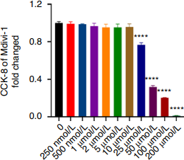
Mdivi-1 purchased from MedChemExpress. Usage Cited in: Bone Res. 2025 Jan 26;13(1):18. [Abstract]
Effect of Mdivi-1 (250 nM; 200 μM) on the proliferation of MC3T3-E1 cells was determined by CCK-8 assay after 2 days of stimulation.
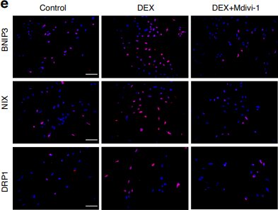
Mdivi-1 purchased from MedChemExpress. Usage Cited in: Bone Res. 2025 Jan 26;13(1):18. [Abstract]
IF staining of BNIP3, NIX, and DRP1 expression in MC3T3-E1 cells treated with DEX and Mdivi1 (5 μM). Scale bar = 50 μm.
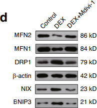
Mdivi-1 purchased from MedChemExpress. Usage Cited in: Bone Res. 2025 Jan 26;13(1):18. [Abstract]
WB analysis of BNIP3, NIX, DRP1, MFN1 and MFN2 expression in MC3T3-E1 cells treated with DEX and Mdivi1 (5 μM).
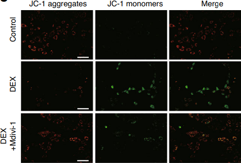
Mdivi-1 purchased from MedChemExpress. Usage Cited in: Bone Res. 2025 Jan 26;13(1):18. [Abstract]
The evaluation MMP was performed using JC-1 staining following DEX and Mdivi1 (5 μM) intervention. Scale bar = 200 μm.
-
Autophagy
Porcine reproductive and respiratory syndrome virus hijacks the non-canonical enzymatic function of PHGDH to arrest autophagic flux for viral replication. [Abstract]2026 Jun 16:1-20. PMID: 42260976 -
Autophagy
Restricting intracellular Salmonella proliferation by coordinating p-TBK1 mediated mitophagy and xenophagy. [Abstract]2026 Mar;22(3):445-467. PMID: 40660474 -
Autophagy
Senecavirus a VP2 protein orchestrates PRDX1 degradation through dual autophagy pathways: macroautophagy and chaperone-mediated autophagy. [Abstract]2026 Jan 1:1-19. PMID: 41479169 -
Autophagy
Viral SAM-binding proteins nsp14 and NP868R reprogram ATG4A-dependent autophagy from antiviral LC3B activity to GABARAP-mediated mitophagy. [Abstract]2026 Jan 1:1-17. PMID: 41432349 -
Autophagy
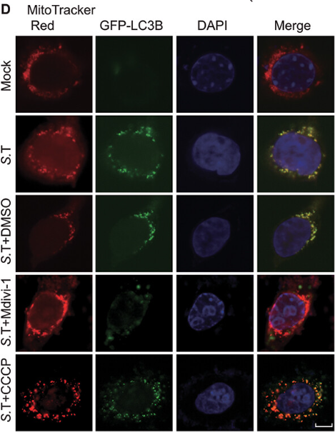
Salmonella Typhimurium persistently infects host via its effector SseJ-induced PHB2-mediated mitophagy. [Abstract]2025 Jun;21(6):1228-1244. PMID: 39902787
Mdivi-1 purchased from MedChemExpress. Usage Cited in: Autophagy. 2025 Jun;21(6):1228-1244. [Abstract]
Mdivi-1 (20 μM) significantly inhibits the S. Typhimurium infection-induced colocalization of mitochondria (labeled by TIMM23) with the autophagy marker LC3B in RAW264.7 cells, indicating that it blocked the occurrence of mitophagy.
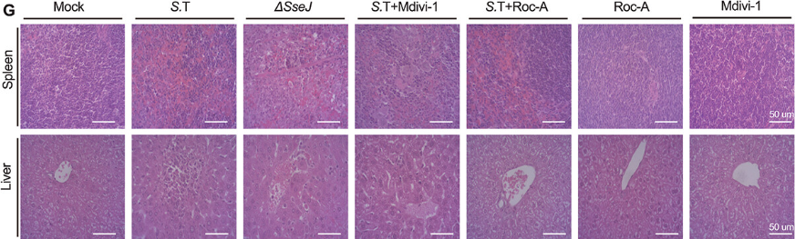
Mdivi-1 purchased from MedChemExpress. Usage Cited in: Autophagy. 2025 Jun;21(6):1228-1244. [Abstract]
Mdivi-1 (50 mg/kg/day; i.p.;Mice infected with S. Typhimurium for 5 consecutive days ). Representative histologicalexaminations and pathological tubular injury scores were performed by H-E staining in mice infected with S. Typhimurium and S.T-ΔSseJ strains and treated withdifferent drugs.
-
Autophagy
Bunyavirus SFTSV nucleoprotein exploits TUFM-mediated mitophagy to impair antiviral innate immunity. [Abstract]2025 Jan;21(1):102-119. PMID: 39189526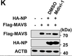
Mdivi-1 purchased from MedChemExpress. Usage Cited in: Autophagy. 2025 Jan;21(1):102-119. [Abstract]
Mdivi-1 (25 μM; 6 h) treatment can rescue the degradation of exogenous and endogenous MAVS induced by SFTSV NP overexpression. 293T cells were co-transfected with Flag-MAVS (2 μg) along with HA-NP plasmid (2 μg) or empty vector (pCAGGS) for 20 h and then treated with Mdivi-1 (25 μM) or vehicle (DMSO) for 6 h. The cell lysates were analyzed with western blot.
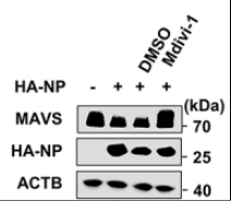
Mdivi-1 purchased from MedChemExpress. Usage Cited in: Autophagy. 2025 Jan;21(1):102-119. [Abstract]
Mdivi-1 (25 μM; 6 h) treatment can rescue the degradation of exogenous and endogenous MAVS induced by SFTSV NP overexpression. 293T cells were co-transfected with HA-NP plasmid (2 μg) or empty vector (pCAGGS) for 20 h and then treated with Mdivi-1 (25 μM) or vehicle (DMSO) for 6 h. The cell lysates were analyzed with western blot.
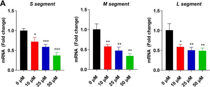
Mdivi-1 purchased from MedChemExpress. Usage Cited in: Autophagy. 2025 Jan;21(1):102-119. [Abstract]
Mdivi-1 (10-25 μM; 24 h) reduces intracellular SFTSV S, M, and L segments mRNA levels in infected HeLa cells.
-
Autophagy
Cannabidiol inhibits human glioma by induction of lethal mitophagy through activating TRPV4. [Abstract]2021 Nov;17(11):3592-3606. PMID: 33629929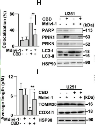
Mdivi-1 purchased from MedChemExpress. Usage Cited in: Autophagy. 2021 Nov;17(11):3592-3606. [Abstract]
Effect of Mdivi-1 on CBD-induced mitophagy in glioma cells. U251 cells were pretreated with mdivi-1 (2.5 μM) for 1 h and then treated with CBD (20 μM) for 24 h. Mitophagy induction was evaluated by immunoblotting of PINK1, PRKN, LC3, TOMM20, and COX4I1.
-
Autophagy
Manipulation of Mitophagy by "All-in-One" nanosensitizer augments sonodynamic glioma therapy. [Abstract]2020 Aug;16(8):1413-1435. PMID: 31674265 -
Nat Commun
CD38 degrades MAVS through mitophagy to inhibit type I interferon secretion in nasopharyngeal carcinoma cells and impairs CD8+T cell-mediated anti-tumor immunity. [Abstract]2026 Feb 9;17(1):2544. PMID: 41663422 -
Nat Commun
Obesity-associated macrophages dictate adipose stem cell ferroptosis and visceral fat dysfunction by propagating mitochondrial fragmentation. [Abstract]2025 Aug 14;16(1):7564. PMID: 40813577 -
Nat Commun
Coronavirus M protein promotes mitophagy over virophagy by recruiting PDPK1 to phosphorylate SQSTM1 at T138. [Abstract]2024 Oct 16;15(1):8927. PMID: 39414765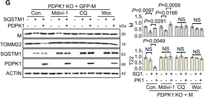
Mdivi-1 purchased from MedChemExpress. Usage Cited in: Nat Commun. 2024 Oct 16;15(1):8927. [Abstract]
Immunoblot analysis of the lysates of PDPK1-KO HEK293T cells transfected with the indicated vectors in the presence or absence of Mdivi-1 (2.5 μM; 4 h), CQ (50 μM; 4 h) or wortmannin (Wor.) (5 μM; 4 h) (n = 3 biological replicates).
-
Nat Commun
Mitochondrial fission induces immunoescape in solid tumors through decreasing MHC-I surface expression. [Abstract]2022 Jul 6;13(1):3882. PMID: 35794100 -
ACS Nano
Gestational Exposure to Black Phosphorus Nanoparticles Induces Placental Trophoblast Dysfunction by Triggering Reactive Oxygen Species-Regulated Mitophagy. [Abstract]2025 Apr 23. PMID: 40264356 -
J Adv Res
Gymconopin C exhibits anti-non-small cell lung cancer effect by regulating miR-6777-5p/ADRB2 pathway to promote mitophagy. [Abstract]2025 Dec 19:S2090-1232(25)01014-8. PMID: 41423048 -
J Adv Res
Peroxisomal dysfunction in cardiac adipose tissue is involved in obesity-associated cardiac hypertrophy. [Abstract]2025 Nov 20:S2090-1232(25)00927-0. PMID: 41274637 -
J Adv Res
Inhibition of mitophagy via the EIF2S1-ATF4-PRKN pathway contributes to viral encephalitis. [Abstract]2025 Jul:73:199-217. PMID: 39103048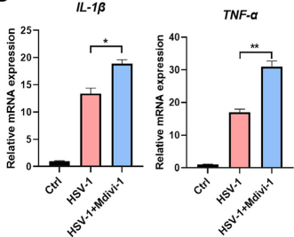
Mdivi-1 purchased from MedChemExpress. Usage Cited in: J Adv Res. 2025 Jul:73:199-217. [Abstract]
Mdivi-1 (10 μM) increases the expression of TNF-α and IL-1β
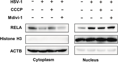
Mdivi-1 purchased from MedChemExpress. Usage Cited in: J Adv Res. 2025 Jul:73:199-217. [Abstract]
Western blot analysis of the cytoplasmic and nuclear proteins in BV2 cells treated with CCCP or Mdivi-1.
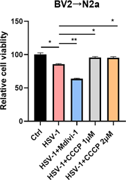
Mdivi-1 purchased from MedChemExpress. Usage Cited in: J Adv Res. 2025 Jul:73:199-217. [Abstract]
N2A cells were treated with conditional medium derived from HSV-1 infected BV2 cells with or without CCCP or Mdivi-1 for 24 h and cell viability was examined.
-
Metabolism
2026 May:178:156567. PMID: 41707754 -
Redox Biol
The UCP2/PINK1/LC3b-mediated mitophagy is involved in the protection of NRG1 against myocardial ischemia/reperfusion injury. [Abstract]2025 Mar:80:103511. PMID: 39874927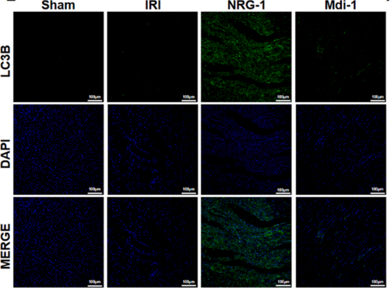
Mdivi-1 purchased from MedChemExpress. Usage Cited in: Redox Biol. 2025 Mar:80:103511. [Abstract]
Mdivi-1 (10 mg/kg; i.p.; single dose) inhibits the NRG-1 postconditioning-induced increase in LC3B fluorescence intensity in rat myocardial tissue.
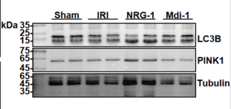
Mdivi-1 purchased from MedChemExpress. Usage Cited in: Redox Biol. 2025 Mar:80:103511. [Abstract]
Mdivi-1 (10 mg/kg; i.p.; single dose) significantly reduces the NRG-1 postconditioning-induced PINK1 protein expression in rat myocardial tissue.
-
Redox Biol
Cardiomyocyte-specific Piezo1 deficiency mitigates ischemia-reperfusion injury by preserving mitochondrial homeostasis. [Abstract]2025 Feb:79:103471. PMID: 39740362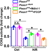
Mdivi-1 purchased from MedChemExpress. Usage Cited in: Redox Biol. 2025 Feb:79:103471. [Abstract]
Cell viability assayed by cellular CCK8 activity of NMCMs subjected to H/R with or without Mdixi-1 or FCCP.
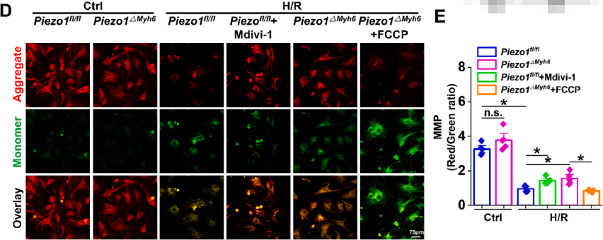
Mdivi-1 purchased from MedChemExpress. Usage Cited in: Redox Biol. 2025 Feb:79:103471. [Abstract]
Mdivi-1 (5 μM). Representative images and statistical data of MMP in NMCMs. Scale bar, 75 μm.
-
Redox Biol
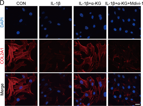
The physiological metabolite α-ketoglutarate ameliorates osteoarthritis by regulating mitophagy and oxidative stress. [Abstract]2023 Jun:62:102663. PMID: 36924682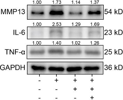
Mdivi-1 purchased from MedChemExpress. Usage Cited in: Redox Biol. 2023 Jun:62:102663. [Abstract]
The effect of Mdivi-1 (50 μM) on α-KG activity in human chondrocytes. The protein expression of MMP13, IL-6, and TNF-α were determined by western blot.
Mdivi-1 purchased from MedChemExpress. Usage Cited in: Redox Biol. 2023 Jun:62:102663. [Abstract]
The effect of Mdivi-1 (50 μM) on the activity of α-KG in human chondrocytes. IF was used to determine protein expression of COL2A1, scale bar = 25 μm, n=3.
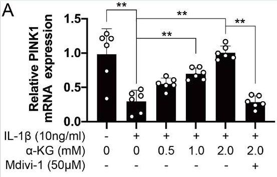
Mdivi-1 purchased from MedChemExpress. Usage Cited in: Redox Biol. 2023 Jun:62:102663. [Abstract]
The effect of Mdivi-1 (50 μM) on α-KG activity in human chondrocytes. RT-qPCR was used to determine the mRNA expression of PINK1.
-
Redox Biol
Targeted up-regulation of Drp1 in dorsal horn attenuates neuropathic pain hypersensitivity by increasing mitochondrial fission. [Abstract]2022 Feb;49:102216. PMID: 34954498 -
Redox Biol
NLRP3 inflammasome-mediated pyroptosis contributes to the pathogenesis of non-ischemic dilated cardiomyopathy. [Abstract]2020 Jul:34:101523. PMID: 32273259 -
Mol Cell
The human antibacterial factor APOL3 couples lysosomal damage to mitochondrial DNA efflux and type I IFN induction. [Abstract]2026 Feb 24:S1097-2765(26)00071-7. PMID: 41742416 -
Gut Microbes
The intestinal microbial metabolite nicotinamide n-oxide prevents herpes simplex encephalitis via activating mitophagy in microglia. [Abstract]Jan-Dec 2022;14(1):2096989. PMID: 35793266 -
J Nanobiotechnology
Lyophilized apoptotic vesicles restore DNA damage and mitochondria dysfunction to ameliorate radiation enteritis. [Abstract]2025 Jul 16;23(1):521. PMID: 40671132 -
J Nanobiotechnology
RGD hydrogel-loaded ADSC extracellular vesicles mitigate uranium-induced renal injury via TLR4/NF-κB pathway inhibition. [Abstract]2025 Feb 17;23(1):114. PMID: 39962465 -
Theranostics
Melanocortin 1 receptor attenuates early brain injury following subarachnoid hemorrhage by controlling mitochondrial metabolism via AMPK/SIRT1/PGC-1α pathway in rats. [Abstract]2021 Jan 1;11(2):522-539. PMID: 33391490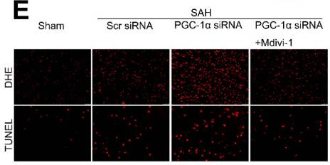
Mdivi-1 purchased from MedChemExpress. Usage Cited in: Theranostics. 2021 Jan 1;11(2):522-539. [Abstract]
Mdivi-1 (1.2 mg/kg; i.v. injection 30 min before SAH) reduces DHE- and TUNEL-positive cells in the SAH+PGC-1α siRNA+Mdivi-1 group when compared with the SAH + PGC-1α group.
-
Theranostics
Berberine Protects Glomerular Podocytes via Inhibiting Drp1-Mediated Mitochondrial Fission and Dysfunction. [Abstract]2019 Feb 28;9(6):1698-1713. PMID: 31037132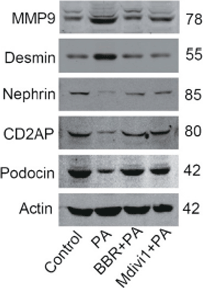
Mdivi-1 purchased from MedChemExpress. Usage Cited in: Theranostics. 2019 Feb 28;9(6):1698-1713. [Abstract]
Western blotting of SDs and markers of podocyte dedifferentiation with the treatment of PA , BBR + PA and Mdivi1+PA.
-
-
Genes Dis
PTEN-induced putative kinase 1 regulates mitochondrial quality control and is essential for the maturation of human induced pluripotent stem cell-derived cardiomyocytes. [Abstract]2022 Sep 10;10(5):2151-2166. PMID: 37492732 -
Adv Sci (Weinh)
Herba Lysimachiae Polysaccharide-Modified Selenium Nanoparticles Alleviate Oxidative Injury in Kidney Stones via TOMM22-Regulated Mitophagy Activation. [Abstract]2026 May 19:e75784. PMID: 42154455 -
Adv Sci (Weinh)
Targeted Extracellular Vesicles Deliver Asiaticoside to Inhibit AURKB/DRP1-Mediated Mitochondrial Fission and Attenuate Hypertrophic Scar Formation. [Abstract]2026 Apr;13(21):e17108. PMID: 41637535 -
Adv Sci (Weinh)
Melatonin Modulates Glucose Metabolism Reprogramming via Targeting G6PD to Alleviate Lead-Induced Hepatocytes Pyroptosis in Common Carp (Cyprinus carpio L.). [Abstract]2025 Aug 11:e01041. PMID: 40788223 -
Cell Rep Med
Screening of patient-derived organoids identifies mitophagy as a cell-intrinsic vulnerability in colorectal cancer during statin treatment. [Abstract]2025 Apr 15;6(4):102039. PMID: 40154491 -
Biomaterials
A sono-piezoelectric scaffold prompts disc regeneration by activating Ca2+/CaMKII/Parkin-mediated mitophagy. [Abstract]2026 May 15:334:124316. PMID: 42155175 -
Cell Death Differ
CEND1 deficiency induces mitochondrial dysfunction and cognitive impairment in Alzheimer's disease. [Abstract]2022 Dec;29(12):2417-2428. PMID: 35732922 -
Carbohydr Polym
Aloe gel glucomannan induced colon cancer cell death via mitochondrial damage-driven PINK1/Parkin mitophagy pathway. [Abstract]2022 Nov 1:295:119841. PMID: 35989033 -
Cell Death Dis
Glutamine metabolic stress induces SLC25A6-dependent mitofission via MIC60-MIC19 complex disassembly in colorectal cancer. [Abstract]2026 Apr 23;17(1):537. PMID: 42020360 -
Pharmacol Res
Mdivi-1 promotes lipid droplets-mitochondria contact and ameliorates cardiac lipotoxicity in high-fat diet-fed mice. [Abstract]2025 Dec:222:108028. PMID: 41205727 -
Environ Sci Technol
2025 Oct 28;59(42):22423-22438. PMID: 41104744 -
Environ Sci Technol
Chronic Dietary Exposure to Environmental Levels of Glyphosate Increases the Risk of Reproductive Dysfunction in Male Mice. [Abstract]2025 Aug 5;59(30):15705-15719. PMID: 40698942 -
Pharmacol Res
NSD2 modulates Drp1-mediated mitochondrial fission in chronic renal allograft interstitial fibrosis by methylating STAT1. [Abstract]2024 Feb:200:107051. PMID: 38190956 -
Pharmacol Res
Targeting P2RX1 alleviates renal ischemia/reperfusion injury by preserving mitochondrial dynamics. [Abstract]2021 Aug:170:105712. PMID: 34091010 -
Cell Death Dis
Inhibition of PINK1/Parkin-dependent mitophagy sensitizes multidrug-resistant cancer cells to B5G1, a new betulinic acid analog. [Abstract]2019 Mar 8;10(3):232. PMID: 30850585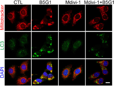
Mdivi-1 purchased from MedChemExpress. Usage Cited in: Cell Death Dis. 2019 Mar 8;10(3):232. [Abstract]
HepG2/ADM cells are pretreated with mdivi-1 (10 μM) for 1 h, followed by treatment with B5G1 (6 μM) for another 24 h. Mitochondrial colocalization with LC3 is detected by immunofluorescence.
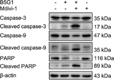
Mdivi-1 purchased from MedChemExpress. Usage Cited in: Cell Death Dis. 2019 Mar 8;10(3):232. [Abstract]
HepG2/ADM cells are pretreated with mdivi-1 (10 μM) for 1 h, followed by treatment with B5G1 (6 μM) for another 48 h. Apoptosis-related proteins expression level is detected by western blotting. β-actin was used as a loading control.
-
Pharmacol Res
Paris Saponin II inhibits colorectal carcinogenesis by regulating mitochondrial fission and NF-κB pathway. [Abstract]2019 Jan:139:273-285. PMID: 30471409 -
Cell Death Dis
2018 Sep 5;9(9):907. PMID: 30185782 -
Small
Optimally Aligned Nerve Scaffolds with Sustained Astaxanthin Release Improve the Inflammatory Microenvironment through Mitophagy Activation. [Abstract]2025 Jul;21(26):e2502939. PMID: 40370272 -
Cancer Lett
Blockade of the lncRNA-PART1-PHB2 axis confers resistance to PARP inhibitor and promotes cellular senescence in ovarian cancer. [Abstract]2024 Oct 10:602:217192. PMID: 39181433 -
Small
Enhanced Chemodynamic Therapy Mediated by a Tumor-Specific Catalyst in Synergy with Mitophagy Inhibition Improves the Efficacy for Endometrial Cancer. [Abstract]2023 Aug;19(33):e2301497. PMID: 37086131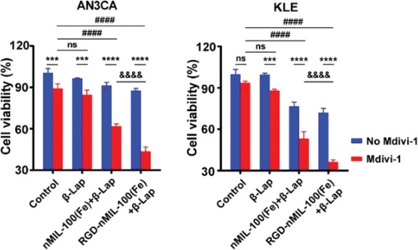
Mdivi-1 purchased from MedChemExpress. Usage Cited in: Small. 2023 Aug;19(33):e2301497. [Abstract]
Effect of different treatment combinations, including nMIL-100(Fe) (12.5 µg/mL, 48 h), RGD-nMIL-100(Fe) (12.5 µg/mL, 48 h), β-Lap (1.0 × 10⁻⁶ m, 4 h) and Mdivi-1(10 × 10−6 m, 1 h) pretreatment on KLE and AN3CA cell viability.
-
Int J Biol Sci
Enhancing mitophagy by ligustilide through BNIP3-LC3 interaction attenuates oxidative stress-induced neuronal apoptosis in spinal cord injury. [Abstract]2024 Aug 12;20(11):4382-4406. PMID: 39247814 -
Int J Biol Sci
Mitochondrial Dysfunction by FADDosome Promotes Gastric Mucosal Injury in Portal Hypertensive Gastropathy. [Abstract]2024 Apr 29;20(7):2658-2685. PMID: 38725851 -
J Neuroinflammation
Drp1 mitochondrial fission in astrocyte modulates behavior and neuroinflammation during morphine addiction. [Abstract]2025 Apr 17;22(1):108. PMID: 40247294 -
J Neuroinflammation
Morphine-induced microglial immunosuppression via activation of insufficient mitophagy regulated by NLRX1. [Abstract]2022 Apr 12;19(1):87. PMID: 35414088 -
Phytomedicine
Kakkalide promotes spinal cord injury repair by regulating microglial M2 polarization via mitophagy. [Abstract]2026 Apr:153:157985. PMID: 41720005 -
Phytomedicine
Anti-tumor effects of Guggulsterone in osteosarcoma: Role of SIRT3-mediated PINK1-Parkin mitophagy activation. [Abstract]2026 Jan 20:153:157860. PMID: 41650525 -
Phytomedicine
Kaempferol attenuated LPS-induced microglial neurotoxicity by promoting mitophagy to inhibit mtDNA-mediated NLRP3 inflammasome activation. [Abstract]2025 Nov 25:148:157486. PMID: 41202376 -
Phytomedicine
Allicin alleviates traumatic brain injury-induced neuroinflammation by enhancing PKC-δ-mediated mitophagy. [Abstract]2025 Feb 21:139:156500. PMID: 39986225 -
Phytomedicine
Paeoniflorin suppresses kidney inflammation by regulating macrophage polarization via KLF4-mediated mitophagy. [Abstract]2023 Jul 25:116:154901. PMID: 37247587 -
Mater Today Bio
Layered double hydroxide nanocarriers loaded with butylphthalide attenuate the AKI-CKD transition by regulating mitophagy. [Abstract]2026 Jun 23;39:103393. PMID: 42440424 -
Adv Healthc Mater
Antigen-Enriched Tumor-Dendritic Cell Fusion Membrane-Coated Metal-Phenolic Nanovaccine Enables Dual T-Cell Activation for Robust Cancer Immunotherapy. [Abstract]2026 Jan 19:e05255. PMID: 41549866 -
Arch Toxicol
2,2',4,4'-tetrabromodiphenyl ether (BDE-47) induces early hearing loss in guinea pigs via activating AhR to trigger mitochondrial and endoplasmic reticulum stress-regulated autophagy. [Abstract]2025 Aug 20. PMID: 40835755 -
Environ Health Perspect
2022 May;130(5):57004. PMID: 35511227 -
J Hazard Mater
CYP450 activation and mitophagy induction mediate Ti3C2 MXene detoxification in RAW 264.7 cells. [Abstract]2025 Dec 5:500:140498. PMID: 41252987 -
J Hazard Mater
Gestational exposure to environmental cadmium induces placental apoptosis and fetal growth restriction via Parkin-modulated MCL-1 degradation. [Abstract]2022 Feb 15;424(Pt A):127268. PMID: 34583167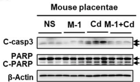
Mdivi-1 purchased from MedChemExpress. Usage Cited in: J Hazard Mater. 2022 Feb 15;424(Pt A):127268. [Abstract]
Mdivi-1 (25 mg/kg, i.p.) significantly reduces the levels of apoptosis-related proteins (including C-Casp3 and C-PARP) in the placentas of Cd-exposed CD-1 mice.
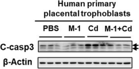
Mdivi-1 purchased from MedChemExpress. Usage Cited in: J Hazard Mater. 2022 Feb 15;424(Pt A):127268. [Abstract]
Mdivi-1 (5 μM; 1 h) rescues environmental cadmium-induced apoptosis in human primary trophoblasts by decreasing C-Casp3 levels.
-
Cell Death Discov
Inhibiting HSP27 activates the XBP1s/CerS1 interplay, which triggers DRP1-driven mitophagy, thereby protecting against cell death and promoting the KSHV lytic cycle in primary effusion lymphoma cells. [Abstract]2026 Feb 26. PMID: 41748537 -
Cell Death Discov
Mitochondrial retrograde signaling initiates HIF-1α/BNIP3/NIX-mediated mitophagy in Tibetan high-altitude adaptation. [Abstract]2026 Jan 6;12(1):81. PMID: 41490888 -
Mol Psychiatry
Early-life inflammation increases aggressive behavior in adult male mice through an astrocyte-neuron signaling. [Abstract]2025 Sep 16. PMID: 40954280 -
Cell Death Discov
Mitochondrial homeostasis regulates definitive endoderm differentiation of human pluripotent stem cells. [Abstract]2022 Feb 17;8(1):69. PMID: 35177589 -
Environ Int
Environmental cadmium exposure induces fetal growth restriction via triggering PERK-regulated mitophagy in placental trophoblasts. [Abstract]2021 Feb:147:106319. PMID: 33348103 -
-
J Transl Med
Human neural stem cell-derived exosomes activate PINK1/Parkin pathway to protect against oxidative stress-induced neuronal injury in ischemic stroke. [Abstract]2025 Apr 5;23(1):402. PMID: 40188077 -
J Transl Med
Staphylococcus aureus induces mitophagy via the HDAC11/IL10 pathway to sustain intracellular survival. [Abstract]2025 Feb 4;23(1):156. PMID: 39905391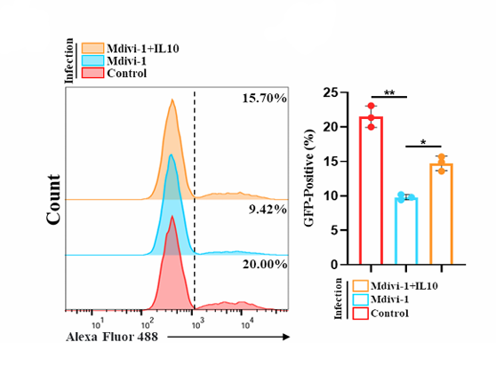
Mdivi-1 purchased from MedChemExpress. Usage Cited in: J Transl Med. 2025 Feb 4;23(1):156. [Abstract]
Flow cytometry analysis of the impact of Mdivi-1 (20µM) pretreatment, with or without added IL10 (1ng/ml), on the bacterial load of BMDMs infected with GFP-S. aureus (MOI = 10) for 24 h. Quantitative analysis of the proportion of BMDMs infected with GFP-S. aureus.
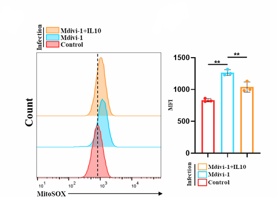
Mdivi-1 purchased from MedChemExpress. Usage Cited in: J Transl Med. 2025 Feb 4;23(1):156. [Abstract]
Flow cytometry analysis of mtROS levels in BMDMs 24 h after infection with S. aureus (MOI = 10) in various treatment groups. Quantitative analysis of intracellular mtROS levels (IL-10:1 ng/mL, 24 h,Mdivi-1:20 µM, 24 h).
-
J Transl Med
Matrix stiffness induces Drp1-mediated mitochondrial fission through Piezo1 mechanotransduction in human intervertebral disc degeneration. [Abstract]2023 Oct 10;21(1):711. PMID: 37817199 -
J Transl Med
Mitochondrial ROS driven by NOX4 upregulation promotes hepatocellular carcinoma cell survival after incomplete radiofrequency ablation by inducing of mitophagy via Nrf2/PINK1. [Abstract]2023 Mar 25;21(1):218. PMID: 36964576 -
J Colloid Interface Sci
Redox-engineered gold single-atom nanozymes orchestrate mitochondria-driven PANoptosis for energy-independent cancer catalytic therapy. [Abstract]2025 Sep 25;703(Pt 1):139108. PMID: 41045899 -
Basic Res Cardiol
Protective effect of HINT2 on mitochondrial function via repressing MCU complex activation attenuates cardiac microvascular ischemia-reperfusion injury. [Abstract]2021 Dec 16;116(1):65. PMID: 34914018 -
Chin Med J (Engl)
Inhibitory effect of blestriarene C on triple-negative breast cancer: Inducing ferroptosis and mitophagy via SESN2/AKT/FOXO4 axis. [Abstract]2026 Mar 5;139(5):699-709. PMID: 41527177 -
Oncogene
2025 Aug;44(32):2893-2906. PMID: 40468051 -
Int J Surg
Targeting SIRT3 to regulate mitophagy-dependent ferroptosis for preventing glucocorticoid-induced osteoporosis. [Abstract]2025 Jul 2. PMID: 40607908 -
Int J Biol Macromol
HSP70 preserves brain microvascular endothelial integrity under heat stress associated with suppressed JNK-mediated apoptosis and mitophagy. [Abstract]2026 Apr:353:151239. PMID: 41791534 -
Int J Biol Macromol
Lentinan rewrites extracellular matrix homeostasis by activating mitophagy via mTOR/PINK1/Parkin pathway in cartilage to alleviating osteoarthritis. [Abstract]2025 Sep;322(Pt 3):146900. PMID: 40819752 -
BMC Med
Enhancing anti-CD3 mAb-mediated diabetes remission in autoimmune diabetes through regulation of dynamin-related protein 1(Drp1)-mediated mitochondrial dynamics in exhausted CD8+T-cell subpopulations. [Abstract]2025 Mar 31;23(1):189. PMID: 40165248 -
Int J Nanomedicine
Enhancing of Nanocatalyst-Driven Chemodynaminc Therapy for Endometrial Cancer Cells Through Inhibition of PINK1/Parkin-Mediated Mitophagy. [Abstract]2021 Sep 29;16:6661-6679. PMID: 34616150 -
-
Mol Med
Activation of the MEK1-CHK2 axis in macrophages by Staphylococcus aureus promotes mitophagy, resulting in a reduction in bactericidal efficacy. [Abstract]2025 May 29;31(1):211. PMID: 40437411 -
Antioxidants (Basel)
Isoquercitrin from Apocynum venetum L. Exerts Antiaging Effects on Yeasts via Stress Resistance Improvement and Mitophagy Induction through the Sch9/Rim15/Msn Signaling Pathway. [Abstract]2023 Oct 31;12(11):1939. PMID: 38001792 -
Antioxidants (Basel)
O-GlcNAcylation Is Required for the Survival of Cerebellar Purkinje Cells by Inhibiting ROS Generation. [Abstract]2023 Mar 26;12(4):806. PMID: 37107182 -
Phytother Res
Paeoniflorin Ameliorates Liver Fibrosis by Inhibiting HIF-1α-Mediated Mitophagy in Hepatic Stellate Cells. [Abstract]2026 Jul;40(7):3990-4004. PMID: 42017683 -
Phytother Res
Resveratrol prevents Drp1-mediated mitochondrial fission in the diabetic kidney through the PDE4D/PKA pathway. [Abstract]2023 Dec;37(12):5916-5931. PMID: 37767771 -
Free Radic Biol Med
PARS2 deficiency impairs mitochondrial homeostasis and activates ferroptotic to drive developmental and epileptic encephalopathy. [Abstract]2026 Jun 7:254:307-322. PMID: 42259431 -
Free Radic Biol Med
DRP1-mediated mitophagy facilitates oral squamous cell carcinoma tumor cell survival under glucose restriction. [Abstract]2026 Jun 27:254:391-404. PMID: 42364552 -
Free Radic Biol Med
Optineurin enhances the chemoresistance by activating mitophagy in acute myeloid leukemia with NPM1 mutation. [Abstract]2026 May:248:162-176. PMID: 41722661 -
Free Radic Biol Med
Lysophosphatidylcholine promotes pulmonary fibrosis following lung injury by facilitating alveolar type 2 cell senescence via Mfsd2a-dependent, Drp1-mediated mitochondrial fission. [Abstract]2026 May:248:255-271. PMID: 41740689 -
Free Radic Biol Med
SIRT1 activation restores PINK1-dependent mitophagy to reverse airway barrier dysfunction in Acinetobacter baumannii infection. [Abstract]2026 Mar 25:250:153-167. PMID: 41895413 -
Free Radic Biol Med
The non-metabolic role of MTHFD2 in regulating mitochondrial fission-dependent mitophagy via stabilizing TOP2A mRNA in glioblastoma. [Abstract]2026 Mar 16:246:93-106. PMID: 41534569 -
Free Radic Biol Med
Nsun2-mediated m5C methylation of Ncor1 exacerbates sepsis-induced cardiomyopathy by promoting mitochondrial dysfunction. [Abstract]2025 Dec 30:S0891-5849(25)01474-1. PMID: 41478419 -
Free Radic Biol Med
Redox switch C674 in SERCA2 triggers Ca2+-calcineurin-MCU-Drp1 cascade and pulmonary vascular remodeling. [Abstract]2025 Dec 16:244:380-394. PMID: 41412527 -
Free Radic Biol Med
TFEB-nuclear translocation promotes BNIP3-mediated mitophagy and alleviates oxidative stress and ferroptosis in acute pancreatitis. [Abstract]2025 Nov 23:243:398-413. PMID: 41290101 -
Free Radic Biol Med
HDAC inhibition protects RPE cells from oxidative stress via enhanced mitochondrial fusion, cytoskeletal repair, and Nrf-2 activation. [Abstract]2025 Aug 13:240:59-70. PMID: 40816649 -
Free Radic Biol Med
Sevoflurane exposure accelerates the onset of cognitive impairment via promoting p-Drp1S616-mediated mitochondrial fission in a mouse model of Alzheimer's disease. [Abstract]2024 Nov 20:225:699-710. PMID: 39490772 -
Free Radic Biol Med
TRPA1 protects against contrast-induced renal tubular injury by preserving mitochondrial dynamics via the AMPK/DRP1 pathway. [Abstract]2024 Nov 1:224:521-539. PMID: 39278575 -
Free Radic Biol Med
The tolerable upper intake level of manganese alleviates Parkinson-like motor performance and neuronal loss by activating mitophagy. [Abstract]2024 Nov 20:225:665-676. PMID: 39401732 -
Free Radic Biol Med
Hydrogen alleviates impaired lung epithelial barrier in acute respiratory distress syndrome via inhibiting Drp1-mediated mitochondrial fission through the Trx1 pathway. [Abstract]2024 Jun:218:132-148. PMID: 38554812 -
Free Radic Biol Med
Quercetin induces ferroptosis in gastric cancer cells by targeting SLC1A5 and regulating the p-Camk2/p-DRP1 and NRF2/GPX4 Axes. [Abstract]2024 Mar:213:150-163. PMID: 38190923 -
Free Radic Biol Med
Crosstalk between reactive oxygen species and Dynamin-related protein 1 in periodontitis. [Abstract]2021 Aug 20:172:19-32. PMID: 34052344 -
Free Radic Biol Med
Mdivi-1 ameliorates early brain injury after subarachnoid hemorrhage via the suppression of inflammation-related blood-brain barrier disruption and endoplasmic reticulum stress-based apoptosis. [Abstract]2017 Nov:112:336-349. PMID: 28790012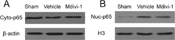
Mdivi-1 purchased from MedChemExpress. Usage Cited in: Free Radic Biol Med. 2017 Nov:112:336-349. [Abstract]
Mdivi-1 administration reduces NF-κB-dependent inflammation. A. Western blot assay and quantitative analysis of Cyto-p65 (A) and Nec-p65 (B) expression. ß-actin and H3 are used as internal controls of cytosolic and nuclear subfractions, respectively.
-
Clin Transl Med
Mitochondrial dysfunction induced by HIF-1α under hypoxia contributes to the development of gastric mucosal lesions. [Abstract]2024 Apr;14(4):e1653. PMID: 38616702 -
J Anim Sci Biotechnol
DRP1 deficiency induces mitochondrial dysfunction and oxidative stress-mediated apoptosis during porcine oocyte maturation. [Abstract]2020 Aug 5:11:77. PMID: 32782788 -
Stem Cell Res Ther
Mesenchymal stem cells inhibit mitochondrial fission by upregulating armadillo repeat containing 1, ameliorating oxidative stress in renal fibrosis. [Abstract]2025 Oct 28;16(1):584. PMID: 41153008 -
Stem Cell Res Ther
Mesenchymal stem cell-derived small extracellular vesicles reduced hepatic lipid accumulation in MASLD by suppressing mitochondrial fission. [Abstract]2025 Mar 5;16(1):116. PMID: 40045380 -
Cell Rep
Maternal hyperglycemia disrupts cardiomyocyte maturation via aberrant nucleotide metabolism and suppression of AMPK signaling. [Abstract]2026 May 26;45(5):117321. PMID: 42090289 -
Aging Cell
β-Hydroxybutyrate enhances chondrocyte mitophagy and reduces cartilage degeneration in osteoarthritis via the HCAR2/AMPK/PINK1/Parkin pathway. [Abstract]2024 Nov;23(11):e14294. PMID: 39126207 -
Cell Rep
CLOCK regulates Drp1 mRNA stability and mitochondrial homeostasis by interacting with PUF60. [Abstract]2022 Apr 12;39(2):110635. PMID: 35417690 -
World J Gastroenterol
PTEN-induced kinase 1-induced dynamin-related protein 1 Ser637 phosphorylation reduces mitochondrial fission and protects against intestinal ischemia reperfusion injury. [Abstract]2020 Apr 21;26(15):1758-1774. PMID: 32351292 -
Cell Prolif
Dissection of Mitochondrial Function via Chemical Perturbation and Single-Cell Profiling. [Abstract]2026 Apr 27:e70216. PMID: 42044679 -
Cell Prolif
Targeting ROCK1/YAP1 Axis Ameliorates Inflammation-Induced Prostatic Hyperplasia via Stabilising SIRT1-Dependent Mitochondrial Dynamics. [Abstract]2025 Jul 4:e70085. PMID: 40616266 -
Cell Prolif
Optineurin Cooperates With NRF2 to Regulate Tooth Root Morphogenesis by Controlling Mitochondrial Dynamics and Apoptosis. [Abstract]2025 May;58(5):e13799. PMID: 39762159 -
Cell Prolif
ERK/Drp1-dependent mitochondrial fission contributes to HMGB1-induced autophagy in pulmonary arterial hypertension. [Abstract]2021 Jun;54(6):e13048. PMID: 33948998 -
J Pineal Res
Melatonin prevents Drp1-mediated mitochondrial fission in diabetic hearts through SIRT1-PGC1α pathway. [Abstract]2018 Sep;65(2):e12491. PMID: 29575122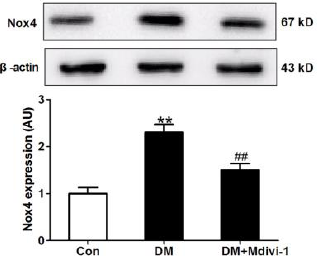
Mdivi-1 purchased from MedChemExpress. Usage Cited in: J Pineal Res. 2018 Sep;65(2):e12491. [Abstract]
Inhibition of mitochondrial fission with mdivi-1 or Drp1 siRNA suppresses hyperglycemia-induced oxidative stress. Protein expression of Nox4.
-
J Pineal Res
Dynamin-related protein 1-mediated mitochondrial fission contributes to post-traumatic cardiac dysfunction in rats and the protective effect of melatonin. [Abstract]2018 Jan;64(1). PMID: 29024001 -
Clin Sci
Glutamine alleviates immunosuppression in polymicrobial sepsis by augmenting bacterial phagocytosis through sustaining the GFAT-DRP1 dependent mitochondrial calcium dynamics. [Abstract]2025 Oct 22;139(20):1163-1185. PMID: 41082631 -
Environ Pollut
Disturbance of mitochondrial dynamics led to spermatogenesis disorder in mice exposed to polystyrene micro- and nanoplastics. [Abstract]2024 Dec 1:362:124935. PMID: 39260550 -
Environ Pollut
TDCPP and TiO2 NPs aggregates synergistically induce SH-SY5Y cell neurotoxicity by excessive mitochondrial fission and mitophagy inhibition. [Abstract]2024 Apr 15:347:123740. PMID: 38462198 -
-
Atherosclerosis
Valine acts as an early biomarker and exacerbates pathological cardiac hypertrophy by impairing mitochondrial quality control. [Abstract]2025 Jun:405:119216. PMID: 40318256 -
Atherosclerosis
Irisin alleviates vascular calcification by inhibiting VSMC osteoblastic transformation and mitochondria dysfunction via AMPK/Drp1 signaling pathway in chronic kidney disease. [Abstract]2022 Apr:346:36-45. PMID: 35255258 -
Cancer Cell Int
Abiraterone and MDV3100 inhibits the proliferation and promotes the apoptosis of prostate cancer cells through mitophagy. [Abstract]2019 Dec 10:19:332. PMID: 31827406 -
Redox Rep
CISD2 regulates oxidative stress and mitophagy to maintain the balance of the follicular microenvironment in PCOS. [Abstract]2024 Dec;29(1):2377870. PMID: 39010730 -
Lipids Health Dis
Dihydromyricetin suppresses endothelial NLRP3 inflammasome activation and attenuates atherogenesis by promoting mitophagy. [Abstract]2024 Sep 3;23(1):279. PMID: 39227809 -
J Ethnopharmacol
Dendrobine ameliorates non-alcoholic fatty liver disease by inhibiting mitochondrial fission through modulation of the Wnt5a/p-CaMKII/p-Drp1 signaling axis. [Abstract]2026 May 23:363:121445. PMID: 41763618 -
J Ethnopharmacol
Realgar transforming solution suppresses KG-1a-derived CD34+CD38- acute myeloid leukemia stem cell-like phenotypes in association with ER-mitochondrial stress and mitophagy-related alterations. [Abstract]2026 May 25:369:121896. PMID: 42184909 -
J Ethnopharmacol
Integrating UPLC-Q-TOF-MS/MS, feature-based molecular networking, network pharmacology, and molecular docking to investigate the mechanism of Persicae Ramulus against myocardial ischemia. [Abstract]2026 Feb 28:357:120899. PMID: 41241210 -
Antioxid Redox Signal
Hydrogen Attenuates Oxidative Damage via NRF2-Mediated Mitophagy after Subarachnoid Hemorrhage. [Abstract]2025 Dec 26. PMID: 41468154 -
Antioxid Redox Signal
Suppression of CDK1/Drp1-mediated mitochondrial fission attenuates dexamethasone-induced extracellular matrix deposition in the trabecular meshwork. [Abstract]2025 Feb;42(4-6):249-264. PMID: 39096204 -
J Agric Food Chem
Magnoflorine Ameliorates Chronic Kidney Disease in High-Fat and High-Fructose-Fed Mice by Promoting Parkin/PINK1-Dependent Mitophagy to Inhibit NLRP3/Caspase-1-Mediated Pyroptosis. [Abstract]2024 Jun 5;72(22):12775-12787. PMID: 38776285 -
J Agric Food Chem
Conjugated Linoleic Acid Ameliorates Hydrogen Peroxide-Induced Mitophagy and Inflammation via the DRP1-mtDNA-STING Pathway in Bovine Hepatocytes. [Abstract]2024 Jan 31;72(4):2120-2134. PMID: 38235560 -
Ecotoxicol Environ Saf
Polystyrene nanoplastics induce cognitive dysfunction and dendritic spine deterioration via excessive mitochondrial fission. [Abstract]2025 Oct 1:304:119133. PMID: 41038023 -
Ecotoxicol Environ Saf
Mechanistic study on nonylphenol-induced liver fibrosis via Pink1/Parkin-mediated mitophagy and lipid droplet degradation in hepatic stellate cells. [Abstract]2025 Oct 15:305:119206. PMID: 41106202 -
Ecotoxicol Environ Saf
2024 Jun 1:277:116392. PMID: 38677065 -
CNS Neurosci Ther
Hippocampal mitophagy contributes to spatial memory via maintaining neurogenesis during the development of mice. [Abstract]2024 Jun;30(6):e14800. PMID: 38887162 -
Biochem Pharmacol
Ginsenoside Rg1 alleviates MASH by targeting GLS2 to enhance PINK1/Parkin-mediated mitophagy and improve mitochondrial function. [Abstract]2026 Jul:249:117891. PMID: 41833827 -
-
Cell Biosci
Selonsertib, a potential drug for liver failure therapy by rescuing the mitochondrial dysfunction of macrophage via ASK1-JNK-DRP1 pathway. [Abstract]2021 Jan 7;11(1):9. PMID: 33413667 -
Precis Clin Med
Agrimol B inhibits pancreatic ductal adenocarcinoma by induction of lethal mitophagy through decreasing mitochondrial transcription termination factor 3. [Abstract]2026 Mar 14;9(2):pbag009. PMID: 41978696 -
Cancer Gene Ther
LAMC2 mitigates ER stress by enhancing ER-mitochondria interaction via binding to MYH9 and MYH10. [Abstract]2024 Jan;31(1):43-57. PMID: 37891404 -
Life Sci
Quercetin alleviates kidney fibrosis by reducing renal tubular epithelial cell senescence through the SIRT1/PINK1/mitophagy axis. [Abstract]2020 Sep 15:257:118116. PMID: 32702447 -
Food Funct
Lactoferrin protects against radiation-induced intestinal injury by regulating pyroptosis and mitophagy. [Abstract]2026 Jan 26;17(2):1045-1060. PMID: 41524100 -
Prog Orthod
2026 Jan 13;27(1):1. PMID: 41528413 -
Food Funct
Iron deficiency exacerbates aortic medial degeneration by inducing excessive mitochondrial fission. [Abstract]2022 Jul 18;13(14):7666-7683. PMID: 35735054 -
-
-
Transl Res
Inhibition of dynamin-related protein 1 has neuroprotective effect comparable with therapeutic hypothermia in a rat model of cardiac arrest. [Abstract]2018 Apr:194:68-78. PMID: 29351829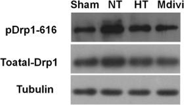
Mdivi-1 purchased from MedChemExpress. Usage Cited in: Transl Res. 2018 Apr:194:68-78. [Abstract]
Representative western blot analysis of pDrp1-616 and total Drp1 protein expression in the hippocampus (left). Tubulin is used as the loading control. Protein expressions are quantified by western blot band intensity (right).
-
Pharm Biol
Harmine derivative H-2-168 induces the death of Echinococcus granulosus by regulating mitochondrial fusion and fission. [Abstract]2025 Dec;63(1):188-200. PMID: 40188381 -
Commun Biol
Elevated levels of S100A8 and S100A9 exacerbate muscle mitochondrial fragmentation in sepsis-induced muscle atrophy. [Abstract]2025 Feb 28;8(1):338. PMID: 40021770 -
Radiother Oncol
Mitophagy induction improves salivary gland stem/progenitor cell function by reducing senescence after irradiation. [Abstract]2024 Jan:190:110028. PMID: 38007043 -
Commun Biol
Activating Parkin-dependent mitophagy alleviates oxidative stress, apoptosis, and promotes random-pattern skin flaps survival. [Abstract]2022 Jun 22;5(1):616. PMID: 35732814 -
Eur J Pharmacol
Acetaldehyde dehydrogenase 2 attenuates lipopolysaccharide -induced endothelial barrier damage by inhibiting mitochondrial fission in sepsis-associated encephalopathy. [Abstract]2025 Jun 15:997:177468. PMID: 40054720 -
Cell Biol Toxicol
NPR1 promotes cisplatin resistance by inhibiting PARL-mediated mitophagy-dependent ferroptosis in gastric cancer. [Abstract]2024 Oct 30;40(1):93. PMID: 39476297 -
Eur J Pharmacol
Chronic mitochondrial dynamic-targeted therapy alleviates left ventricular dysfunction by reducing multiple programmed cell death in post-myocardial infarction rats. [Abstract]2024 Aug 15:977:176736. PMID: 38878877 -
Respir Res
Macrophage migration inhibitory factor exacerbates asthmatic airway remodeling via dynamin-related protein 1-mediated autophagy activation. [Abstract]2023 Sep 6;24(1):216. PMID: 37674165 -
Eur J Pharmacol
Empagliflozin ameliorates cardiac dysfunction in heart failure mice via regulating mitochondrial dynamics. [Abstract]2023 Mar 5:942:175531. PMID: 36690056 -
Cell Biol Toxicol
Inhibition of calpain reduces cell apoptosis by suppressing mitochondrial fission in acute viral myocarditis. [Abstract]2022 Jun;38(3):487-504. PMID: 34365571 -
Biomolecules
Malic Enzyme 2 Regulates Dynamin-Related Protein 1-Dependent Mitochondrial Fission and Mitochondria-Associated Membranes to Drive Odontogenic Differentiation: An In Vitro and In Vivo Study. [Abstract]2026 Apr 30;16(5):664. PMID: 42194014 -
Biomolecules
Mitophagy Activation via the YAP/Parkin Pathway Underlies the Neuroprotective Action of Tetramethylpyrazine in Cerebral Ischemia/Reperfusion Injury. [Abstract]2026 Mar 13;16(3):429. PMID: 41897365 -
Int Immunopharmacol
DPEP1 mediates regulation of mitochondrial quality control via FOXO1/ALDH1L2 axis to attenuate ferroptosis in pulmonary endothelial cells to alleviate sepsis-associated acute lung injury. [Abstract]2026 Feb 1:170:116049. PMID: 41421227 -
Int J Mol Sci
Endoplasmic Reticulum Stress Induced by Turbulence of Mitochondrial Fusion and Fission Was Involved in Isoproterenol-Induced H9c2 Cell Injury. [Abstract]2026 Jan 30;27(3):1390. PMID: 41683814 -
Int Immunopharmacol
GLP-1/GIP dual agonist tirzepatide alleviates mice model of Parkinson's disease by promoting mitochondrial homeostasis. [Abstract]2025 Nov 14:165:115443. PMID: 40886502 -
Int Immunopharmacol
Aβ impairs bone vascular homeostasis in APP/PS1 mice via disrupting the mitochondrial fission-efferocytosis axis in macrophages. [Abstract]2025 Sep 10:165:115526. PMID: 40934542 -
Int Immunopharmacol
Mitochondrial calcium overload contributes to mechanical allodynia in neuropathic pain via inducing mitochondrial dynamic imbalance. [Abstract]2025 Jun 17:158:114863. PMID: 40359888 -
Int Immunopharmacol
Ginsenoside Rh1 attenuates chondrocyte senescence and osteoarthritis via AMPK/PINK1/Parkin-mediated mitophagy. [Abstract]2025 Jun 26:159:114911. PMID: 40409109 -
Int Immunopharmacol
Dendrobine attenuates sepsis-associated acute kidney injury by promoting PINK1/PARKIN-mediated mitophagy. [Abstract]2025 Jun 5:157:114741. PMID: 40306112 -
Int J Mol Sci
Vital Role of PINK1/Parkin-Mediated Mitophagy of Pulmonary Epithelial Cells in Severe Pneumonia Induced by IAV and Secondary Staphylococcus aureus Infection. [Abstract]2025 Apr 27;26(9):4162. PMID: 40362402 -
Biomolecules
Mechanism of Mitophagy to Protect Yak Kidney from Hypoxia-Induced Fibrosis Damage by Regulating Ferroptosis Pathway. [Abstract]2025 Apr 9;15(4):556. PMID: 40305351 -
Int Immunopharmacol
AdipoRon exerts an antidepressant effect by inhibiting NLRP3 inflammasome activation in microglia via promoting mitophagy. [Abstract]2024 Nov 15:141:113011. PMID: 39213872 -
Int Immunopharmacol
Targeting TGR5 to mitigate liver fibrosis: Inhibition of hepatic stellate cell activation through modulation of mitochondrial fission. [Abstract]2024 Oct 25:140:112831. PMID: 39111149 -
Int Immunopharmacol
Oxidative stress induces ferroptosis in tendon stem cells by regulating mitophagy through cGAS-STING pathway. [Abstract]2024 Sep 10:138:112652. PMID: 38986301 -
Int Immunopharmacol
GDF11 promotes osteogenic/odontogenic differentiation of dental pulp stem cells to accelerate dentin restoration via modulating SIRT3/FOXO3-mediated mitophagy. [Abstract]2024 Sep 23;142(Pt B):113092. PMID: 39317051 -
Int J Mol Sci
Mitophagy Regulates the Circadian Rhythms by Degrading NR1D1 in Simulated Microgravity and Isolation Environments. [Abstract]2024 Apr 29;25(9):4853. PMID: 38732079 -
Int J Mol Sci
Mitochondrial Trafficking of MLKL, Bak/Bax, and Drp1 Is Mediated by RIP1 and ROS which Leads to Decreased Mitochondrial Membrane Integrity during the Hyperglycemic Shift to Necroptosis. [Abstract]2023 May 11;24(10):8609. PMID: 37239951 -
Int J Mol Sci
2023 Mar 20;24(6):5896. PMID: 36982968 -
Int J Mol Sci
Map of Enteropathogenic Escherichia coli Targets Mitochondria and Triggers DRP-1-Mediated Mitochondrial Fission and Cell Apoptosis in Bovine Mastitis. [Abstract]2022 Apr 28;23(9):4907. PMID: 35563295 -
Mol Neurobiol
Inhibition of SIRT1/PGC-1α Axis Exacerbates Fluorine and Aluminium Induced Neurotoxicity via Drp1-dependent Aggravated Mitochondrial Fission. [Abstract]2025 Nov 26;63(1):177. PMID: 41296099 -
Mol Neurobiol
Inhibition of DRP1-mediated Mitochondrial Fission and NRF2/HO-1/GPX4-mediated Ferroptosis by Mdivi-1 Protects Against Vascular Cognitive Impairment. [Abstract]2025 Nov 19;63(1):76. PMID: 41254362 -
Mol Neurobiol
2025 Mar;62(3):3125-3142. PMID: 39230869 -
Mol Neurobiol
Acetaldehyde Induces Cytotoxicity via Triggering Mitochondrial Dysfunction and Overactive Mitophagy. [Abstract]2022 Jun;59(6):3933-3946. PMID: 35438433 -
mBio
The intracellular agent of Q fever, Coxiella burnetii, alters human alveolar macrophage metabolism and mitochondrial physiology. [Abstract]2025 Sep 10;16(9):e0143625. PMID: 40823830 -
Am J Physiol Cell Physiol
Thrombopoietin Mitigates Neuronal Death by Enhancing Mitophagy and Suppressing NLRP3 Inflammasome Activation Under Oxygen-Glucose Deprivation Conditions. [Abstract]2025 Aug 20. PMID: 40833847 -
Am J Physiol Cell Physiol
CD147 mitochondria translocation induced airway remodeling in asthmatic mouse models by regulating M2 macrophage polarization via ANT1-mediated mitophagy. [Abstract]2024 Dec 31. PMID: 39740799 -
Inflammation
LncRNA Tug1 Regulates Post-Stroke Microglial Pyroptosis via PINK1/Parkin-Mediated Mitophagy. [Abstract]2024 Dec 30. PMID: 39739230 -
mBio
The nonstructural protein 1 of respiratory syncytial virus hijacks host mitophagy as a novel mitophagy receptor to evade the type I IFN response in HEp-2 cells. [Abstract]2023 Dec 19;14(6):e0148023. PMID: 37909764 -
Front Pharmacol
2021 Mar 9;12:616803. PMID: 33767625 -
Front Pharmacol
PINK1/Parkin-Mediated Mitophagy Regulation by Reactive Oxygen Species Alleviates Rocaglamide A-Induced Apoptosis in Pancreatic Cancer Cells. [Abstract]2019 Sep 3:10:968. PMID: 31551778 -
Int Dent J
Inhibition of Dynamin-Related Protein 1-Dependent Mitochondrial Fission Ameliorates Apical Periodontitis by Attenuating NLRP3 Inflammasome-Mediated M1 Macrophage Polarisation. [Abstract]2025 Jun 14;75(4):100853. PMID: 40517458 -
Chem Biol Interact
Trifluoperazine activates AMPK / mTOR / ULK1 signaling pathway to induce mitophagy in osteosarcoma cells. [Abstract]2024 Apr 1:392:110904. PMID: 38360085 -
Chem Biol Interact
Hexavalent chromium disrupts the skin barrier by targeting ROS-mediated mitochondrial pathway apoptosis in keratinocytes. [Abstract]2023 Jul 1:379:110523. PMID: 37146930 -
Toxicology
Impaired Autophagy and Mitochondrial Dynamics Are Involved in Sorafenib-Induced Cardiomyocyte Apoptosis. [Abstract]2022 Nov:481:153348. PMID: 36209947 -
Toxicology
2022 Jan 30:466:153082. PMID: 34952138 -
Exp Gerontol
Metformin attenuates lens epithelial cell senescence by suppressing cGAS-STING via SIRT1-PGC-1α-mediated mitochondrial fission. [Abstract]2026 Jun 15:219:113157. PMID: 42069238 -
J Physiol Biochem
Renal denervation ameliorates atrial remodeling in type 2 diabetic rats by regulating mitochondrial dynamics. [Abstract]2024 Nov;80(4):935-948. PMID: 39436584 -
Eur J Pharm Sci
Penehyclidine hydrochloride regulates mitochondrial dynamics and apoptosis through p38MAPK and JNK signal pathways and provides cardioprotection in rats with myocardial ischemia-reperfusion injury. [Abstract]2018 Aug 30:121:243-250. PMID: 29860115 -
Biochim Biophys Acta Mol Basis Dis
Urolithin A protects against calcium oxalate-induced crystal formation and kidney injury by regulating PCK1 to restore mitophagy function in kidney stone disease. [Abstract]2025 Nov 19;1872(3):168106. PMID: 41265017 -
Biochim Biophys Acta Mol Basis Dis
SIRT1-PINK1-Parkin axis orchestrated mitophagy and renal repair by dapagliflozin in diabetic nephropathy. [Abstract]2025 Oct 15;1872(2):168074. PMID: 41106506 -
Hypertens Res
Mitochondrial fission inhibition protects against hypertension induced by angiotensin II. [Abstract]2024 May;47(5):1338-1349. PMID: 38383894 -
J Mol Med (Berl)
Tyrosine kinase Fyn promotes apoptosis after intracerebral hemorrhage in rats by activating Drp1 signaling. [Abstract]2021 Mar;99(3):359-371. PMID: 33409551 -
Sci Rep
RANBP3 promotes mitophagy through the CCAR2/SIRT1 pathway to alleviate pyroptosis in macrophages in TBTB. [Abstract]2026 Jun 11. PMID: 42277087 -
Sci Rep
Curcumin ameliorates hepatic insulin resistance by activating PINK1/Parkin-mediated mitophagy. [Abstract]2026 Apr 8;16(1):16667. PMID: 41951718 -
PLoS Pathog
The UBC/SIRT5/DRP1 axis regulates mitochondrial dynamics to alleviate Staphylococcus aureus-induced oxidative stress and senescence in bovine mammary epithelial cells. [Abstract]2026 Feb 12;22(2):e1013975. PMID: 41678546 -
Mediators Inflamm
Sirtuin 4 Knockout Aggravates Sepsis-Induced Acute Liver Injury by Enhancing Mitochondrial Fission and Mitophagy in Hepatocytes. [Abstract]2026 Jan 8:2026:7600668. PMID: 41523985 -
Sci Rep
Mechanistic study of modulating mitochondrial fission and fusion to ameliorate neuropathic pain in mice. [Abstract]2025 May 4;15(1):15571. PMID: 40320455 -
Sci Rep
Curcumin liposomes alleviate senescence of bone marrow mesenchymal stem cells by activating mitophagy. [Abstract]2024 Dec 28;14(1):31291. PMID: 39732809 -
Sci Rep
Microbial metabolite sodium butyrate enhances the anti-tumor efficacy of 5-fluorouracil against colorectal cancer by modulating PINK1/Parkin signaling and intestinal flora. [Abstract]2024 Jun 6;14(1):13063. PMID: 38844824 -
PLoS Pathog
MoDnm1 Dynamin Mediating Peroxisomal and Mitochondrial Fission in Complex with MoFis1 and MoMdv1 Is Important for Development of Functional Appressorium in Magnaporthe oryzae. [Abstract]2016 Aug 24;12(8):e1005823. PMID: 27556292 -
Cancers (Basel)
Synthesis and Biological Evaluation of a Caffeic Acid Phenethyl Ester Derivatives as Anti-Hepatocellular Carcinoma Agents via Inhibition of Mitochondrial Respiration and Disruption of Cellular Metabolism. [Abstract]2025 Dec 27;18(1):92. PMID: 41514605 -
Exp Neurol
RIP1-RIP3-DRP1 pathway regulates NLRP3 inflammasome activation following subarachnoid hemorrhage. [Abstract]2017 Sep:295:116-124. PMID: 28579326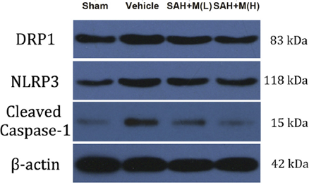
Mdivi-1 purchased from MedChemExpress. Usage Cited in: Exp Neurol. 2017 Sep:295:116-124. [Abstract]
Mdivi-1 treatment inhibits the expression of DRP1, alleviates ROS and reduces the expression of NLRP3 and cleaved caspase-1 at 24 h after SAH. Representative Western blots showing levels of DRP1.
-
Cell Signal
PDCD4 exacerbates atrial fibrillation by inducing mitochondrial fission and activating Ca2+/CaMKII signaling via ROS. [Abstract]2026 Oct:146:112679. PMID: 42297062 -
Mol Cell Biochem
The role of mitochondrial regulation in macrophage polarization by Ganoderma lucidum polysaccharide for the treatment of colitis-associated colorectal cancer. [Abstract]2026 Jun 2. PMID: 42228269 -
Cell Signal
2025 Sep:133:111868. PMID: 40373838 -
J Cell Mol Med
Celastrol alleviates atopic dermatitis by regulating Ezrin-mediated mitochondrial fission and fusion. [Abstract]2024 Jul;28(14):e18375. PMID: 39039796 -
J Cell Mol Med
QiDongNing induces lung cancer cell apoptosis via triggering P53/DRP1-mediated mitochondrial fission. [Abstract]2024 May;28(9):e18353. PMID: 38682742 -
Virol Sin
2023 Aug;38(4):520-530. PMID: 37156297 -
J Cell Mol Med
PINK1/Parkin-mediated mitophagy enhances the survival of Staphylococcus aureus in bovine macrophages. [Abstract]2023 Feb;27(3):412-421. PMID: 36625039 -
Cell Signal
FOXO3a-dependent up-regulation of HSP90 alleviates cisplatin-induced apoptosis by activating FUNDC1-mediated mitophagy in hypoxic osteosarcoma cells. [Abstract]2023 Jan:101:110500. PMID: 36270475 -
J Cell Mol Med
Interfering with mitochondrial dynamics sensitizes glioblastoma multiforme to temozolomide chemotherapy. [Abstract]2022 Feb;26(3):893-912. PMID: 34964241 -
Oncol Rep
Suppression of DRP1‑mediated mitophagy increases the apoptosis of hepatocellular carcinoma cells in the setting of chemotherapy. [Abstract]2020 Mar;43(3):1010-1018. PMID: 32020220 -
J Inflamm Res
Metformin Attenuates Osteoarthritis Progression by Modulating Mitochondrial Dynamics via Activation of the AMPK/Drp1 Pathway. [Abstract]2026 Feb 2:19:540176. PMID: 41884158 -
J Inflamm Res
Apigenin Suppresses NLRP3 Inflammasome Activation and Pyroptosis and Promotes Functional Recovery by Promoting Mitophagy in Experimental Spinal Cord Injured Rats. [Abstract]2025 Oct 8:18:13965-13984. PMID: 41084615 -
J Neurochem
Continued P2X7 activation leads to mitochondrial fission and compromising microglial phagocytosis after subarachnoid haemorrhage. [Abstract]2022 Dec;163(5):419-437. PMID: 36269673 -
Brain Res Bull
Excessive mitochondrial fission induces postoperative delirium in mice undergoing high-altitude deacclimatization by exacerbating neuroinflammation and synaptic injury. [Abstract]2026 Jun 10:243:111997. PMID: 42269727 -
Brain Res Bull
Vitexin alleviates cerebral ischemia/reperfusion injury by regulating mitophagy via the SIRT1/PINK1/Parkin pathway. [Abstract]2025 Jul:227:111404. PMID: 40441663 -
Poult Sci
Diethylhexyl phthalate exposure promotes mitophagy through Drp1-mediated mitochondrial fission in neural crest cells of chick embryos. [Abstract]2025 Jun 26;104(9):105491. PMID: 40582160 -
iScience
Circ-CIMIRC inhibition alleviates CIH-induced myocardial damage via FbxL4-mediated ubiquitination of PINK1. [Abstract]2024 Jan 20;27(2):108982. PMID: 38333696 -
iScience
Fucoxanthin ameliorates traumatic brain injury by suppressing the blood-brain barrier disruption. [Abstract]2023 Oct 20;26(11):108270. PMID: 37965135 -
iScience
Protective roles of MITOL against myocardial senescence and ischemic injury partly via Drp1 regulation. [Abstract]2022 Jun 11;25(7):104582. PMID: 35789860 -
Neurochem Res
Remote Ischemic Postconditioning Inhibited Mitophagy to Achieve Neuroprotective Effects in the Rat Model of Cardiac Arrest. [Abstract]2021 Mar;46(3):573-583. PMID: 33409854 -
-
FASEB J
Hirsutine Ameliorates High-Fat Diet-Induced Cardiomyopathy Through Promoting LRPPRC-Regulated Mitophagy via the PINK1/Parkin Axis in Mice. [Abstract]2026 Jun 30;40(12):e72050. PMID: 42290393 -
Biol Trace Elem Res
Realgar Transforming Solution as a Novel Arsenic Agent Triggers PINK1/Parkin-Dependent Mitophagy and Apoptosis in the Molm-13 Acute Myeloid Leukemia Cell Line. [Abstract]2026 Mar 27. PMID: 41896471 -
-
Biochim Biophys Acta Mol Cell Res
MARCH5 ameliorates aortic valve calcification via RACGAP1-DRP1 associated mitochondrial quality control. [Abstract]2025 Mar;1872(3):119911. PMID: 39880131 -
FASEB J
Hypoxia-inducible factor prolyl hydroxylase inhibitor alleviates heatstroke-induced acute kidney injury by activating BNIP3-mediated mitophagy. [Abstract]2024 Jun 30;38(12):e23723. PMID: 38865198 -
Biochim Biophys Acta Mol Cell Res
Age-related cataract: GSTP1 ubiquitination and degradation by Parkin inhibits its anti-apoptosis in lens epithelial cells. [Abstract]2023 Apr;1870(4):119450. PMID: 36871745 -
Fish Shellfish Immunol
2026 Jul 6:177:111583. PMID: 42409135 -
Fish Shellfish Immunol
Aeromonas hydrophila induces mitophagy for intracellular survival through its secreted effectors in the splenic macrophages of grass carp (Ctenopharyngodon idella). [Abstract]2026 Jun:173:111248. PMID: 41786106 -
FEBS J
Radiation resistance of cancer cells caused by mitochondrial dysfunction depends on SIRT3-mediated mitophagy. [Abstract]2023 Jul;290(14):3629-3645. PMID: 36871142 -
Drug Dev Res
Ilexgenin A inhibits mitochondrial fission and promote Drp1 degradation by Nrf2-induced PSMB5 in endothelial cells. [Abstract]2019 Jun;80(4):481-489. PMID: 30762899 -
J Virol
PEDV Nsp14 induces mitophagy-mediated degradation of MAVS to antagonize host innate immunity and facilitate viral proliferation. [Abstract]2025 Aug 19;99(8):e0049825. PMID: 40631894 -
Cancer Res Commun
Urolithin-A Promotes CD8+ T Cell-mediated Cancer Immunosurveillance via FOXO1 Activation. [Abstract]2024 May 3;4(5):1189-1198. PMID: 38626334 -
Environ Microbiol
Transcription factor MoMsn2 targets the putative 3-methylglutaconyl-CoA hydratase-encoding gene MoAUH1 to govern infectious growth via mitochondrial fusion/fission balance in Magnaporthe oryzae. [Abstract]2021 Feb;23(2):774-790. PMID: 32431008 -
-
Biochim Biophys Acta Mol Cell Biol Lipids
Palmitic acid activates NLRP3 inflammasome through NF-κB and AMPK-mitophagy-ROS pathways to induce IL-1β production in large yellow croaker (Larimichthys crocea). [Abstract]2024 Mar;1869(2):159428. PMID: 38029958 -
Biochim Biophys Acta Mol Cell Biol Lipids
Docosahexaenoic acid (DHA) inhibits abdominal fat accumulation by promoting adipocyte apoptosis though PPARγ-LC3-BNIP3 pathway-mediated mitophagy. [Abstract]2024 Jan;1869(1):159425. PMID: 37952912 -
J Orthop Surg Res
2023 Aug 24;18(1):620. PMID: 37620972 -
ESC Heart Fail
The protective effect of LCZ696 in coxsackievirus B3-induced acute viral myocarditis mice. [Abstract]2023 Feb;10(1):366-376. PMID: 36245336 -
Viruses
Porcine Circovirus 2 Induction of ROS Is Responsible for Mitophagy in PK-15 Cells via Activation of Drp1 Phosphorylation. [Abstract]2020 Mar 6;12(3):289. PMID: 32155766 -
J Anim Sci
Klebsiella pneumoniae causes mammary gland damage via FNIP1-mediated mitochondrial dysfunction. [Abstract]2025 Jan 4:103:skaf384. PMID: 41206543 -
Mol Immunol
Gelatin enhances bacteria-phagocytosis via a ROS-mitochondria-STING axis in differentiated human macrophage-like U937 cells. [Abstract]2025 Oct:186:48-62. PMID: 40815890 -
J Med Virol
Age-Dependent Pathogenesis of Influenza A Virus H7N9 Mediated Through PB1-F2-Induced Mitochondrial DNA Release and Activation of cGAS-STING-NF-κB Signaling. [Abstract]2024 Nov;96(11):e70062. PMID: 39569434 -
Toxicol Appl Pharmacol
Alleviation of cigarette smoke-induced cellular senescence in BALB/c mice by the Lipoxin A4 receptor agonist BML-111 is associated with mitophagy. [Abstract]2026 May:510:117772. PMID: 41724215 -
Toxicol Appl Pharmacol
p38 MAPK-DRP1 signaling is involved in mitochondrial dysfunction and cell death in mutant A53T α-synuclein model of Parkinson's disease. [Abstract]2020 Feb 1:388:114874. PMID: 31881179 -
Appl Biochem Biotechnol
FAM72A Knockdown Drives Cell Mitophagy and Pyroptosis in Ovarian Cancer Via the PINK1/Parkin Pathway. [Abstract]2026 May 20. PMID: 42159844 -
Int J Biochem Cell Biol
PROTAC-mediated degradation of Class I HDACs by JPS016 alleviates septic cardiomyopathy via mitophagy-driven exopher formation and mitochondrial quality control. [Abstract]2026 Mar:192:106910. PMID: 41687730 -
Cardiovasc Drugs Ther
Empagliflozin Supplementation in Cardioplegic Solution Improves Donor Heart Preservation by Maintaining Mitochondrial Homeostasis. [Abstract]2026 Jan 12. PMID: 41525029 -
Arch Biochem Biophys
Mitochondrial dynamic modulators attenuate iron overload-mediated cardiac toxicity via decreased mitochondrial fission, mitophagy/autophagy, and apoptosis in iron-overloaded rats. [Abstract]2025 May:767:110354. PMID: 39988036 -
Int J Biochem Cell Biol
BNIP3 in hypoxia-induced mitophagy: novel insights and promising target for non-alcoholic fatty liver disease. [Abstract]2024 Mar:168:106517. PMID: 38216085 -
Arch Biochem Biophys
Increased mitochondrial fission induces NLRP3/cGAS-STING mediated pro-inflammatory pathways and apoptosis in UVB-irradiated immortalized human keratinocyte HaCaT cells. [Abstract]2023 Apr:738:109558. PMID: 36878340 -
Arch Biochem Biophys
Mdivi-1 alleviates cardiac fibrosis post myocardial infarction at infarcted border zone, possibly via inhibition of Drp1-Activated mitochondrial fission and oxidative stress. [Abstract]2022 Mar 30;718:109147. PMID: 35143784 -
Int J Biochem Cell Biol
Activation of CD137 signaling promotes macrophage apoptosis dependent on p38 MAPK pathway-mediated mitochondrial fission. [Abstract]2021 Jul:136:106003. PMID: 33971320 -
Arch Biochem Biophys
Silibinin-induced mitochondria fission leads to mitophagy, which attenuates silibinin-induced apoptosis in MCF-7 and MDA-MB-231 cells. [Abstract]2020 May 30:685:108284. PMID: 32014401 -
Arch Biochem Biophys
2019 Aug 15:671:42-51. PMID: 31085166 -
Reprod Toxicol
Melatonin attenuates di-(2-ethylhexyl) phthalate-induced apoptosis of human granulosa cells by inhibiting mitochondrial fission. [Abstract]2022 Oct:113:18-29. PMID: 35952901 -
Cell Immunol
Silibinin restricts mitochondria-associated inflammatory pathways in LPS-stimulated murine microglia BV2 through TREM2. [Abstract]2026 Jul:425:105102. PMID: 41996954 -
Food Chem Toxicol
TDCIPP induces placental dysfunction and fetal growth restriction via oxidative stress-mediated PINK1/Parkin mitophagy. [Abstract]2026 Jul:213:116103. PMID: 42001928 -
Immunol Lett
Aerobic exercise attenuates airway inflammation in allergic asthma by regulating M2 macrophage polarization via drp1-mediated mitophagy. [Abstract]2026 Jun 6:281:107198. PMID: 42251903 -
Food Chem Toxicol
Deficiency of aldehyde dehydrogenase 2 aggravates ethanol-induced cytotoxicity in N2a cells via CaMKII/Drp1-mediated mitophagy. [Abstract]2023 Dec:182:114129. PMID: 37967785 -
IUBMB Life
Induced effect of zinc oxide nanoparticles on human acute myeloid leukemia cell apoptosis by regulating mitochondrial division. [Abstract]2022 Jun;74(6):519-531. PMID: 35383422 -
Brain Res
Dephosphorylation by calcineurin regulates translocation of dynamin-related protein 1 to mitochondria in hepatic ischemia reperfusion induced hippocampus injury in young mice. [Abstract]2019 May 15:1711:68-76. PMID: 30659828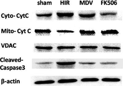
Mdivi-1 purchased from MedChemExpress. Usage Cited in: Brain Res. 2019 May 15:1711:68-76. [Abstract]
Levels of CytC in cytosol (Cyto-CytC), CytC in mitochondria (Mito- CytC) and cleaved caspase-3 in hippocampus are determined by western blot with the treatment of sham, HIR, MDV or FK506.
-
Kaohsiung J Med Sci
Terazosin Attenuates Neuronal Pyroptosis by Regulating the Mitochondrial ROS/NLRP3 Inflammasome Axis Through Mitophagy in Cerebral Ischemia-Reperfusion Injury. [Abstract]2026 Jun 9:e70242. PMID: 42266013 -
Toxicol Lett
The mitochondrial fission-mitophagy axis drives neuronal S-phase arrest and hippocampal damage in co-exposure to polystyrene nanoplastics and lead. [Abstract]2026 May 15:420:111896. PMID: 41990978 -
3 Biotech
Curcumin improves diabetic vascular aging rats and high glucose-induced cells aging by promoting mitophagy. [Abstract]2025 Oct;15(10):341. PMID: 40951878 -
Exp Eye Res
Drp1-dependent mitochondrial fragmentation mediates photoreceptor abnormalities in type 1 diabetic retina. [Abstract]2024 May:242:109860. PMID: 38467174 -
Toxicol Lett
Bavachin induces liver injury and cell apoptosis by targeting Wnt/β-catenin/DRP1 signaling pathway mediated mitochondrial dysfunction. [Abstract]2023 Sep 15:387:1-13. PMID: 37748667 -
Exp Eye Res
Inhibition of Drp1 ameliorates diabetic retinopathy by regulating mitochondrial homeostasis. [Abstract]2022 Apr 28;220:109095. PMID: 35490835 -
Expert Rev Anticancer Ther
Knockdown of TFB2M induces ferroptosis in lung adenocarcinoma via mitophagy-mediated GPX4 degradation. [Abstract]2025 Sep 3:1-10. PMID: 40878482 -
Theriogenology
Non-thermal plasma enhances boar sperm quality via FOXO3-PINK1/Parkin-mediated mitophagy. [Abstract]2026 Jul 1:258:117890. PMID: 41806618 -
Biochim Biophys Acta Gen Subj
Unraveling the power of TOMM40: Driving PHB1-mediated mtDNA release and mitophagy to fuel breast cancer progression. [Abstract]2025 Oct;1869(11):130851. PMID: 40816542 -
Behav Brain Res
Two polyphenols isolated from Corallodiscus flabellata B. L. Burtt ameliorate amyloid β-protein induced Alzheimer's disease neuronal injury by improving mitochondrial homeostasis. [Abstract]2023 Feb 25:440:114264. PMID: 36535434 -
Toxicol Res (Camb)
Drp1-mediated mitochondrial fission promotes carbon tetrachloride-induced hepatic fibrogenesis in mice. [Abstract]2022 May 23;11(3):486-497. PMID: 35782650 -
Vet Microbiol
2025 Sep 6:310:110721. PMID: 40925072 -
Vet Microbiol
Staphylococcus aureus induces mitophagy to promote its survival within bovine mammary epithelial cells. [Abstract]2023 May:280:109697. PMID: 36827937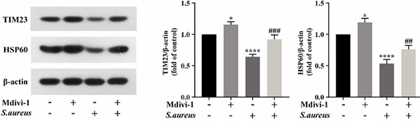
Mdivi-1 purchased from MedChemExpress. Usage Cited in: Vet Microbiol. 2023 May:280:109697. [Abstract]
S.aureus-induced reduction of mitochondrial proteins is blocked by Mdivi-1 (20 μM; 2 h) in BMECs.
-
Clin Rheumatol
High glucose-induced mitophagy accelerates premature aging of T cells in patients with rheumatoid arthritis. [Abstract]2025 Nov 20. PMID: 41261244 -
Toxicol In Vitro
Mitochondrial division inhibitor (mdivi-1) inhibits proliferation and epithelial-mesenchymal transition via the NF-κB pathway in thyroid cancer cells. [Abstract]2023 Apr:88:105552. PMID: 36621616 -
Steroids
Vigilin interacts with ER-β to protect against palmitic acid-induced granulosa cells apoptosis via inhibiting calcineurin-mediated Drp1 signaling pathway. [Abstract]2020 Nov:163:108699. PMID: 32687845 -
Biochem Biophys Res Commun
2026 May 7:812:153592. PMID: 41833073 -
Biochem Biophys Res Commun
PKA-mediated Drp1 Ser637 phosphorylation modulates mitochondrial function in the placenta of gestational diabetes mellitus. [Abstract]2025 Nov 19:790:152919. PMID: 41197405 -
Biochem Biophys Res Commun
Icariin alleviates cellular injury induced by cardiac ischemia-reperfusion injury by inhibiting IRE1/JNK-induced ferroptosis. [Abstract]2024 Nov 12:733:150716. PMID: 39321486 -
J Mol Neurosci
Drp1-Mediated Mitochondrial Fission Is Essential for Chemical-Induced Neuronal Transdifferentiation from Human Primary Fibroblasts. [Abstract]2025 Jun 13;75(2):75. PMID: 40512396 -
Dev Comp Immunol
Molecular cloning, characterization and expression analysis of PINK1 (Phosphatase and tensin homolog-induced putative kinase 1) and Parkin (parkin RBR E3 ubiquitin protein ligase) in grass carp (Ctenopharyngodon idellus). [Abstract]2025 May:166:105349. PMID: 40044098 -
Exp Ther Med
PIN1‑silencing mitigates keratinocyte proliferation and the inflammatory response in psoriasis by activating mitochondrial autophagy. [Abstract]2024 Aug 19;28(4):402. PMID: 39234585 -
Neurosci Lett
FUNDC1 inhibits NLRP3-mediated inflammation after intracerebral hemorrhage by promoting mitophagy in mice. [Abstract]2021 Jun 21:756:135967. PMID: 34022268 -
Exp Ther Med
Mitochondrial separation protein inhibitor inhibits cell apoptosis in rat lungs during intermittent hypoxia. [Abstract]2019 Mar;17(3):2349-2358. PMID: 30867720 -
Inhal Toxicol
GRP78 mediates mitochondrial fusion and fission in cigarette smoke-induced inflammatory responses in airway epithelial cells. [Abstract]2024 Oct-Nov;36(9-10):511-520. PMID: 39565149 -
In Vitro Cell Dev Biol Anim
Adipose-derived stem cells regulate mitochondrial dynamics to alleviate the aging of HFF-1 cells. [Abstract]2025 Mar;61(3):357-367. PMID: 39871034 -
J Physiol Investig
Apelin-13 Ameliorates Sepsis-induced Brain Injury by Activating Phosphatase and Tensin Homolog-induced Putative Kinase 1/Parkin-mediated Mitophagy and Modulating Nucleotide-binding Oligomerization Domain-like Receptor Pyrin Domain-Containing 3-driven Pyroptosis in Rats. [Abstract]2025 Jan 1;68(1):31-42. PMID: 39774002 -
-
Int Heart J
USP30 Knockdown Drives Mitophagy and Suppresses Pyroptosis in Heart Failure by Activating the PINK1/Parkin Pathway. [Abstract]2025;66(6):1002-1014. PMID: 41320325 -
-
bioRxiv
It's Not Rewarding for Mitochondria: Dopamine-Induced Mitochondrial Dysfunction Activates cGAS-STING to Drive IL-6 Secretion in Macrophages. [Abstract]2026 Apr 25:2026.04.23.719926. PMID: 42079251 -
-
-
-
-
-
bioRxiv
The antibacterial factor APOL3 couples lysosomal damage to mitochondrial DNA efflux and type I IFN induction. [Abstract]2025 May 19:2025.05.16.654477. PMID: 40475483 -
-
-
-
-
Biomed Pharmacother
2024 Sep:178:117279. PMID: 39121587 -
-
Sci Total Environ
Cr(VI) induces ferroptosis in DF-1 cells by simultaneously perturbing iron homeostasis of ferritinophagy and mitophagy. [Abstract]2024 May 15:925:171818. PMID: 38508245 -
-
Sci Total Environ
Mystery of bisphenol F-induced nonalcoholic fatty liver disease-like changes: Roles of Drp1-mediated abnormal mitochondrial fission in lipid droplet deposition. [Abstract]2023 Dec 15:904:166831. PMID: 37683851 -
Biomed Pharmacother
Inhibition of mitochondrial fusion via SIRT1/PDK2/PARL axis breaks mitochondrial metabolic plasticity and sensitizes cancer cells to glucose restriction therapy. [Abstract]2023 Oct:166:115342. PMID: 37633053 -
-
Sci Total Environ
The lysosome-mitochondrion crosstalk engaged in silver nanoparticles-disturbed mitochondrial homeostasis. [Abstract]2023 Sep 1:889:164078. PMID: 37209729 -
-
-
Oxid Med Cell Longev
Repurposing Dihydroartemisinin to Combat Oral Squamous Cell Carcinoma, Associated with Mitochondrial Dysfunction and Oxidative Stress. [Abstract]2023 Feb 16:2023:9595201. PMID: 37273554 -
-
-
Ann Transl Med
Levosimendan protects from sepsis-inducing cardiac dysfunction by suppressing inflammation, oxidative stress and regulating cardiac mitophagy via the PINK-1-Parkin pathway in mice. [Abstract]2022 Feb;10(4):212. PMID: 35280364 -
-
-
Aging (Albany NY)
CHIP protects against MPP+/MPTP-induced damage by regulating Drp1 in two models of Parkinson's disease. [Abstract]2021 Jan 2;13(1):1458-1472. PMID: 33472166 -
-
Oxid Med Cell Longev
Salidroside Ameliorates Mitochondria-Dependent Neuronal Apoptosis after Spinal Cord Ischemia-Reperfusion Injury Partially through Inhibiting Oxidative Stress and Promoting Mitophagy. [Abstract]2020 Jul 23:2020:3549704. PMID: 32774670 -
Aging (Albany NY)
Melatonin regulates mitochondrial dynamics and alleviates neuron damage in prion diseases. [Abstract]2020 Jun 10;12(11):11139-11151. PMID: 32526704


Solvent & Solubility
DMSO : 110 mg/mL (311.42 mM; Need ultrasonic; Hygroscopic DMSO has a significant impact on the solubility of product, please use newly opened DMSO)
Please refer to the solubility information to select the appropriate solvent. Once prepared, please aliquot and store the solution to prevent product inactivation from repeated freeze-thaw cycles.
Storage method and period of stock solution: -80°C, 6 months; -20°C, 1 month. When stored at -80°C, please use it within 6 months. When stored at -20°C, please use it within 1 month.
Concentration (start) × Volume (start) = Concentration (final) × Volume (final)
Select the appropriate dissolution method based on your experimental animal and administration route.
- For the following dissolution methods, please ensure to first prepare a clear stock solution using an In Vitro approach and then sequentially add co-solvents:
- To ensure reliable experimental results, the clarified stock solution can be appropriately stored based on storage conditions. As for the working solution for In Vivo experiments, it is recommended to prepare freshly and use it on the same day.
- The percentages shown for the solvents indicate their volumetric ratio in the final prepared solution. If precipitation or phase separation occurs during preparation, heat and/or sonication can be used to aid dissolution.
Add each solvent one by one: 10% DMSO 40% PEG300 5% Tween-80 45% Saline
Solubility: ≥ 4 mg/mL (11.32 mM); Clear solution
This protocol yields a clear solution of ≥ 4 mg/mL (saturation unknown).
Taking 1 mL working solution as an example, add 100 μL DMSO stock solution (40.0 mg/mL) to 400 μL PEG300, and mix evenly; then add 50 μL Tween-80 and mix evenly; then add 450 μL Saline to adjust the volume to 1 mL.
Preparation of Saline: Dissolve 0.9 g sodium chloride in ddH₂O and dilute to 100 mL to obtain a clear Saline solution.
For the following dissolution methods, please prepare the working solution directly:
It is recommended to prepare fresh solutions and use them promptly within a short period of time.
The percentages shown for the solvents indicate their volumetric ratio in the final prepared solution. If precipitation or phase separation occurs during preparation, heat and/or sonication can be used to aid dissolution.
Add each solvent one by one: 0.5% CMC-Na/saline water
Solubility: 2.5 mg/mL (7.08 mM); Suspended solution; Need ultrasonic
Please enter the basic information of animal experiments:
-
-
-
-
Recommended: Prepare an additional quantity of animals to account for potential losses during experiments.
Please enter your animal formula composition:
-
%DMSO +
Recommended: Keep the proportion of DMSO in working solution below 2% if your animal is weak.
-
%+
-
+%Tween-80 + +
-
%Saline +
The co-solvents required include: DMSO, . All of co-solvents are available by MedChemExpress (MCE). , Tween 80. All of co-solvents are available by MedChemExpress (MCE).
Working solution concentration: 0.22 mg/mL
Method for preparing stock solution: mg drug dissolved in μL DMSO. Stock solution concentration: mg/mL.
1. Take μL DMSO stock solution;
2. Add μL .
μL , mix evenly;
3. Then add μL Tween 80, mix evenly;
4. Then add μL
Please ensure that the stock solution in the first step is dissolved to a clear state, and add co-solvents in sequence. You can use ultrasonic heating (ultrasonic cleaner, recommended frequency 20-40 kHz), vortexing, etc. to assist dissolution.

Protocol
All GTPase assay reactions are started in a 200 μL volume, of which 150 μL is placed into the well of a 96-well plate. Depletion of NADH, as monitored by reading the A340 of the reaction, is measured every 20 s for a total of 40 min using a SpectraMAX 250 96-well plate reader. Spectrophotometric data are transferred to Excel and the measured steady state depletion of NADH over time is converted to protein activity.
MedChemExpress (MCE) has not independently confirmed the accuracy of these methods. They are for reference only.
YPGlycerol plates are topped with 10 mL YPGlycerol containing 1% low melt agar and 75 μM mdivi-1, and cells are spotted 12 hours later using a 48 well pinning device. After pinning cells, plates are incubated at 24°C or 37°C and imaged using an Eagle Eye II imaging system.
MedChemExpress (MCE) has not independently confirmed the accuracy of these methods. They are for reference only.


Purity & Documentation
-
Data Sheet (279 KB)
-
SDS (396 KB)
- English - EN (396 KB)
- Français - FR (396 KB)
- Deutsch - DE (396 KB)
- Norwegian - NO (396 KB)
- Español - ES (396 KB)
- Swedish - SV (396 KB)
- Italian - IT (396 KB)
- Korean - KR (396 KB)
- Portuguese - PT (396 KB)
-
Handling Instructions (2659 KB)
References
[1]. Cassidy-Stone A, et al. Chemical inhibition of the mitochondrial division dynamin reveals its role in Bax/Bak-dependent mitochondrial outer membrane permeabilization. Dev Cell. 2008 Feb;14(2):193-204. [Content Brief]
[2]. Tanaka A, et al. A chemical inhibitor of DRP1 uncouples mitochondrial fission and apoptosis. Mol Cell. 2008 Feb 29;29(4):409-10. [Content Brief]
[3]. Park SW, et al. A selective inhibitor of drp1, mdivi-1, increases retinal ganglion cell survival in acute ischemic mouse retina. Invest Ophthalmol Vis Sci. 2011 Apr 27;52(5):2837-43. [Content Brief]


Complete Stock Solution Preparation Table
Please refer to the solubility information to select the appropriate solvent. Once prepared, please aliquot and store the solution to prevent product inactivation from repeated freeze-thaw cycles.
Storage method and period of stock solution: -80°C, 6 months; -20°C, 1 month. When stored at -80°C, please use it within 6 months. When stored at -20°C, please use it within 1 month.
| Optional Solvent | Concentration Solvent Mass | 1 mg | 5 mg | 10 mg | 25 mg |
|---|---|---|---|---|---|
| DMSO | 1 mM | 2.8311 mL | 14.1555 mL | 28.3110 mL | 70.7774 mL |
| 5 mM | 0.5662 mL | 2.8311 mL | 5.6622 mL | 14.1555 mL | |
| 10 mM | 0.2831 mL | 1.4155 mL | 2.8311 mL | 7.0777 mL | |
| 15 mM | 0.1887 mL | 0.9437 mL | 1.8874 mL | 4.7185 mL | |
| 20 mM | 0.1416 mL | 0.7078 mL | 1.4155 mL | 3.5389 mL | |
| 25 mM | 0.1132 mL | 0.5662 mL | 1.1324 mL | 2.8311 mL | |
| 30 mM | 0.0944 mL | 0.4718 mL | 0.9437 mL | 2.3592 mL | |
| 40 mM | 0.0708 mL | 0.3539 mL | 0.7078 mL | 1.7694 mL | |
| 50 mM | 0.0566 mL | 0.2831 mL | 0.5662 mL | 1.4155 mL | |
| 60 mM | 0.0472 mL | 0.2359 mL | 0.4718 mL | 1.1796 mL | |
| 80 mM | 0.0354 mL | 0.1769 mL | 0.3539 mL | 0.8847 mL | |
| 100 mM | 0.0283 mL | 0.1416 mL | 0.2831 mL | 0.7078 mL |


 Powered by Bioz