



Squalamine
Based on 1 publication(s) in Google Scholar
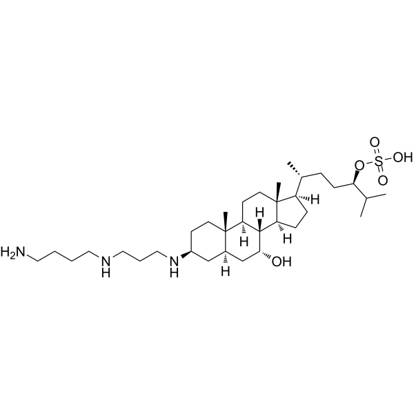
Squalamine (MSI-1256) is an aminosterol compound with broad-spectrum antiviral activity. Squalamine makes cells less conducive to certain viral replication by altering the electrostatic interactions in the inner membrane of host cells. Squalamine also has antibacterial and antitumor activities. Squalamine has broad-spectrum antibacterial activity against Gram-negative and Gram-positive bacteria, fungi and protozoa. Squalamine inhibits tumor-related angiogenesis and the growth of human breast cancer cells. Squalamine restores the function of enteric nervous system in Parkinson,s disease mouse models.
For research use only. We do not sell to patients.
- Purity : 98.38%
- CAS No.: 148717-90-2
- Formula: C34H65N3O5S
- Molecular Weight:627.96
-
Storage:Powder -20°C, 3 years , 4°C, 2 years ; In solvent -80°C, 2 years , -20°C, 1 year

To place orders, for customer services and technical support, please contact: MedChemExpress USA
Tel: 609-228-6898 E-mail: [email protected] [email protected]


-
 Biological Activity
-
Chemical Information
-
Solvent & Solubility
- Protocols
- Purity & Documentation
- References
-
 Help & FAQs


-
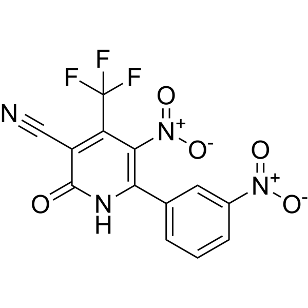
HY-124876
SynuClean-Dα-synuclein Aggregation Inhibitor -
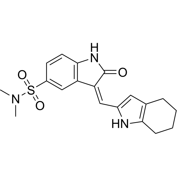
HY-B0789
SU6656Src Inhibitor -
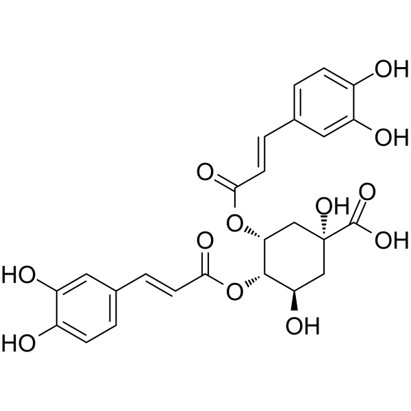
HY-N0058
4,5-Dicaffeoylquinic acidAntioxidant -
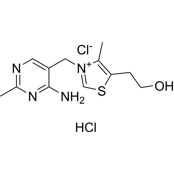
HY-N0680
Thiamine hydrochlorideVitamin B1 -
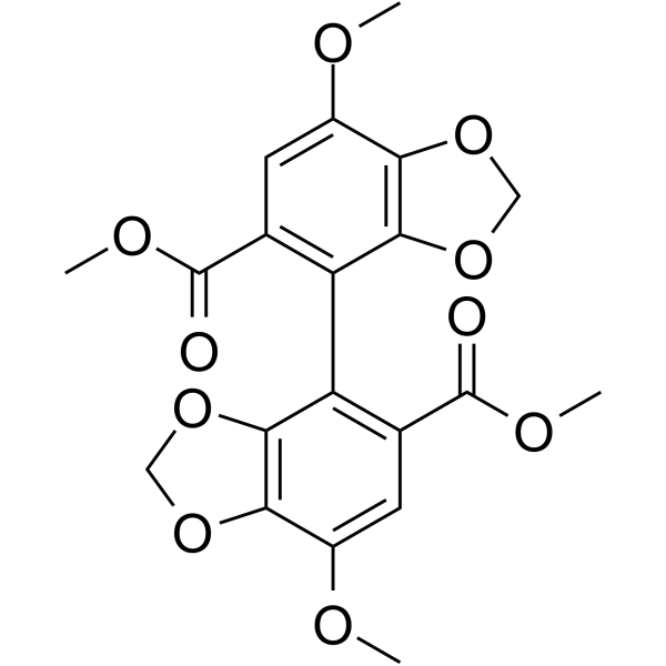
HY-W018791
BifendateAntioxidant, Anti-HBV Agent -
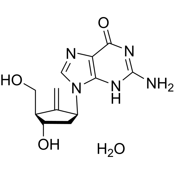
HY-13623A
Entecavir monohydrateHBV Inhibitor -
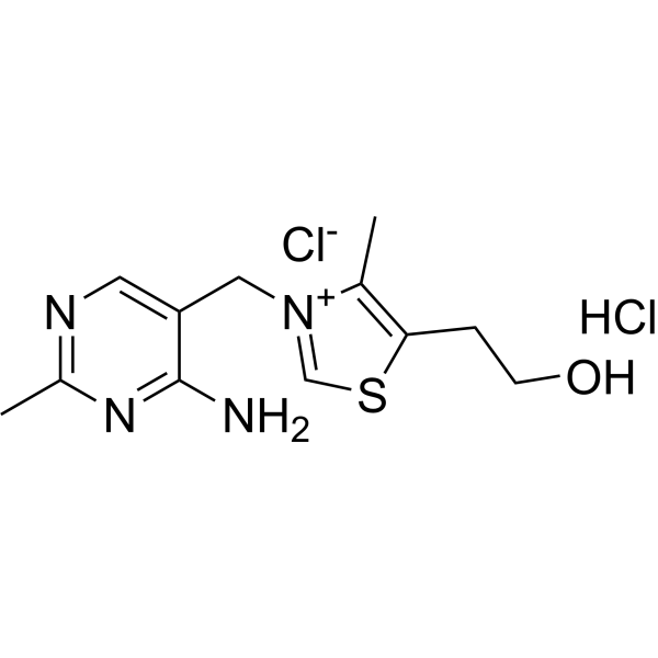
HY-N0680R
Thiamine hydrochloride (Standard)Vitamin B1 -
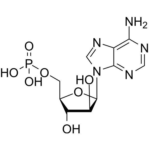
HY-B0277A
Vidarabine phosphateAntiviral Agent -

HY-N0058R
4,5-Dicaffeoylquinic acid (Standard)Antioxidant -

HY-W018791R
Bifendate (Standard)Antioxidant, Anti-HBV Agent -

HY-13623AR
Entecavir monohydrate (Standard)HBV Inhibitor -

HY-B0277AR
Vidarabine phosphate (Standard)Standard of Vidarabine phosphate
Publications Citing Use of MedChemExpress (MCE) Squalamine
More

Biological Activity
Description
In Vitro
Squalamine (0-100 μg/mL; 2 h) inhibits dengue virus (Den V2) infection in HMEC-1 cells, and dengue infection is suppressed by about 60% at 40 μg/mL and completely suppressed at 100 μg/mL[1].
Squalamine (0-20 μg/mL; 16 h) effectively inhibits the replication of human hepatitis B virus (HBV) in human primary hepatocytes[1].
Squalamine (0.16-3.2 μM; 8 days) inhibits VEGF-induced proliferation of vascular endothelial cells HUVEC in a dose-dependent manner, but has no direct inhibitory effect on the proliferation of human breast MCF-7 cancer cells[2].
Squalamine (0.001-1 μM; 1 h) blocks FAK phosphorylation in HUVECs exposed to VEGF[2].
MedChemExpress (MCE) has not independently confirmed the accuracy of these methods. They are for reference only.
-
Cell Line:HUVEC cells
-
Concentration:0.001, 0.01, 0.1 and 1 μM
-
Incubation Time:1 h
-
Result:Inhibited the phosphorylation levels of FAK in a concentration‐dependent manner
In Vivo
Squalamine (20 mg; Single application; 0-3 days) has a decolonizing effect on S. aureus on the skin in a mouse model[3].
Squalamine (20-120 mg/kg; Oral gavage; 5 days) restores the function of the mesenteric nervous system in PD mouse models[4].
MedChemExpress (MCE) has not independently confirmed the accuracy of these methods. They are for reference only.
-
Animal Model:Female athymic mice aged 6 weeks old bearing breast tumor xenografts[2]
-
Dosage:2 mg/kg
-
Administration:Intraperitoneal injection (i.p.); 28 days
-
Result:Significantly retarded the growth of tumors, but the combination with Trastuzumab (HY-P9907) was more effective.
Suppressed MCF-7/HER-2 breast xenograft-associated angiogenesis.
-
Animal Model:10 μL S. aureus suspension(108 cfu/mL) was applied to the female BALB/c mice skin[3]
-
Dosage:20 mg
-
Administration:Single application; 3 days
-
Result:Reduced S. aureus viable cells by up to 4 log after two days compared with the control.
-
Animal Model:PD mice models ( hSNCAA53T mice and PrP-A53T human α-syn overexpressing transgenic mice)[4]
-
Dosage:20, 40, 80, or 120 mg/kg
-
Administration:Oral gavage (i.g.); 5 days
-
Result:Effectively restored disordered colonic motility in PD mice models.
Increased colonic transit in PrP-A53T mice.
Reduced myenteric intrinsic primary afferent neuron excitability in hSNCAA53T mice.
- Research Protocol for Infectious Diseases
- Human pluripotent stem cell endothelial-cell differentiation
- Vascular/Branching Fractal Analysis
- Endothelial Tube Formation Assay
- Gram Staining of Tissue Sections
- Bacterial live/dead nucleic-acid viability staining
- Breast Cancer Modeling
- Endothelial Cell Migration/Angiogenic Sprouting Assay


Chemical Information
-
CAS No. 148717-90-2
-
Appearance Solid
-
Molecular Weight 627.96
-
Formula C34H65N3O5S
-
Color White to light yellow
-
SMILES
C[C@@]12[C@](C[C@@H](O)[C@]3([H])[C@]2([H])CC[C@@]4(C)[C@@]3([H])CC[C@]4([H])[C@@H](CC[C@H](C(C)C)OS(=O)(O)=O)C)([H])C[C@@H](NCCCNCCCCN)CC1
-
Synonyms
MSI-1256; ENT-01 free acid
-
Structure Classification
-
Initial Source
-
Shipping
Room temperature in continental US; may vary elsewhere.
-
Storage
Powder -20°C 3 years 4°C 2 years In solvent -80°C 2 years -20°C 1 year


Publications (1)
-
Journal Impact Factor
-
Most Recent
-
J Enzyme Inhib Med Chem
Identification of chemical scaffolds for targeting ubiquitin-specific protease 11 (USP11) through high-throughput virtual screening. [Abstract]2025 Dec;40(1):2518191. PMID: 40588719


Solvent & Solubility
In Vitro:
DMSO : 100 mg/mL (159.25 mM; Need ultrasonic; Hygroscopic DMSO has a significant impact on the solubility of product, please use newly opened DMSO)
Please refer to the solubility information to select the appropriate solvent. Once prepared, please aliquot and store the solution to prevent product inactivation from repeated freeze-thaw cycles.
Storage method and period of stock solution: -80°C, 2 years; -20°C, 1 year. When stored at -80°C, please use it within 2 years. When stored at -20°C, please use it within 1 year.
Please refer to the solubility information to select the appropriate solvent. Once prepared, please aliquot and store the solution to prevent product inactivation from repeated freeze-thaw cycles.
Storage method and period of stock solution: -80°C, 2 years; -20°C, 1 year. When stored at -80°C, please use it within 2 years. When stored at -20°C, please use it within 1 year.
Concentration (start) × Volume (start) = Concentration (final) × Volume (final)
In Vivo:
Select the appropriate dissolution method based on your experimental animal and administration route.
- For the following dissolution methods, please ensure to first prepare a clear stock solution using an In Vitro approach and then sequentially add co-solvents:
- To ensure reliable experimental results, the clarified stock solution can be appropriately stored based on storage conditions. As for the working solution for In Vivo experiments, it is recommended to prepare freshly and use it on the same day.
- The percentages shown for the solvents indicate their volumetric ratio in the final prepared solution. If precipitation or phase separation occurs during preparation, heat and/or sonication can be used to aid dissolution.
Add each solvent one by one: 10% DMSO 40% PEG300 5% Tween-80 45% Saline
Solubility: ≥ 2.75 mg/mL (4.38 mM); Clear solution
This protocol yields a clear solution of ≥ 2.75 mg/mL (saturation unknown).
Taking 1 mL working solution as an example, add 100 μL DMSO stock solution (27.5 mg/mL) to 400 μL PEG300, and mix evenly; then add 50 μL Tween-80 and mix evenly; then add 450 μL Saline to adjust the volume to 1 mL.
Preparation of Saline: Dissolve 0.9 g sodium chloride in ddH₂O and dilute to 100 mL to obtain a clear Saline solution.
Add each solvent one by one: 10% DMSO 90% (20% SBE-β-CD in Saline)
Solubility: ≥ 2.75 mg/mL (4.38 mM); Clear solution
This protocol yields a clear solution of ≥ 2.75 mg/mL (saturation unknown).
Taking 1 mL working solution as an example, add 100 μL DMSO stock solution (27.5 mg/mL) to 900 μL 20% SBE-β-CD in Saline, and mix evenly.
Preparation of 20% SBE-β-CD in Saline (4°C, storage for one week): 2 g SBE-β-CD powder is dissolved in 10 mL Saline, completely dissolve until clear.
In Vivo Dissolution Calculator
Please enter the basic information of animal experiments:
-
-
-
-
Recommended: Prepare an additional quantity of animals to account for potential losses during experiments.
Please enter your animal formula composition:
-
%DMSO +
Recommended: Keep the proportion of DMSO in working solution below 2% if your animal is weak.
-
%+
-
+%Tween-80 + +
-
%Saline +
The co-solvents required include: DMSO, . All of co-solvents are available by MedChemExpress (MCE). , Tween 80. All of co-solvents are available by MedChemExpress (MCE).
Working solution concentration: 0.22 mg/mL
Method for preparing stock solution: mg drug dissolved in μL DMSO. Stock solution concentration: mg/mL.
1. Take μL DMSO stock solution;
2. Add μL .
μL , mix evenly;
3. Then add μL Tween 80, mix evenly;
4. Then add μL
Please ensure that the stock solution in the first step is dissolved to a clear state, and add co-solvents in sequence. You can use ultrasonic heating (ultrasonic cleaner, recommended frequency 20-40 kHz), vortexing, etc. to assist dissolution.


Purity & Documentation
-
Data Sheet (278 KB)
-
SDS (252 KB)
- English - EN (252 KB)
- Français - FR (252 KB)
- Deutsch - DE (252 KB)
- Norwegian - NO (252 KB)
- Español - ES (252 KB)
- Swedish - SV (252 KB)
- Italian - IT (252 KB)
- Korean - KR (252 KB)
- Portuguese - PT (252 KB)
-
Handling Instructions (2659 KB)
References
[1]. Zasloff M, et al. Squalamine as a broad-spectrum systemic antiviral agent with therapeutic potential. Proc Natl Acad Sci U S A. 2011 Sep 20;108(38):15978-83. [Content Brief]
[2]. Márquez-Garbán DC, et al. Squalamine blocks tumor-associated angiogenesis and growth of human breast cancer cells with or without HER-2/neu overexpression. Cancer Lett. 2019 May 1;449:66-75. [Content Brief]
[3]. Djouhri-Bouktab L, et al. Squalamine ointment for Staphylococcus aureus skin decolonization in a mouse model. J Antimicrob Chemother. 2011 Jun;66(6):1306-10. [Content Brief]
[4]. West CL, et al. Squalamine Restores the Function of the Enteric Nervous System in Mouse Models of Parkinson's Disease. J Parkinsons Dis. 2020;10(4):1477-1491. [Content Brief]
[5]. Moore KS, et al. Squalamine: an aminosterol antibiotic from the shark. Proc Natl Acad Sci U S A. 1993 Feb 15;90(4):1354-8. [Content Brief]


Complete Stock Solution Preparation Table
Please refer to the solubility information to select the appropriate solvent. Once prepared, please aliquot and store the solution to prevent product inactivation from repeated freeze-thaw cycles.
Storage method and period of stock solution: -80°C, 2 years; -20°C, 1 year. When stored at -80°C, please use it within 2 years. When stored at -20°C, please use it within 1 year.
| Optional Solvent | Concentration Solvent Mass | 1 mg | 5 mg | 10 mg | 25 mg |
|---|---|---|---|---|---|
| DMSO | 1 mM | 1.5925 mL | 7.9623 mL | 15.9246 mL | 39.8115 mL |
| 5 mM | 0.3185 mL | 1.5925 mL | 3.1849 mL | 7.9623 mL | |
| 10 mM | 0.1592 mL | 0.7962 mL | 1.5925 mL | 3.9811 mL | |
| 15 mM | 0.1062 mL | 0.5308 mL | 1.0616 mL | 2.6541 mL | |
| 20 mM | 0.0796 mL | 0.3981 mL | 0.7962 mL | 1.9906 mL | |
| 25 mM | 0.0637 mL | 0.3185 mL | 0.6370 mL | 1.5925 mL | |
| 30 mM | 0.0531 mL | 0.2654 mL | 0.5308 mL | 1.3270 mL | |
| 40 mM | 0.0398 mL | 0.1991 mL | 0.3981 mL | 0.9953 mL | |
| 50 mM | 0.0318 mL | 0.1592 mL | 0.3185 mL | 0.7962 mL | |
| 60 mM | 0.0265 mL | 0.1327 mL | 0.2654 mL | 0.6635 mL | |
| 80 mM | 0.0199 mL | 0.0995 mL | 0.1991 mL | 0.4976 mL | |
| 100 mM | 0.0159 mL | 0.0796 mL | 0.1592 mL | 0.3981 mL |


Squalamine Related Classifications
HY-16468 Related Classifications
 Powered by Bioz