1. Resources
  2. Articles
  3. Wnt/β-catenin and tumor EMT
Wnt/β-catenin and tumor EMT
EMT ≈ Cancer?

When we mention Epithelial-to-mesenchymal transition (EMT), most people will immediately think of tumors. We often say that tumors have EMT characteristics. However, the EMT is an indispensable mechanism during morphogenesis, as without mesenchymal cells, tissues and organs will never be formed[1].

The EMT process describes the differentiation of stationary epithelial cells towards a mesenchymal, motile phenotype, which was initially observed in early development[1]. Later, it was found that EMT is not only critical in development and wound healing, but also represents a salient property of primary tumor formation and metastasis[2].

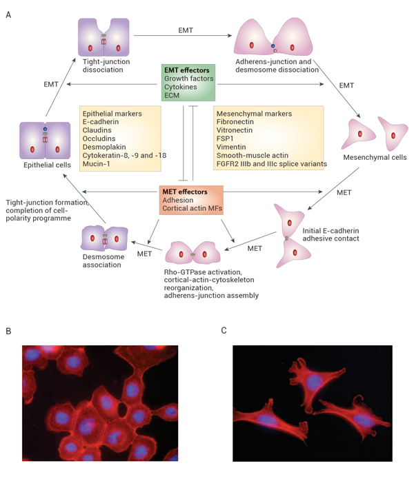
Fig 1. The cycle of epithelial-cell plasticity
Fig 1. The cycle of epithelial-cell plasticity[1].
(A) Epithelial cells can convert into mesenchymal cells by EMT, which epithelial cells lose many of their epithelial characteristics and take on properties that are typical of mesenchymal cells. EMT regulates important processes during the early stages of development of most organisms. Development cannot proceed past the blastula stage in the absence of EMT. SCp2 murine mammary cells (B) were treated with Matrix metalloproteinase-3 (C) to induce EMT.

The EMT process can promote cancer stem-like properties, immune evasion, multi-drug resistance, invasive phenotype, mediate the plasticity of cancer cells, allowing them to persistently and irreversibly adapt to changing conditions. EMT is inseparably linked with tumor invasion, metastasis, drug resistance, and immune escape[3][4][5].

EMT and Wnt/β-catenin Pathway

EMT is regulated by various signaling pathways such as the TGF-β, Wnt/β-catenin, Hedgehog, and Notch signaling pathway. These pathways trigger EMT by stimulating transcription factors including Snail, Twist, and ZEB1/2. Among all the signaling pathways, the Wnt/β-catenin pathway shows its pivotal role in the regulation of EMT[6].

• Activation of Wnt/β-catenin induces EMT

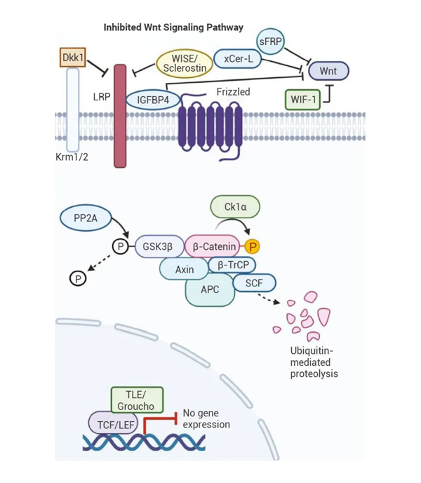
Under normal circumstances, negative regulators of the Wnt/β-catenin signaling pathway, such as adenomatous polyposis coli protein (APC), glycogen synthase kinase 3 protein (GSK-3), and Axin, can bind with β-catenin to form a complex resulting in phosphorylation, and further degradation.

Fig 2. The inhibited Wnt signaling cascade
Fig 2. The inhibited Wnt signaling cascade[7].

However, mutations and deletions of these negative regulator genes of the Wnt/β-catenin pathway have been found in various cancer cells. β-catenin accumulates in large quantities in the cytoplasm and then moves into the nucleus to form complexes with transcription factors such as T cell factor/Lymphoid enhancing factor (TCF/LEF), activating downstream target genes to promote cell cycle development or produce abnormal proteins, inducing EMT, and leading to cell carcinogenesis.

• Pathways of Wnt activation

The activation of Wnt is achieved by different Wnt-protein ligands binding to the Frizzled family cell surface receptor, thereby transmitting biological signals into the cell, including three pathways.

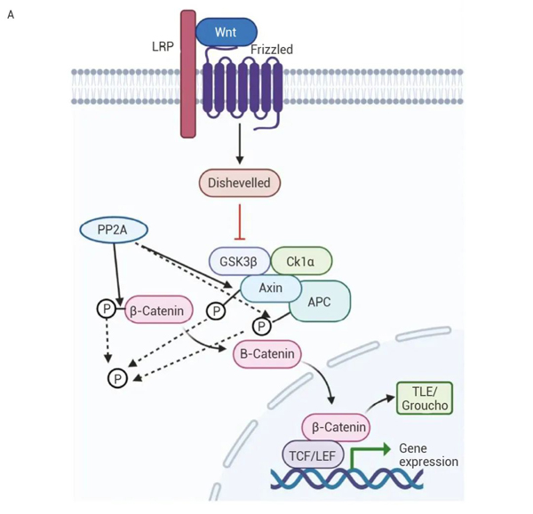
(1) Canonical Wnt pathway (Wnt/β-catenin): The Wnt gene is abnormally activated in tumor cells, activating the phosphorylation of Dsh protein in the cytoplasm, inhibiting the key component GSK3β activity in the GSK3β/APC/Axin complex, preventing GSK3β's phosphorylation and ubiquitination of β-catenin, reducing the phosphorylation degradation of β-catenin[9]. β-catenin accumulates into the cytosol and then translocates to the nucleus to bind the LEF-TCF co-transcription factors, thereby affecting cell adhesion, tissue morphogenesis, and tumor development[10].

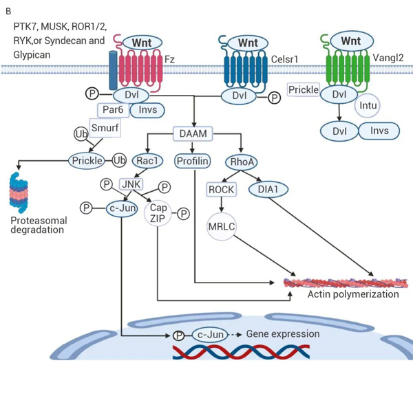
Fig 3. Canonical Wnt/β-catenin (A) and noncanonical Wnt/PCP signaling pathways(B)
Fig 3. Canonical Wnt/β-catenin (A) and noncanonical Wnt/PCP signaling pathways(B)[7].

(2) Non-canonical Wnt/PCP pathway: On one hand, the Fz receptor binds to the Wnt ligand and causes Dvl to be phosphorylated. On the other hand, Smurf ubiquitinates Prickle (a protein that typically inhibits Wnt/PCP signaling). The degradation of Prickle allows Dvl to bind with DAAM, activating Rac1, Profilin, and RhoA. Rac1 activates JNK, which in turn phosphorylates c-Jun and CapZIP. Then c-Jun enters the cell nucleus to stimulate gene transcription. Meanwhile, RhoA activates DIA1 and ROCK, which in turn activates MRLC. CapZIP, MRLC, DIA1 and Profilin all stimulate actin polymerization, thereby affecting cell polarity and migration[7].

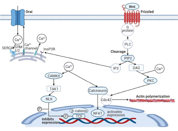
(3) Non-canonical Wnt/Ca2+ pathway: The binding of Wnt to the Fz receptor leads to G protein-mediated activation of PLC, stimulating Ca2+ release. DAG and Ca2+ together activate protein kinase C (PKC) to stimulate Cdc42, leading to actin polymerization, promoting cell polarization and migration. At the same time, the binding of IP3 with InsP3R leads to an increase in cytoplasmic Ca2+, calcineurin activates the nuclear factor of activated T cells (NFAT), which causes gene transcription[7].

Note: The experiment should be set up negative control, positive control (if any) and blank control.
Fig 4. Noncanonical Wnt/Ca2+ pathway
Fig 4. Noncanonical Wnt/Ca2+ pathway[7].
Wnt/β-catenin, who is in control?

A large amount of research shows that dysregulation of the Wnt/β-catenin signaling pathway can lead to EMT, but who is involved in the regulation of Wnt/β-catenin?

• miRNA targets Wnt/β-catenin to regulate EMT

The core of Wnt/β-catenin signal transduction activation is the accumulation of β-catenin in the cytoplasm. Therefore, miRNA targeting β-catenin may inhibit EMT by targeting the Wnt signaling pathway or its downstream transcription factors.

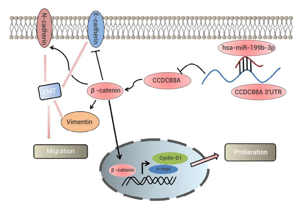
In August of this year, Dongsheng Zhu and others revealed that miR-199b-3p plays a key role in the formation and progression of osteosarcoma (OS). miR-199b-3p can bind to the 3' untranslated region (UTR) of CCDC88A, downregulating the expression level of CCDC88A, inhibiting EMT and the Wnt/β-catenin signaling pathway, thereby mediating its tumour-suppressive effect on OS cell proliferation and invasion[11].

Fig 5. The miR-199b-3p/CCDC88A axis regulates the malignant behavior of OS cells in vitro through the Wnt/β-catenin pathway and EMT process
Fig 5. The miR-199b-3p/CCDC88A axis regulates the malignant behavior of OS cells in vitro through the Wnt/β-catenin pathway and EMT process[11].

Additionally, research by Ling has proven that miR-145 inhibits EMT in lung cancer cells via targeting the Oct4 mediated Wnt/β‑catenin signaling pathway[12]. MiR-33b also binds to ZEB1 3'-UTR region and suppresses the activity of WNT/β-catenin signaling in lung adenocarcinoma cells and in turn suppressed tumor cell growth and EMT in vitro and in vivo[13].

• WNT3A-RIP1-β-catenin pathway induces EMT

In July 2023, A-Ram Kang revealed new potential roles of RIP1 in controlling WNT/β-catenin canonical signaling to enhance metastasis of colorectal cancer (CRC)[14].

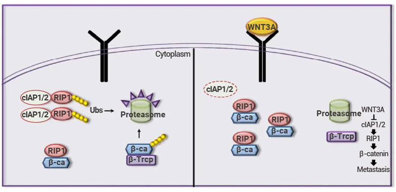
In the absence of WNT ligands, phosphorylated β-catenin is recognized and bound by β-TrCP. Ubiquitination of β-catenin and regulation of RIP1 ubiquitination by cIAP1/2 subsequently lead to the degradation of β-catenin protein.

However, WNT3A treatment induced cIAP1/2 degradation, abrogated the recruitment of β-TrCP to β-catenin and sequentially blocked β-catenin ubiquitination. RIP1 and β-catenin binded and stabilized one another. This binding of RIP1 and β-catenin also stimulated the dissociation of the β-catenin–β-TrCP complex (but does not change the protein levels of β-catenin and β-TrCP) and the inhibition of β-catenin ubiquitination, thereby stimulating EMT induction, enhancing the in vitro migration and invasion ability of CRC cells[14].

Fig 6. Model of RIP1's role in CRC metastasis
Fig 6. Model of RIP1's role in CRC metastasis[14].

In addition, many oncogenic signals, such as receptor tyrosine kinase (RTKs) family, PI(3)K/Akt, MAPK etc., inhibit GSK-3β activity, and thus leading to a large accumulation of β-catenin and triggering cell migration and EMT[15]. The Wnt family will produce crosstalk with Ras/Raf/MEK/ERK, TGFβ/Smad and other pathways, mutually influencing and cooperatively regulating genes related to cell invasion and metastasis, jointly participating in mediating the EMT process of tumor cells.

There are also many protein targets that affect the EMT process of tumor cells by participating in the regulation of Wnt/β-catenin. For example, IL-1β secreted by TAMs increases the availability of β-catenin in colorectal cancer cells by phosphorylating GSK3β, hindering the function of the β-catenin destruction complex[16]. MRGBP promotes CRC progression via DKK1/Wnt/β-catenin and NF-kB/p65 pathways mediated EMT[17].

Conclusion
In this issue, we introduced how the activation of the Wnt/β-catenin pathway can induce tumor EMT, the activation pathways of the Wnt pathway, and the related regulation of the Wnt/β-catenin pathway. The regulation of cell dynamic plasticity is also an important breakthrough point for researchers seeking cures for diseases. Those who are involved in related topics can like and bookmark this~

Related Products

IWP-2

Inhibitor of Wnt processing and secretion, targets the porcupine.

XAV939

Tankyrase inhibitor, targets Axin and inhibits Wnt signal transduction.

IWR-1

Targets Axin and inhibits the Wnt/β-catenin pathway.

SB-216763

ATP-competitive GSK-3 inhibitor.

ICG-001

Inhibitor of β-catenin/TCF mediated transcription binding to CREB protein.

MSAB

Selective inhibitor of Wnt/β-catenin signaling.

NSC668036

Frizzled-Dvl domain inhibitor, blocks Wnt signaling.

Box5

Wnt5a antagonist.

TWS119

Inhibitor of GSK-3β, activates the wnt/β-catenin pathway.

SKL2001

Agonist of the Wnt/β-catenin pathway, disrupts the Axin/β-catenin interaction.

References
[1] Thiery JP, et al. Complex networks orchestrate epithelial-mesenchymal transitions. Nat Rev Mol Cell Biol. 2006 Feb, 7(2):131-42.
[2] Bakir B, et al. EMT, MET, Plasticity, and Tumor Metastasis. Trends Cell Biol. 2020 Oct, 30(10):764-776.
[3] Yeung KT, et al. Epithelial-mesenchymal transition in tumor metastasis. Mol Oncol. 2017 Jan, 11(1): 28-39.
[4] Brabletz S, et al. Dynamic EMT: a multi-tool for tumor progression. EMBO J. 2021 Sep 15, 40(18): e108647.
[5] Hay ED. An overview of epithelio-mesenchymal transformation. Acta Anat (Basel). 1995, 154(1): 8-20.
[6] Lei Y, et al. MicroRNAs target the Wnt/β‑catenin signaling pathway to regulate epithelial‑mesenchymal transition in cancer (Review). Oncol Rep. 2020 Oct, 44(4): 1299-1313.
[7] Qin K, et al. Canonical and noncanonical Wnt signaling: Multilayered mediators, signaling mechanisms and major signaling crosstalk. Genes Dis. 2023 Mar 24, 11(1): 103-134.
[8] Buyuk B, et al. Epithelial-to-Mesenchymal Transition Signaling Pathways Responsible for Breast Cancer Metastasis. Cell Mol Bioeng. 2021 Sep 2, 15(1): 1-13.
[9] Yu F, et al. Wnt/β-catenin signaling in cancers and targeted therapies. Signal Transduct Target Ther. 2021 Aug 30, 6(1): 307.
[10] Lecarpentier Y, et al. Multiple Targets of the Canonical WNT/β-Catenin Signaling in Cancers. Front Oncol. 2019 Nov 18, 9: 1248.
[11] Zhu D, et al. hsa-miR-199b-3p suppresses osteosarcoma progression by targeting CCDC88A, inhibiting epithelial-to-mesenchymal transition, and Wnt/beta-catenin signaling pathway. Sci Rep. 2023 Aug 2, 13(1): 12544.
[12] Ling DJ, et al. MicroRNA-145 inhibits lung cancer cell metastasis. Mol Med Rep. 2015 Apr, 11(4): 3108-14.
[13] Qu J, et al. MicroRNA-33b inhibits lung adenocarcinoma cell growth, invasion, and epithelial-mesenchymal transition by suppressing Wnt/β-catenin/ZEB1 signaling. Int J Oncol. 2015 Dec, 47(6): 2141-52.
[14] Kang AR, et al. A novel RIP1-mediated canonical WNT signaling pathway that promotes colorectal cancer metastasis via β -catenin stabilization-induced EMT. Cancer Gene Ther. 2023 Jul 27.
[15] Zhou B., et al. Dual regulation of Snail by GSK-3β-mediated phosphorylation in control of epithelial–mesenchymal transition. Nat Cell Biol. 2004, 6: 931-940.
[16] Kaler P, et al. Macrophage-derived IL-1beta stimulates Wnt signaling and growth of colon cancer cells: a crosstalk interrupted by vitamin D3. Oncogene. 2009 Nov 5, 28(44): 3892-3902.
[17] Long X, et al. MRGBP promotes colorectal cancer metastasis via DKK1/Wnt/β-catenin and NF-kB/p65 pathways mediated EMT. Exp Cell Res. 2022 Dec 1, 421(1): 113375.