



AGER Protein, Human (HEK293, hFc)

Based on 1 Customer Validation
AGER proteins are cell surface pattern recognition receptors that expertly sense endogenous stress signals utilizing an extensive library of ligands, including advanced glycation end products, S100 proteins, high mobility Group Box 1 proteins/HMGB1, starch Like protein β/APP oligomers, nucleic acids, phospholipids, and glycosaminoglycans. AGER Protein, Human (HEK293, hFc) is the recombinant human-derived AGER protein, expressed by HEK293 , with C-hFc labeled tag.
- Species: Human
- Source: HEK293
-
Storage:Stored at -20°C for 2 years from date of receipt. After reconstitution, it is stable at 4°C for 1 week or -20°C for longer (with carrier protein). It is recommended to freeze aliquots at -20°C or -80°C for extended storage.


To place orders, for customer services and technical support, please contact: MedChemExpress USA
Tel: 609-228-6898 E-mail: [email protected] [email protected]


-
 Biological Activity
-
Technical Parameters
-
Product Properties
- Documentation
- Help & FAQs




Biological Activity
Description
AGER proteins are cell surface pattern recognition receptors that expertly sense endogenous stress signals utilizing an extensive library of ligands, including advanced glycation end products, S100 proteins, high mobility Group Box 1 proteins/HMGB1, starch Like protein β/APP oligomers, nucleic acids, phospholipids, and glycosaminoglycans. AGER Protein, Human (HEK293, hFc) is the recombinant human-derived AGER protein, expressed by HEK293 , with C-hFc labeled tag.
Background
AGER Protein, a cell surface pattern recognition receptor, adeptly senses endogenous stress signals with a wide-ranging ligand repertoire, encompassing advanced glycation end products, S100 proteins, high-mobility group box 1 protein/HMGB1, amyloid beta/APP oligomers, nucleic acids, phospholipids, and glycosaminoglycans. Accumulation of advanced glycosylation end products, especially prevalent in aging and diabetes, triggers inflammatory responses at various disease sites, including diabetes, vascular complications, neurodegenerative disorders, and cancers. RAGE, upon ligand binding, utilizes TIRAP and MYD88 as adapters to transduce signals, ultimately inducing inflammatory cytokines IL6, IL8, and TNFalpha through NF-kappa-B activation. Noteworthy interactions include S100A12-triggered cellular activation, S100B-induced apoptosis post-myocardial infarction, and the facilitation of amyloid-beta peptide translocation in cortical neurons. AGER also plays a role in endothelial albumin transcytosis with HMGB1 through the RAGE/SRC/Caveolin-1 pathway, leading to endothelial hyperpermeability, and mediates the loading of HMGB1 in extracellular vesicles for hepatocyte pyroptosis via the NLRP3 inflammasome. Additionally, it promotes extracellular hypomethylated DNA uptake for the activation of inflammatory responses. The constitutive homodimeric and oligomeric forms, along with interactions with S100 proteins, APP, TIRAP, and HMGB1, highlight the intricate involvement of AGER Protein in various cellular processes and pathological conditions.
Verified Bioactivity
1.Immobilized Human AGER at 20 μg/mL (100 μL/well) can bind Biotinylated Human HMGB1.The ED50 for this effect is 30.85 ng/mL, corresponding to a specific activity is 3.24×10⁴ Unit/mg.
2.Measured by its binding ability in a functional ELISA. Immobilized human S100A12 at 2 μg/mL (100 μl/well) can bind recombinant human AGER with a linear range of 0.032-20 μg/mL.
MCE Validation Data
-
Purity - SDS-PAGE
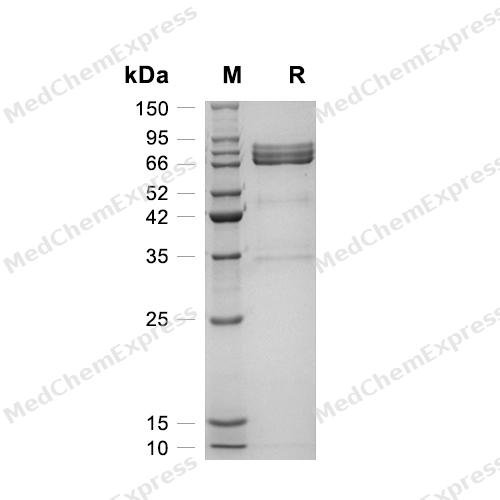
Purity - SDS-PAGE
-
Bioactivity - ELISA
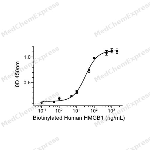
Bioactivity - ELISA


Technical Parameters
-
Species Human
-
Source HEK293
-
Tag C-hFc
-
Accession
Q15109 (Q24-A344)
-
Molecular Construction
-
N-term
-
AGER (Q24-A344)
Accession # Q15109 -
hFc
-
C-term
-
-
Protein Length
Partial Extracellular Domain
-
Synonyms
AGER; Receptor For Advanced Glycosylation End-Products Deletion Exon3-10 Variant; Advanced Glycosylation End-Product Specific Receptor; Receptor For Advanced Glycosylation End-Products Deletion Exon2-6 Variant; RAGE; Advanced Glycosylation End Product-Spe
-
AA Sequence
QNITARIGEPLVLKCKGAPKKPPQRLEWKLNTGRTEAWKVLSPQGGGPWDSVARVLPNGSLFLPAVGIQDEGIFRCQAMNRNGKETKSNYRVRVYQIPGKPEIVDSASELTAGVPNKVGTCVSEGSYPAGTLSWHLDGKPLVPNEKGVSVKEQTRRHPETGLFTLQSELMVTPARGGDPRPTFSCSFSPGLPRHRALRTAPIQPRVWEPVPLEEVQLVVEPEGGAVAPGGTVTLTCEVPAQPSPQIHWMKDGVPLPLPPSPVLILPEIGPQDQGTYSCVATHSSHGPQESRAVSISIIEPGEEGPTAGSVGGSGLGTLALA
-
Molecular Weight
Approximately 80-90 kDa, based on SDS-PAGE under reducing conditions, due to the glycosylation.
-
Glycosylation
Yes
-
Purity
≥ 90%, as determined by reducing SDS-PAGE.


Product Properties
Lyophilized powder.
1.Lyophilized from a 0.22 μm filtered solution of PBS, pH 7.4, 5% trehalose, 5% mannitol, 0.01% Tween 80.
2.Lyophilized from a 0.22 μm filtered solution of 20 mM PB, 150 mM NaCl, pH 7.4.
Please refer to the lot-specific COA for specific buffer information.
<1 EU/μg, determined by LAL method.
It is not recommended to reconstitute to a concentration less than 100 μg/mL in ddH2O.
Stored at -20°C for 2 years from date of receipt. After reconstitution, it is stable at 4°C for 1 week or -20°C for longer (with carrier protein). It is recommended to freeze aliquots at -20°C or -80°C for extended storage.
Room temperature in continental US; may vary elsewhere.


Documentation
-
Data Sheet (238 KB)
-
SDS (252 KB)
- English - EN (252 KB)
- Français - FR (252 KB)
- Deutsch - DE (252 KB)
- Norwegian - NO (252 KB)
- Español - ES (252 KB)
- Swedish - SV (252 KB)
- Italian - IT (252 KB)
- Korean - KR (252 KB)
- Portuguese - PT (252 KB)
-
Handling Instructions (2659 KB)

Calculators
Concentration (start) × Volume (start) = Concentration (final) × Volume (final)


 Powered by Bioz

