



Fibrillin-1/Asprosin Protein, Human (HEK293, His)
Based on 3 publication(s) in Google Scholar
Fibrillin-1 and Asprosin are two proteins generated from the same precursor protein, which is encoded by the Fibrillin 1 gene, through proteolytic cleavage. Fibrillin-1 protein, a member of the fibrillin family, is a crucial structural protein in the formation of microfibrils in the extracellular matrix, maintaining tissue homeostasis and cell adhesion, regulating the availability of growth factors, and inhibiting osteoclastogenesis. In contrast, Asprosin is a hormone primarily secreted by white adipose tissue and is involved in regulating glucose metabolism within the body. Fibrillin-1/Asprosin protein, Human (HEK293, His) is a recombinant protein expressed in HEK293 cells with an N-terminal His tag, comprising a full length of 140 amino acids (S2732-H2871).
- Species: Human
- Source: HEK293
-
Storage:Stored at -20°C for 2 years from date of receipt. After reconstitution, it is stable at 4°C for 1 week or -20°C for longer (with carrier protein). It is recommended to freeze aliquots at -20°C or -80°C for extended storage.


To place orders, for customer services and technical support, please contact: MedChemExpress USA
Tel: 609-228-6898 E-mail: [email protected] [email protected]


-
 Biological Activity
-
Technical Parameters
-
Product Properties
- Documentation
- References
- Help & FAQs




Biological Activity
Description
Fibrillin-1 and Asprosin are two proteins generated from the same precursor protein, which is encoded by the Fibrillin 1 gene, through proteolytic cleavage. Fibrillin-1 protein, a member of the fibrillin family, is a crucial structural protein in the formation of microfibrils in the extracellular matrix, maintaining tissue homeostasis and cell adhesion, regulating the availability of growth factors, and inhibiting osteoclastogenesis. In contrast, Asprosin is a hormone primarily secreted by white adipose tissue and is involved in regulating glucose metabolism within the body. Fibrillin-1/Asprosin protein, Human (HEK293, His) is a recombinant protein expressed in HEK293 cells with an N-terminal His tag, comprising a full length of 140 amino acids (S2732-H2871)[1][2][3][4][5][6][7][8][9][10][11].
Background
Fibrillin-1 is a key structural protein in the formation of extracellular matrix microfibrils. It plays a critical role in maintaining the structural integrity and functional stability of connective tissues, not only providing physical support but also participating in the regulation of tissue homeostasis and bone metabolism through interactions with various growth factors, integrins, and heparin.
1.
Physical Structural Support:
(1) In tissues such as the lung, blood vessels, and skin, Fibrillin-1 supports the formation of elastic fibers[1].
(2) In tissues lacking elastin, such as the ciliary zonule, tendon, cornea, and glomerulus, Fibrillin-1 provides structural support[1].
2. Homeostasis Regulation and Metabolic Control:
(1) Fibrillin-1 maintains tissue structural stability through specific interactions with growth factors (such as bone morphogenetic proteins (BMPs), growth and differentiation factors (GDFs), latent transforming growth factor beta-binding proteins (LTBPs)), cell surface integrins, and other extracellular matrix proteins and proteoglycan components[1].
(2) Fibrillin-1 inhibits osteoclastogenesis by binding to the osteoclast differentiation factor TNFSF11 and suppressing the TNFSF11-mediated nuclear translocation and activation of the transcription factor NFATC1, thereby influencing bone metabolism[2].
Fibrillin-1 mediates cell adhesion by binding to cell surface receptors integrins ITGAV and ITGA5[3][4]. It can also bind heparin, ensuring the proper assembly of microfibrils[5].
Asprosin is a hormone secreted by white adipose tissue that plays a crucial role in regulating energy balance in the body, particularly in maintaining blood glucose levels during fasting. Asprosin acts on the liver during fasting by binding to the olfactory receptor OR4M1 on hepatocytes, activating protein kinase A (PKA), and triggering the rapid release of glucose into the bloodstream. Asprosin also promotes rapid glucose release by activating the G protein-cAMP-PKA pathway[6][7][8][9].
In Vitro
Fibrillin-1 protein (Human) (0-20 μg/mL, 48 h) promotes αvβ3 and α5β1 integrin-mediated adhesion of human umbilical vein endothelial cells in a dose-dependent manner and enhances their proliferation and migration at a dose of 1 μg/mL[10].
In Vivo
Asprosin protein (human) (2 mg/kg, i.p., single dose, with blood samples collected 48 hours or 7 days after administration) has a cardioprotective effect in Isoprenaline hydrochloride (HY-B0468)-induced myocardial infarction rat models[11].
Verified Bioactivity
1.Measured by its binding ability in a functional ELISA. Immobilized Human MFAP4 at 0.5 μg/mL (100 μL/well) can bind Biotinylated Human Fibrillin-1. The ED50 for this effect is ≤0.8473 μg/mL, corresponding to a specific activity is ≥1.18×10³ Unit/mg.
2.Measured by its ability to induce TNF-ɑ secretion by macrophages produced by THP-1 cells treated with dihydroxyvitamin D3.
MCE Validation Data
-
Purity - SDS-PAGE
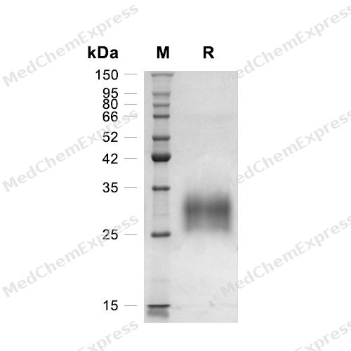
Purity - SDS-PAGE
-
Bioactivity - ELISA
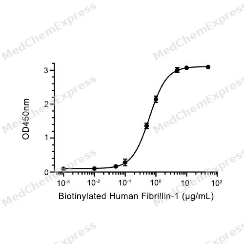
Bioactivity - ELISA


Publications (3)
-
Journal Impact Factor
-
Most Recent
-
Cardiovasc Diabetol
Asprosin induces vascular endothelial-to-mesenchymal transition in diabetic lower extremity peripheral artery disease. [Abstract]2022 Feb 15;21(1):25. PMID: 35168605 -
Antioxid Redox Signal
Intervention of asprosin attenuates oxidative stress and neointima formation in vascular injury. [Abstract]2024 Sep;41(7-9):488-504. PMID: 38814824 -
Clin Immunol
Elevated asprosin expression exacerbates synovial inflammation by PPAR-γ-dependent mechanisms in rheumatoid arthritis. [Abstract]2026 Apr:284:110677. PMID: 41638328


Technical Parameters
-
Species Human
-
Source HEK293
-
Tag N-His
-
Accession
P35555 (S2732-H2871)
-
Molecular Construction
-
N-term
-
8*His
-
Asprosin (S2732-H2871)
Accession # P35555 -
C-term
-
-
Protein Length
Full Length of Asprosin Chain
-
Synonyms
FBN1; Epididymis Secretory Sperm Binding Protein; Prev. FBN; Alternative Protein FBN1; Prev. MFS1; Marfan Syndrome; Prev. WMS; Fibrillin 15; Fibrillin-1; GPHYSD2; MASS; ACMICD; OCTD; ECTOL1; SGS; MFLS; Fibrillin-1 Preproprotein; SSKS; Asprosin; WMS2; Fibr
-
AA Sequence
STNETDASNIEDQSETEANVSLASWDVEKTAIFAFNISHVSNKVRILELLPALTTLTNHNRYLIESGNEDGFFKINQKEGISYLHFTKKKPVAGTYSLQISSTPLYKKKELNQLEDKYDKDYLSGELGDNLKMKIQVLLH
-
Molecular Weight
Approximately 25-35 kDa, based on SDS-PAGE under reducing conditions.
-
Glycosylation
Yes
-
Purity
≥ 95%, as determined by reducing SDS-PAGE.


Product Properties
Lyophilized powder
Lyophilized from a 0.2 μm filtered solution of PBS or 20 mM PB, 150 mM NaCl, pH 7.4 or PBS, pH 7.4, 8% trehalose.
<1 EU/μg, determined by LAL method.
It is not recommended to reconstitute to a concentration less than 100 μg/mL in ddH2O. For long term storage it is recommended to add a carrier protein (0.1% BSA, 5% HSA, 10% FBS or 5% Trehalose).
Stored at -20°C for 2 years from date of receipt. After reconstitution, it is stable at 4°C for 1 week or -20°C for longer (with carrier protein). It is recommended to freeze aliquots at -20°C or -80°C for extended storage.
Room temperature in continental US; may vary elsewhere.


Documentation
-
Data Sheet (265 KB)
-
SDS (252 KB)
- English - EN (252 KB)
- Français - FR (252 KB)
- Deutsch - DE (252 KB)
- Norwegian - NO (252 KB)
- Español - ES (252 KB)
- Swedish - SV (252 KB)
- Italian - IT (252 KB)
- Korean - KR (252 KB)
- Portuguese - PT (252 KB)
-
Handling Instructions (2659 KB)


References
[1]. Jensen SA, et al. New insights into the structure, assembly and biological roles of 10-12 nm connective tissue microfibrils from fibrillin-1 studies. Biochem J. 2016 Apr 1;473(7):827-38. [Content Brief]
[2]. Tiedemann K, et al. Fibrillin-1 directly regulates osteoclast formation and function by a dual mechanism. J Cell Sci. 2013 Sep 15;126(Pt 18):4187-94. [Content Brief]
[3]. Bax DV, et al. Cell adhesion to fibrillin-1 molecules and microfibrils is mediated by alpha 5 beta 1 and alpha v beta 3 integrins. J Biol Chem. 2003 Sep 5;278(36):34605-16. [Content Brief]
[4]. Jovanovic J, et al. alphaVbeta6 is a novel receptor for human fibrillin-1. Comparative studies of molecular determinants underlying integrin-rgd affinity and specificity. J Biol Chem. 2007 Mar 2;282(9):6743-51. [Content Brief]
[5]. Tiedemann K, et al. Interactions of fibrillin-1 with heparin/heparan sulfate, implications for microfibrillar assembly. J Biol Chem. 2001 Sep 21;276(38):36035-42. [Content Brief]
[6]. Romere C, et al. Asprosin, a Fasting-Induced Glucogenic Protein Hormone. Cell. 2016 Apr 21;165(3):566-79. [Content Brief]
[7]. Lee T, et al. Asprosin impairs insulin secretion in response to glucose and viability through TLR4/JNK-mediated inflammation. Mol Cell Endocrinol. 2019 Apr 15;486:96-104. [Content Brief]
[8]. Li E, et al. OLFR734 Mediates Glucose Metabolism as a Receptor of Asprosin. Cell Metab. 2019 Aug 6;30(2):319-328.e8. [Content Brief]
[9]. Li E, et al. OLFR734 Mediates Glucose Metabolism as a Receptor of Asprosin. Cell Metab. 2019 Aug 6;30(2):319-328.e8. [Content Brief]
[10]. Mariko B, et al. Microfibrils and fibrillin-1 induce integrin-mediated signaling, proliferation and migration in human endothelial cells. Am J Physiol Cell Physiol. 2010 Nov;299(5):C977-87. [Content Brief]
[11]. Onat E, et al. The protective effects of humanin in rats with experimental myocardial infarction: The role of asprosin and spexin. Heliyon. 2023 Jul 26;9(8):e18739. [Content Brief]

Calculators
Concentration (start) × Volume (start) = Concentration (final) × Volume (final)


 Powered by Bioz

