



MIG/CXCL9 Protein, Mouse (HEK293, His)
Based on 2 publication(s) in Google Scholar
CXCL9, also known as MIG, is one member of the ELR-negative CXC chemokine subfamily, and can be induced by IFN-γ. CXCL9 binds to its receptor CXCR3 and can recruit CXCR3+ cells, such as effector T cells, regulatory T cells (Tregs) and CD8+ cytotoxic T cells. CXCL9 is involved in immunoregulatory and inflammatory processes, but it also play a key role in tumor growth, angiogenesis, and metastasis. MIG/CXCL9 Protein, Mouse (HEK293, His) is produced in HEK293 cells with six C-Terminal His-tags.
- Species: Mouse
- Source: HEK293
-
Storage:Stored at -20°C for 2 years from date of receipt. After reconstitution, it is stable at 4°C for 1 week or -20°C for longer (with carrier protein). It is recommended to freeze aliquots at -20°C or -80°C for extended storage.


To place orders, for customer services and technical support, please contact: MedChemExpress USA
Tel: 609-228-6898 E-mail: [email protected] [email protected]


-
 Biological Activity
-
Technical Parameters
-
Product Properties
- Documentation
- References
- Help & FAQs




Biological Activity
Description
CXCL9, also known as MIG, is one member of the ELR-negative CXC chemokine subfamily, and can be induced by IFN-γ. CXCL9 binds to its receptor CXCR3 and can recruit CXCR3+ cells, such as effector T cells, regulatory T cells (Tregs) and CD8+ cytotoxic T cells. CXCL9 is involved in immunoregulatory and inflammatory processes, but it also play a key role in tumor growth, angiogenesis, and metastasis[1][2]. MIG/CXCL9 Protein, Mouse (HEK293, His) is produced in HEK293 cells with six C-Terminal His-tags.
Background
CXCL9 is a member of the CXC family and has an important role in the chemotaxis of immune cells. It is secreted by various cell types including immune cells (T lymphocytes, NK cells, dendritic cells, macrophages, eosinophils, etc.), and non-immune cells (hepatic stellate cells, preadipocytes, thyrocytes, endothelial cell, tumor cells, and fibroblasts, etc)[1].
The amino acid sequence of human CXCL9 protein has low homology between mouse and rat CXCL9 protein.
CXCL9 is one of the ligands of chemokine receptor CXCR3 that mediates the infiltration of lymphocytes to focal sites and suppresses tumor growth. CXCL9 attracts CXCR3- (CXCR3-A and CXCR3-B) T lymphocytes, is involved in the pathogenesis of a variety of physiologic diseases during their initiation and their maintenance. The transcriptional regulation of CXCL9 is a multistep process involving many transcription factors, of which STAT1 and NF-κB are two most well-characterized members. Both the gene mutation of STAT1 and the blocking of the JA/STAT1 pathway can reduce CXCL9 expression induced by IFN-γ. Moreover, CXCL9 expression can be suppressed by reducing the levels of components of the STAT1-IRF-1 transcriptional activation pathway by Porphyromonas gingivalis that leads to the immune function decline. Lipopolysaccharide (LPS) and D-galactosamine could induce the phosphorylation of STAT1 and enhance the transcription of CXCL9 leading to the enhancement of liver inflammation, and even liver apoptosis and injury[1][2][3].
CXCL9 could promote cancer metastasis via enhanced migration and invasion of tumor cells, and breaking of the endothelial cells monolayer. However, as a tumor suppressor, it mainly recruited tumor-infiltrating CD8+ T cells and NK cells, and inhibited tumor angiogenesis. In Addition, IL-12 and Th1-derived IFN-γ exerted antitumor effects through the inhibitory effects of endogenous CXCL9 on tumor vasculature in human Burkitt's lymphoma. In cutaneous T-cell lymphoma, expression of CXCL9 was found at early stage but low at advanced stage. CXCL9 is also associated with human hepatic fibrosis and anti-fibrosis in mice. Furthermore, CXCL9 is highly expressed in atherosclerotic plaques of coronary arteries and specifically recruits CXCR3-bearing Th1 cells that increase the risk of plaque progression and the occurrences of myocardial infarction[1][2][3][4].
In Vitro
Recombinant mouse CXCL9 (10 mg/kg; for 4 weeks) is intraperitoneally injected three times a week in the orthotopic murine pancreatic adenocarcinoma (PAAD) model. CXCL9 could significant accelerate the growth of PAAD tumour. CXCL9 promotes PAAD progression and suppresses CD8+ cytotoxic T cells in tumour tissues[4].
In Vivo
Recombinant mouse CXCL9 (100 ng/mL) displays strong antiproliferative and antimigratory effects on VEGF-stimulated endothelial cells and stellate cells by way of reduced VEGFR2 (KDR), phospholipase Cγ (PLCγ), and extracellular signal-regulated kinase (ERK) phosphorylation[5].
Verified Bioactivity
1.Measured by its ability to chemoattract THP-1 human peripheral blood T-lymphocytes. The ED50 for this effect is 85.56 ng/mL, corresponding to a specific activity is 1.169×10⁴ U/mg.
2. Measured by its ability to chemoattract BaF3 mouse pro-B cells transfected with Mouse CXCR3. The ED50 for this effect is 0.1-0.3 μg/mL.
MCE Validation Data
-
Purity - SDS-PAGE
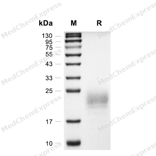
Purity - SDS-PAGE
-
Bioactivity - Cell-Based Assay
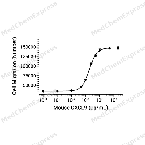
Bioactivity - Cell-Based Assay


Publications (2)
-
Journal Impact Factor
-
Most Recent
-
Immunity
Spatiotemporal dynamics of CXCL10 encode contextual immune information revealed by the genetically encoded fluorescent sensor. [Abstract]2025 Sep 9;58(9):2320-2335.e9. PMID: 40818452 -
J Exp Clin Cancer Res
Augmented ERO1α upon mTORC1 activation induces ferroptosis resistance and tumor progression via upregulation of SLC7A11. [Abstract]2024 Apr 13;43(1):112. PMID: 38610018


Technical Parameters
-
Species Mouse
-
Source HEK293
-
Tag C-6*His
-
Accession
P18340 (T22-T126)
-
Molecular Construction
-
N-term
-
CXCL9 (T22-T126)
Accession # P18340 -
6*His
-
C-term
-
-
Protein Length
Full Length of Mature Protein
-
Synonyms
CXCL9; Monokine Induced By Interferon-Gamma; Prev. CMK; Chemokine (C-X-C Motif) Ligand 9; Prev. MIG; Small-Inducible Cytokine B9; SCYB9; C-X-C Motif Chemokine 9; Crg-10; Alternative Protein CXCL9; Humig; HuMIG; Monokine Induced By Gamma Interferon; C-X-C
-
AA Sequence
TLVIRNARCSCISTSRGTIHYKSLKDLKQFAPSPNCNKTEIIATLKNGDQTCLDPDSANVKKLMKEWEKKISQKKKQKRGKKHQKNMKNRKPKTPQSRRRSRKTT
-
Predicted Molecular Mass
12.2 kDa
-
Molecular Weight
Approximately 18-28 kDa, based on SDS-PAGE under reducing conditions, due to the glycosylation.
-
Glycosylation
Yes
-
Purity
≥ 95%, as determined by reducing SDS-PAGE.


Product Properties
Lyophilized powder
Lyophilized from a 0.2 μm filtered solution of PBS, pH 7.4.
<1 EU/μg, determined by LAL method.
It is not recommended to reconstitute to a concentration less than 100 μg/mL in ddH2O. For long term storage it is recommended to add a carrier protein (0.1% BSA, 5% HSA, 10% FBS or 5% Trehalose).
Stored at -20°C for 2 years from date of receipt. After reconstitution, it is stable at 4°C for 1 week or -20°C for longer (with carrier protein). It is recommended to freeze aliquots at -20°C or -80°C for extended storage.
Room temperature in continental US; may vary elsewhere.


Documentation
-
Data Sheet (265 KB)
-
SDS (252 KB)
- English - EN (252 KB)
- Français - FR (252 KB)
- Deutsch - DE (252 KB)
- Norwegian - NO (252 KB)
- Español - ES (252 KB)
- Swedish - SV (252 KB)
- Italian - IT (252 KB)
- Korean - KR (252 KB)
- Portuguese - PT (252 KB)
-
Handling Instructions (2659 KB)


References
[1]. Qiang Ding, et al. CXCL9: evidence and contradictions for its role in tumor progression. Cancer Med. 2016 Nov;5(11):3246-3259. [Content Brief]
[2]. Weigang Xiu, et al. CXCL9 secreted by tumor-associated dendritic cells up-regulates PD-L1 expression in bladder cancer cells by activating the CXCR3 signaling. BMC Immunol. 2021 Jan 6;22(1):3. [Content Brief]
[3]. Chao-Feng Lin, et al. Potential Effects of CXCL9 and CCL20 on Cardiac Fibrosis in Patients with Myocardial Infarction and Isoproterenol-Treated Rats. J Clin Med. 2019 May 11;8(5):659. [Content Brief]
[4]. Hui-Feng Gao, et al. CXCL9 chemokine promotes the progression of human pancreatic adenocarcinoma through STAT3-dependent cytotoxic T lymphocyte suppression. Aging (Albany NY). 2020 Jan 8;12(1):502-517. [Content Brief]
[5]. Hacer Sahin, et al. Chemokine Cxcl9 attenuates liver fibrosis-associated angiogenesis in mice. Hepatology. 2012 May;55(5):1610-9. [Content Brief]

Calculators
Concentration (start) × Volume (start) = Concentration (final) × Volume (final)


 Powered by Bioz

