


Y-33075
Based on 14 publication(s) in Google Scholar
Y-33075 is a selective ROCK inhibitor derived from Y-27632, and is more potent than Y-27632, with an IC50 of 3.6 nM.
For research use only. We do not sell to patients.
- Purity: 98.44%
- CAS No.: 199433-58-4
- Formula: C16H16N4O
- Molecular Weight:280.33
-
Storage:Powder -20°C, 3 years , 4°C, 2 years ; In solvent -80°C, 2 years , -20°C, 1 year

To place orders, for customer services and technical support, please contact: MedChemExpress USA
Tel: 609-228-6898 E-mail: [email protected] [email protected]


-
 Biological Activity
-
Chemical Information
-
Solvent & Solubility
- Protocol
- Purity & Documentation
- References
-
 Help & FAQs
-


Cell Cycle/DNA Damage Compound Library
HY-L004
-
Kinase Inhibitor Library
HY-L009
-
Stem Cell Signaling Compound Library
HY-L017
-
TGF-beta/Smad Compound Library
HY-L018
-
Anti-Cancer Compound Library
HY-L025
-
Clinical Compound Library
HY-L026
-
Anti-Aging Compound Library
HY-L034
-
Drug Repurposing Compound Library
HY-L035
-
Reprogramming Compound Library
HY-L039
-
Cytoskeleton Compound Library
HY-L060
-
Cancer Stem Cells Compound Library
HY-L135
-
Heterocyclic Compound Library
HY-L138
-
Highly Selective Inhibitors Library
HY-L158
-
Serine/Threonine Kinase Inhibitor Library
HY-L164
-
Extracellular Vesicles (EVs) Compound Library
HY-L168
-
Multi-Target Compound Library
HY-L176
-
Bioactive Compound Library Max
HY-L181
-
MCE Bioactive Compound Library
HY-L001V
-
Drug Repurposing Compound Library Plus
HY-L035P
-
Clinical Compound Library Plus
HY-L026P
-
Bioactive Compound Library
HY-L001
-
High-Throughput Bioactive Compound Library
HY-L205
-
Posttranslational Modification Library
HY-L226
-
3D Diverse Fragment Library
HY-L903


Publications Citing Use of MedChemExpress (MCE) Y-33075
More- Science. 2017 Dec 1;358(6367):eaan4368. [Abstract]
- Cell. 2018 Jul 26;174(3):636-648.e18. [Abstract]
- Nat Commun. 2020 Jan 3;11(1):88. [Abstract]
- Adv Sci (Weinh). 2022 May;9(13):e2104682. [Abstract]
- J Adv Res. 2024 Sep:63:117-128. [Abstract]
- Cell Death Dis. 2024 Dec 26;15(12):932. [Abstract]
- Acta Neuropathol Commun. 2024 Sep 14;12(1):150. [Abstract]
- Cells. 2024 Nov 18;13(22):1907. [Abstract]
- Stem Cell Rep. 2020 Jan 14;14(1):49-59. [Abstract]
- J Cell Physiol. 2021 Dec;236(12):8226-8238. [Abstract]
- Neurotox Res. 2013 Apr;23(3):238-48. [Abstract]
- PLoS One. 2023 Jan 31;18(1):e0270288. [Abstract]
- bioRxiv. 2025 June 03.
- Patent. US20170349879A1.
 Customer Validation & Images

-
WB
-
Cell Proliferation/Viability Assay


Biological Activity
|
ROCK 3.6 nM (IC50) |
PKC 420 nM (IC50) |
CaMKII 810 nM (IC50) |
Y-33075 (Y-39983) is a potent ROCK inhibitor, with an IC50 of 3.6 nM. Y-33075 also inhibits PKC and CaMKII more potently than Y-27632, and the IC50s of Y-27632 and Y-33075 for PKC are 9.0 μM and 0.42 μM, respectively, whereas the IC50s of Y-27632 and Y-33075 for CaMKII are 26 μM and 0.81 μM, respectively. The IC50s of Y-27632 and Y-33075 for PKC is 82 and 117 times those for ROCK, respectively, whereas the IC50s of Y-27632 and Y-33075 for CaMKII is 236 and 225 times those for ROCK, respectively[1]. Y-33075 (Y-39983, 10 μM) extends neurites in the retinal ganglion cells (RGCs) compared with those in RGCs treated without Y-39983[2]. Y-33075 (Y-39983, 1 μM) inhibits the contraction of rabbit ciliary artery segments evoked by histamine in Ca2+-free solutions. Y-33075 (10 μM) shows no effect on the [Ca2+]i increase with the high-potassium (high-K) solution[3].
MedChemExpress (MCE) has not independently confirmed the accuracy of these methods. They are for reference only.
MedChemExpress (MCE) has not independently confirmed the accuracy of these methods. They are for reference only.


Chemical Information
-
CAS No. 199433-58-4
-
Appearance Solid
-
Molecular Weight 280.33
-
Formula C16H16N4O
-
Color White to off-white
-
SMILES
O=C(NC1=C2C(NC=C2)=NC=C1)C3=CC=C(C=C3)[C@H](N)C
-
Synonyms
Y-39983 free base
-
Shipping
Room temperature in continental US; may vary elsewhere.
-
Storage
Powder -20°C 3 years 4°C 2 years In solvent -80°C 2 years -20°C 1 year


Publications (14)
-
Journal Impact Factor
-
Most Recent
-
Science
2017 Dec 1;358(6367):eaan4368. PMID: 29191878 -
Cell
2018 Jul 26;174(3):636-648.e18. PMID: 30017246 -
Nat Commun
2020 Jan 3;11(1):88. PMID: 31900402 -
Adv Sci (Weinh)
Pharmacological Perturbation of Mechanical Contractility Enables Robust Transdifferentiation of Human Fibroblasts into Neurons. [Abstract]2022 May;9(13):e2104682. PMID: 35240008 -
J Adv Res
Leucine zipper protein 1 attenuates pressure overload-induced cardiac hypertrophy through inhibiting Stat3 signaling. [Abstract]2024 Sep:63:117-128. PMID: 37806546 -
Cell Death Dis
TRIM59/RBPJ positive feedback circuit confers gemcitabine resistance in pancreatic cancer by activating the Notch signaling pathway. [Abstract]2024 Dec 26;15(12):932. PMID: 39725730 -
Acta Neuropathol Commun
Evaluation of Rho kinase inhibitor effects on neuroprotection and neuroinflammation in an ex-vivo retinal explant model. [Abstract]2024 Sep 14;12(1):150. PMID: 39300576 -
Cells
Thrombospondin 1 Mediates Autophagy Upon Inhibition of the Rho-Associated Protein Kinase Inhibitor. [Abstract]2024 Nov 18;13(22):1907. PMID: 39594655 -
Stem Cell Rep
iPSC-Derived Platelets Depleted of HLA Class I Are Inert to Anti-HLA Class I and Natural Killer Cell Immunity. [Abstract]2020 Jan 14;14(1):49-59. PMID: 31883921 -
J Cell Physiol
2021 Dec;236(12):8226-8238. PMID: 34180057 -
Neurotox Res
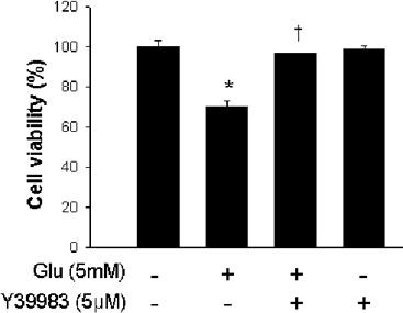
The Rho-kinase (ROCK) inhibitor Y-27632 protects against excitotoxicity-induced neuronal death in vivo and in vitro. [Abstract]2013 Apr;23(3):238-48. PMID: 22810835
Y-33075 purchased from MedChemExpress. Usage Cited in: Neurotox Res. 2013 Apr;23(3):238-48. [Abstract]
Effect of Y39983 on glutamate-treated HT22 cell viability.
-
PLoS One
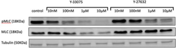
The non-selective Rho-kinase inhibitors Y-27632 and Y-33075 decrease contraction but increase migration in murine and human hepatic stellate cells. [Abstract]2023 Jan 31;18(1):e0270288. PMID: 36719899
Y-33075 purchased from MedChemExpress. Usage Cited in: PLoS One. 2023 Jan 31;18(1):e0270288. [Abstract]
Y-27632 (10, 100 nM and 10, 100 µM; 24 h) decreases phosphorylation of MLC starting at a concentration of 100 nM in HSCs.
-
-


Solvent & Solubility
DMSO : 50 mg/mL (178.36 mM; Need ultrasonic and warming; Hygroscopic DMSO has a significant impact on the solubility of product, please use newly opened DMSO)
H2O : < 0.1 mg/mL (insoluble)
Please refer to the solubility information to select the appropriate solvent. Once prepared, please aliquot and store the solution to prevent product inactivation from repeated freeze-thaw cycles.
Storage method and period of stock solution: -80°C, 2 years; -20°C, 1 year. When stored at -80°C, please use it within 2 years. When stored at -20°C, please use it within 1 year.
Concentration (start) × Volume (start) = Concentration (final) × Volume (final)
Select the appropriate dissolution method based on your experimental animal and administration route.
- For the following dissolution methods, please ensure to first prepare a clear stock solution using an In Vitro approach and then sequentially add co-solvents:
- To ensure reliable experimental results, the clarified stock solution can be appropriately stored based on storage conditions. As for the working solution for In Vivo experiments, it is recommended to prepare freshly and use it on the same day.
- The percentages shown for the solvents indicate their volumetric ratio in the final prepared solution. If precipitation or phase separation occurs during preparation, heat and/or sonication can be used to aid dissolution.
Add each solvent one by one: 10% DMSO 40% PEG300 5% Tween-80 45% Saline
Solubility: ≥ 2.5 mg/mL (8.92 mM); Clear solution
This protocol yields a clear solution of ≥ 2.5 mg/mL (saturation unknown).
Taking 1 mL working solution as an example, add 100 μL DMSO stock solution (25.0 mg/mL) to 400 μL PEG300, and mix evenly; then add 50 μL Tween-80 and mix evenly; then add 450 μL Saline to adjust the volume to 1 mL.
Preparation of Saline: Dissolve 0.9 g sodium chloride in ddH₂O and dilute to 100 mL to obtain a clear Saline solution.
Add each solvent one by one: 10% DMSO 90% (20% SBE-β-CD in Saline)
Solubility: ≥ 2.5 mg/mL (8.92 mM); Clear solution
This protocol yields a clear solution of ≥ 2.5 mg/mL (saturation unknown).
Taking 1 mL working solution as an example, add 100 μL DMSO stock solution (25.0 mg/mL) to 900 μL 20% SBE-β-CD in Saline, and mix evenly.
Preparation of 20% SBE-β-CD in Saline (4°C, storage for one week): 2 g SBE-β-CD powder is dissolved in 10 mL Saline, completely dissolve until clear.
Please enter the basic information of animal experiments:
-
-
-
-
Recommended: Prepare an additional quantity of animals to account for potential losses during experiments.
Please enter your animal formula composition:
-
%DMSO +
Recommended: Keep the proportion of DMSO in working solution below 2% if your animal is weak.
-
%+
-
+%Tween-80 + +
-
%Saline +
The co-solvents required include: DMSO, . All of co-solvents are available by MedChemExpress (MCE). , Tween 80. All of co-solvents are available by MedChemExpress (MCE).
Working solution concentration: 0.22 mg/mL
Method for preparing stock solution: mg drug dissolved in μL DMSO. Stock solution concentration: mg/mL.
1. Take μL DMSO stock solution;
2. Add μL .
μL , mix evenly;
3. Then add μL Tween 80, mix evenly;
4. Then add μL
Please ensure that the stock solution in the first step is dissolved to a clear state, and add co-solvents in sequence. You can use ultrasonic heating (ultrasonic cleaner, recommended frequency 20-40 kHz), vortexing, etc. to assist dissolution.

Protocol
Recombinant ROCK (ROK α/ROCK II), purified protein kinase C (PKC: mixture of α, β, γ isoforms), and recombinant calmodulin-dependent protein kinase II (CaMK II) are used in the assay. ROCK (0.2 U/mL) is incubated with 1 μM [γ-32P] ATP and 10 μg/mL histone as substrates in the absence or presence of various concentrations of Y-27632, Y-33075, or staurosporine at room temperature for 20 minutes in 20 mM MOPS (3-(N-morpholino)propanesulfonic acid) buffer (pH 7.2) containing 0.1 mg/mL bovine serum albumin (BSA), 5 mM dithiothreitol [DTT], 10 mM β-glycerophosphate, 50 μM Na3VO4, and 10 mM MgCl2 in a total volume of 100 μL. PKC (10 ng/mL) is incubated with 1 μM [γ-32P] ATP and 20 μM PKC substrate in the absence or presence of various concentrations of Y-27632, Y-33075, or staurosporine at room temperature for 30 minutes in 20 mM MOPS buffer (pH 7.5) containing 0.1 mg/mL BSA, 10 mM DTT, 10 mM β-glycerophosphate, 50 μM Na3VO4, 2 mM CaCl2, 20 μg/mL phosphatidyl-l-serine, and 10 mM MgCl2 in a total volume of 100 μL. CaMK II (125 U/mL) is incubated with 1 μM [γ-32P] ATP, 10 μM calmodulin, and 20 μM CaMK II substrate, in the absence or presence of various concentrations of Y-27632, Y-33075, or staurosporine at room temperature for 30 minutes in 20 mM MOPS buffer (pH 7.5) containing 0.2 mg/mL BSA, 0.5 mM DTT, 0.1 mM β-glycerophosphate, 50 μM Na3VO4, 1 mM CaCl2, and 5 mM MgCl2 in a total volume of 100 μL. Incubation is terminated by the addition of 100 μL of 0.7% phosphoric acid. A 160 μL portion of the mixture is transferred to Multiscreen-PH plate. A positively charged phosphocellulose filter absorbs the substrate that binds 32P. The filter is washed with 300 μL of 0.5% phosphoric acid and then twice with purified water and then dried. The radioactivity of the dried filter is measured with a liquid scintillation counter. Results are presented as 50% inhibitory concentrations and 95% confidence intervals (CIs)[1].
MedChemExpress (MCE) has not independently confirmed the accuracy of these methods. They are for reference only.
In brief, retinal cell suspensions are obtained from dissected retinas of Wistar rats by papain treatment. Retinal ganglion cells (RGCs) are purified by the panning method using anti-rat CD11 antibody for removal of microglia cells and anti-rat Thy-1 antibody for isolation of ganglion cells. The purified RGCs (5000 cells/plate) are seeded into 24-well plates coated by 50 μg/mL of poly-l-lysine and 2 μg/mL of merosin, and are cultured in serum-free neurobasal medium supplemented with 2% B27 supplement, 50 ng/mL BDNF, 50 ng/mL CNTF, 5 μM forskolin, and 1 mM glutamine under a 95% air-5% CO2 atmosphere at 37°C. After completion of 24-hour cultivation, RGCs are cultured in medium with or without 10 μM Y-33075 as the final concentration for 24 hours and morphologically observed by phase-contrast microscopy. The concentration used is determined based on the effect of Y-33075 on trabecular meshwork contraction in vitro. Since this study is conducted in order to confirm whether Y-33075 has a potential of effect on axonal regeneration of RGCs, the effect is unquantitateively evaluated[2].
MedChemExpress (MCE) has not independently confirmed the accuracy of these methods. They are for reference only.
In brief, SD rats is anesthetized with an intraperitoneal injection of sodium pentobarbital (0.4 mg/kg body weight), and the optic nerve of one eye is transected 4 to 6 mm posterior to the eyeball, taking care to avoid injury to the ophthalmic artery. The anterior branch of the sciatic nerve is excised and sutured autologously to the optic nerve stump with nylon sutures. The other end of the graft is sutured to the temporalis muscle. A small piece (3 mm × 3 mm) of gelatin sponge soaked with 10 μM Y-33075 or saline as a control is implanted in the space behind the optic stump after optic nerve transection in intact animals. Five μL of 0.12 mM or 1.2 mM Y-33075 solution or saline is administered into the vitreous body to final concentrations of 10 μM or 100 μM, respectively. The concentrations of Y-33075 used is determined as 10 μM that is effective in the in vitro study on axonal regeneration of RGCs, and also as 100 μM in order to confirm the dose response of Y-33075. Six weeks after surgery, rats is anesthetized with an intraperitoneal injection of sodium pentobarbital (0.4 mg/kg body weight), and 4-Di-10ASP is embedded in the transplanted sciatic nerve to retrogradely label RGCs with axonal regeneration into the sciatic nerve. Three days after dye embedding, rats is euthanized and the eyes is enucleated for preparation of retinal flat-mounts. The posterior eyecup is then separated from the vitreous body and postfixed with 4% paraformaldehyde solution in phosphate buffer for around 1 hour at room temperature. Fluorescence micrographs of the labeled cells is imported using a fluorescence microscope connected to a computer. Labeled cells is counted using image analysis software. As a normal group, the subsequent procedure for retrograde labeling with 4-Di-10ASP is performed without grafting sciatic nerve and administering the test drug. Statistical analysis is performed using logarithmically transformed values due to differences in variance among the groups. The statistical significance of differences between the normal and saline groups and the saline and Y-33075 groups is examined by t-test (onesided) and William’s test (one-sided). Findings of p < 0.05 is considered significant[2].
MedChemExpress (MCE) has not independently confirmed the accuracy of these methods. They are for reference only.


Purity & Documentation
-
Data Sheet (288 KB)
-
SDS (393 KB)
- English - EN (393 KB)
- Français - FR (393 KB)
- Deutsch - DE (393 KB)
- Norwegian - NO (393 KB)
- Español - ES (393 KB)
- Swedish - SV (393 KB)
- Italian - IT (393 KB)
- Korean - KR (393 KB)
- Portuguese - PT (393 KB)
-
Handling Instructions (2659 KB)
References
[1]. Hideki Tokushige, et al. Effects of Topical Administration of Y-39983, a Selective Rho-Associated Protein Kinase Inhibitor, on Ocular Tissues in Rabbits and Monkeys Invest. Ophthalmol. Vis. Sci. July 2007 vol. 48no. 7 3216-3222 [Content Brief]
[2]. Tokushige H, et al. Effects of Y-39983, a selective Rho-associated protein kinase inhibitor, on blood flow in optic nerve head in rabbits and axonal regeneration of retinal ganglion cells in rats. Curr Eye Res. 2011 Oct;36(10):964-70. [Content Brief]
[3]. Watabe H, et al. Effects of Rho-associated protein kinase inhibitors Y-27632 and Y-39983 on isolated rabbit ciliary arteries.Jpn J Ophthalmol. 2011 Jul;55(4):411-7. Epub 2011 Jun 11. [Content Brief]


Complete Stock Solution Preparation Table
Please refer to the solubility information to select the appropriate solvent. Once prepared, please aliquot and store the solution to prevent product inactivation from repeated freeze-thaw cycles.
Storage method and period of stock solution: -80°C, 2 years; -20°C, 1 year. When stored at -80°C, please use it within 2 years. When stored at -20°C, please use it within 1 year.
| Optional Solvent | Concentration Solvent Mass | 1 mg | 5 mg | 10 mg | 25 mg |
|---|---|---|---|---|---|
| DMSO | 1 mM | 3.5672 mL | 17.8361 mL | 35.6721 mL | 89.1803 mL |
| 5 mM | 0.7134 mL | 3.5672 mL | 7.1344 mL | 17.8361 mL | |
| 10 mM | 0.3567 mL | 1.7836 mL | 3.5672 mL | 8.9180 mL | |
| 15 mM | 0.2378 mL | 1.1891 mL | 2.3781 mL | 5.9454 mL | |
| 20 mM | 0.1784 mL | 0.8918 mL | 1.7836 mL | 4.4590 mL | |
| 25 mM | 0.1427 mL | 0.7134 mL | 1.4269 mL | 3.5672 mL | |
| 30 mM | 0.1189 mL | 0.5945 mL | 1.1891 mL | 2.9727 mL | |
| 40 mM | 0.0892 mL | 0.4459 mL | 0.8918 mL | 2.2295 mL | |
| 50 mM | 0.0713 mL | 0.3567 mL | 0.7134 mL | 1.7836 mL | |
| 60 mM | 0.0595 mL | 0.2973 mL | 0.5945 mL | 1.4863 mL | |
| 80 mM | 0.0446 mL | 0.2230 mL | 0.4459 mL | 1.1148 mL | |
| 100 mM | 0.0357 mL | 0.1784 mL | 0.3567 mL | 0.8918 mL |


 Powered by Bioz