



CHRNA7 Antibody (YA3499)
(Synonyms: 7nAChR,CHRFAM7A,CHRNA7 (cholinergic receptor; nicotinic; alpha 7; exons 5-10) and FAM7A (family with sequence similarity 7A; exons A-E) fusion; D-10; CHRNA7; CHRNA7-DR1)
Based on 1 Customer Validation
CHRNA7 Antibody (YA3499) is a Mouse-derived and non-conjugated IgG1 monoclonal antibody, targeting to CHRNA7.
-
Host:
Mouse
-
Isotype:
IgG1
-
Application:
IHC-P, FC, ELISA
-
Reactivity :
Human
-
Formulation:
Supplied in in PBS with 0.05% sodium azide.
-
Conjugation:
Non-conjugated


To place orders, for customer services and technical support, please contact: MedChemExpress USA
Tel: 609-228-6898 E-mail: [email protected] [email protected]





Applications
| Application |
IHC-P

IHC-P: Immunohistochemistry-Paraffin
|
FC
FC: Flow Cytometry
|
ELISA
ELISA: Enzyme Linked Immunosorbent Assay
|
|---|---|---|---|
| Dilution Ratio | 1:100-1:200 | 1:50-1:100 | 1:10000 |


Product Details
CHRNA7 Antibody (YA3499) is a Mouse-derived and non-conjugated IgG1 monoclonal antibody, targeting to CHRNA7.
-
Host Mouse

-
Clonality Monoclonal

-
Species ReactivityHuman
-
Observed Molecular WeightObserved band size: 56 kDa
Note: Due to possible protein modifications or aggregation, the molecular weight should be confirmed by actual measurement, and the predicted value is for reference only.

-
Calculated Molecular Weight Predicted band size: 56 kDa

Purified recombinant fragment of human CHRNA7 expressed in E. Coli.
Endogenous
affinity purified
Non-conjugated
Unmodified
IgG1


Product Properties
-
Appearance
Solution
-
Formulation
Supplied in in PBS with 0.05% sodium azide.
-
Concentration
Batch-dependent, Please check the COA for the concentration of each lot. Check Lot Concentration
-
Storage & Stability
Stored at -20°C for 1 year. Avoid repeated freeze / thaw cycles.
-
Shipping
Shipping with blue ice.


Verification Images
-
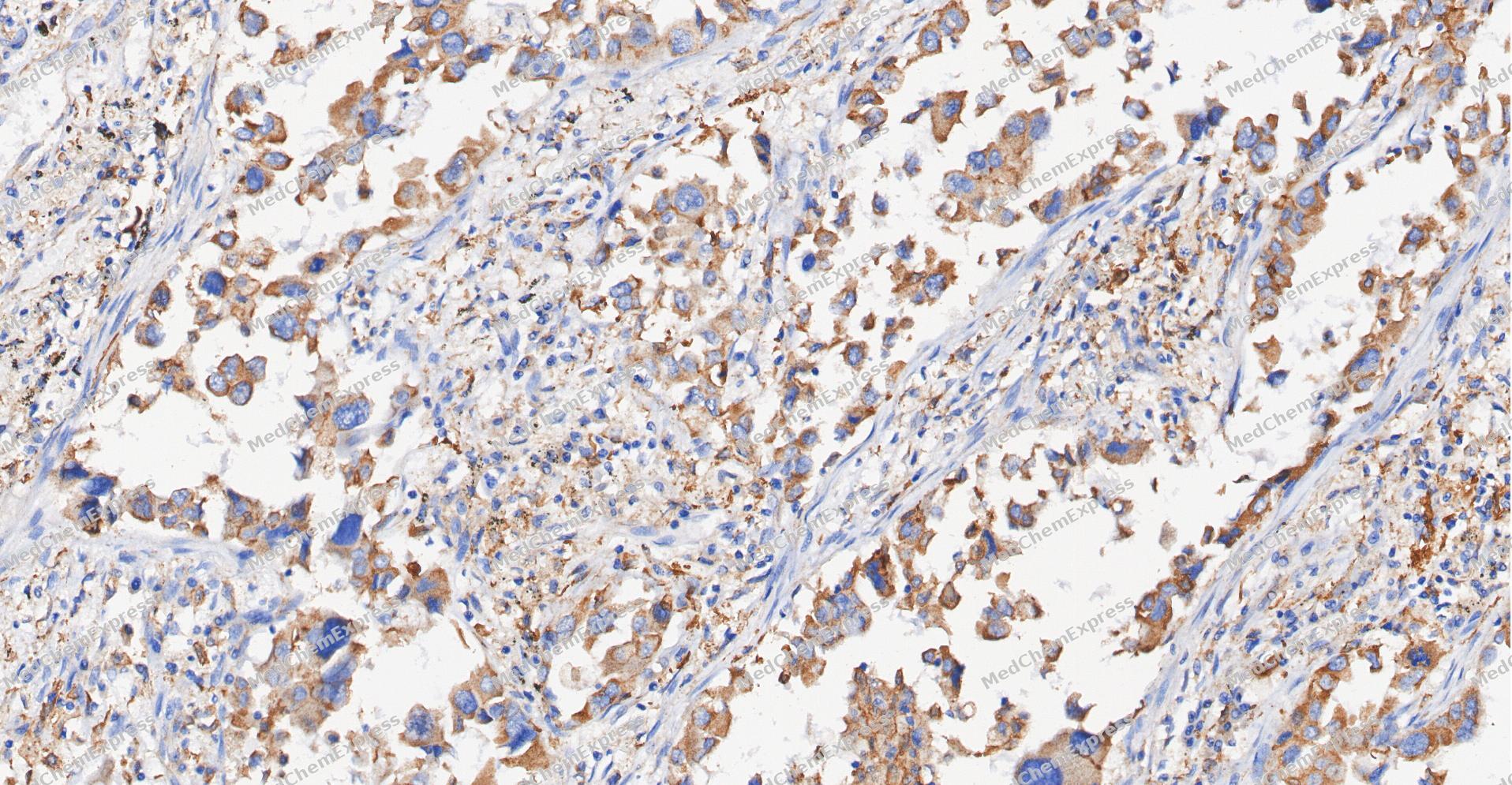 Immunohistochemical analysis of paraffin-embedded human Lung Adenocarcinoma tissue using CHRNA7 antibody was performed. The section was pretreated using high-temperature and high-pressure mediated EDTA antigen retrieval buffer (pH 9.0), for 5 minutes. The tissues were incubated with primary antibody (HY-P83668, 1:500 dilution) at room temperature for 60 minutes. Detection was performed using an HRP conjugated compact polymer system. DAB was used as the chromogen. The tissues were counterstained with hematoxylin and mounted with neutral balsam mounting medium.
 -
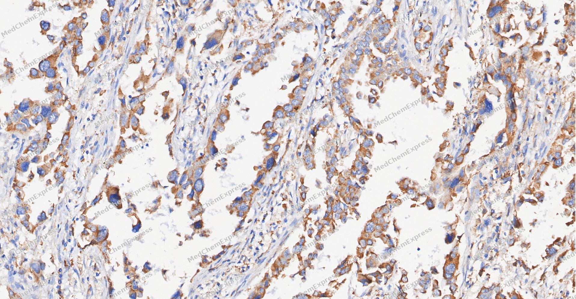 Immunohistochemical analysis of paraffin-embedded human Lung Adenocarcinoma tissue using CHRNA7 antibody was performed. The section was pretreated using high-temperature and high-pressure mediated EDTA antigen retrieval buffer (pH 9.0), for 5 minutes. The tissues were incubated with primary antibody (HY-P83668, 1:500 dilution) at room temperature for 60 minutes. Detection was performed using an HRP conjugated compact polymer system. DAB was used as the chromogen. The tissues were counterstained with hematoxylin and mounted with neutral balsam mounting medium.
 -
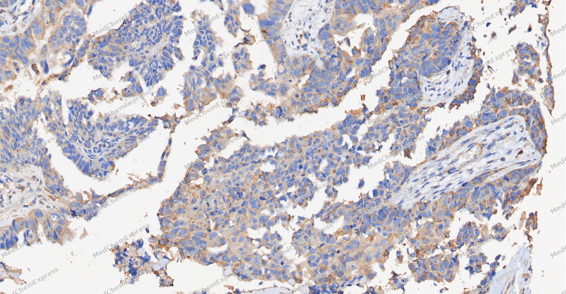 Immunohistochemical analysis of paraffin-embedded human Ovarian Cancer tissue using CHRNA7 antibody was performed. The section was pretreated using high-temperature and high-pressure mediated EDTA antigen retrieval buffer (pH 9.0), for 5 minutes. The tissues were incubated with primary antibody (HY-P83668, 1:500 dilution) at room temperature for 60 minutes. Detection was performed using an HRP conjugated compact polymer system. DAB was used as the chromogen. The tissues were counterstained with hematoxylin and mounted with neutral balsam mounting medium.
 -
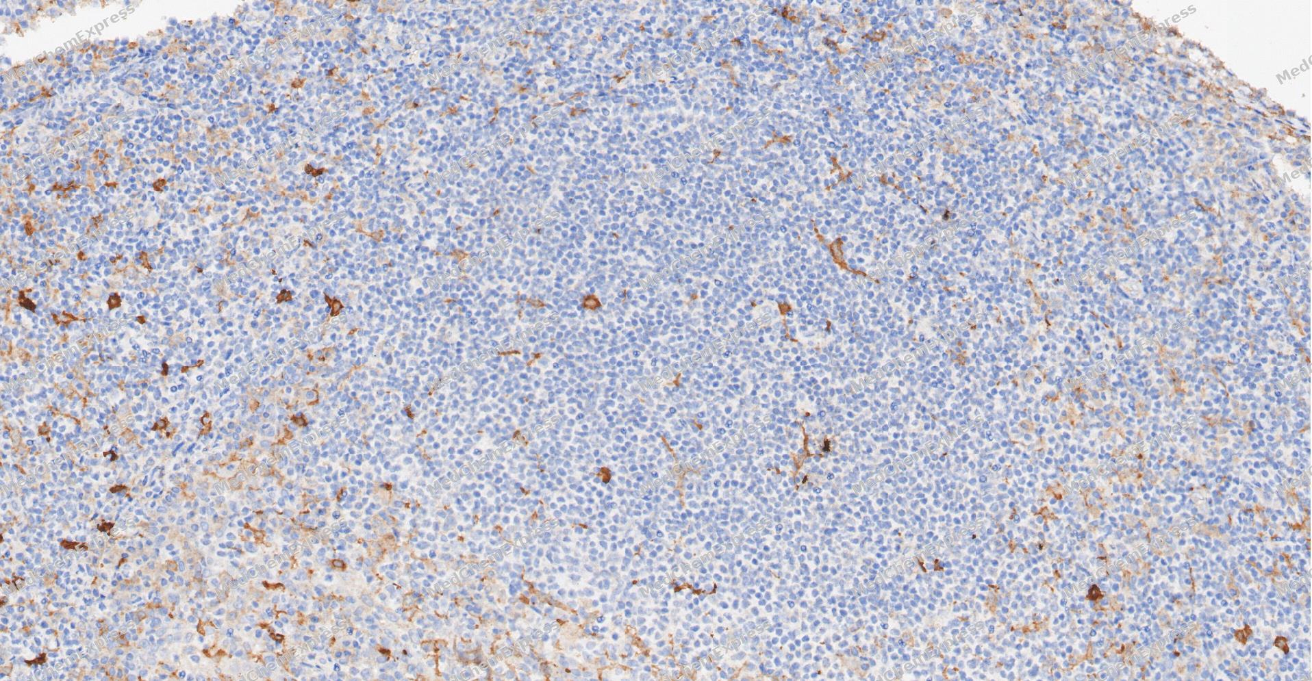 Immunohistochemical analysis of paraffin-embedded human Tonsil tissue using CHRNA7 antibody was performed. The section was pretreated using high-temperature and high-pressure mediated EDTA antigen retrieval buffer (pH 9.0), for 5 minutes. The tissues were incubated with primary antibody (HY-P83668, 1:500 dilution) at room temperature for 60 minutes. Detection was performed using an HRP conjugated compact polymer system. DAB was used as the chromogen. The tissues were counterstained with hematoxylin and mounted with neutral balsam mounting medium.
 -
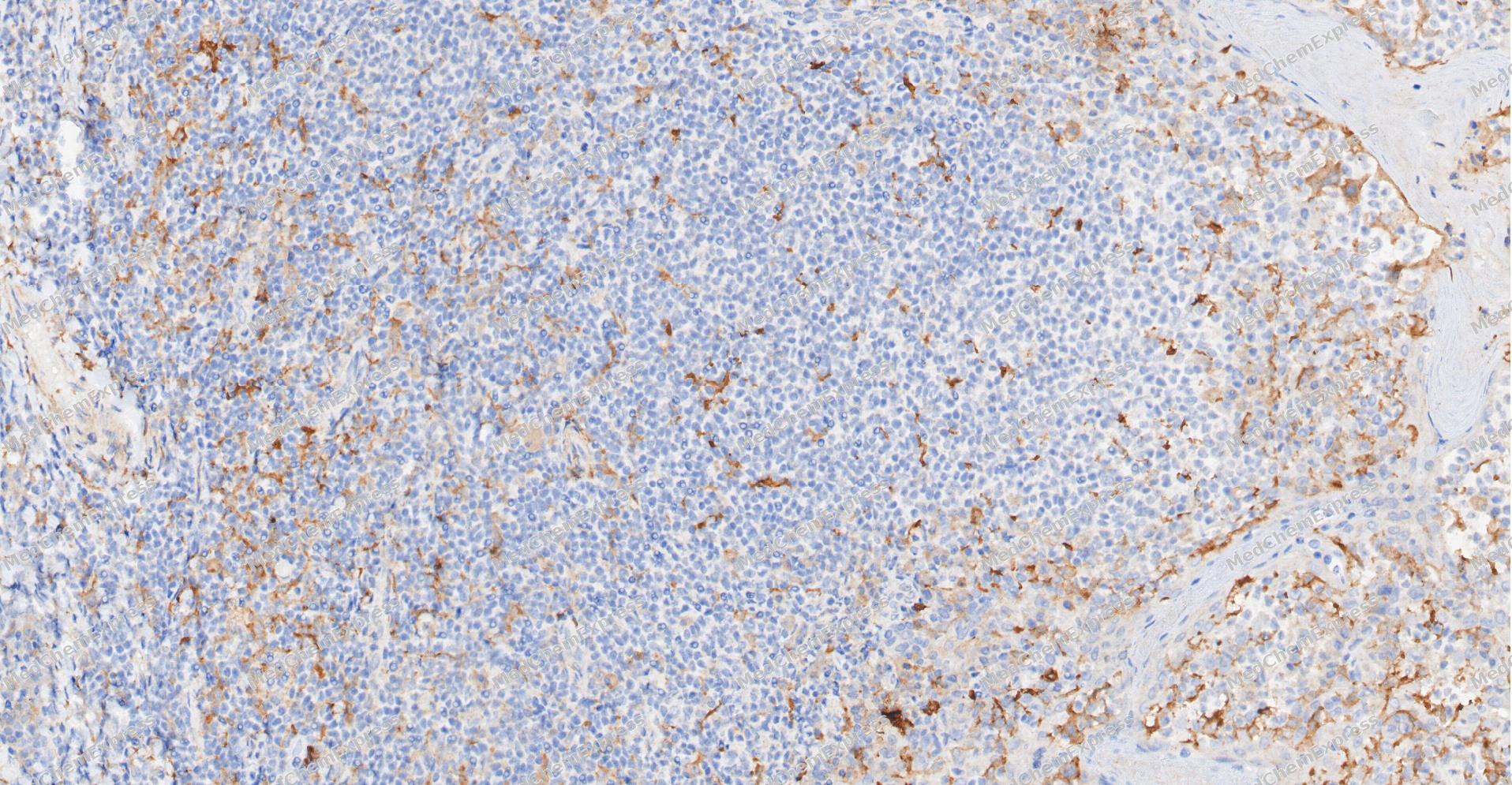 Immunohistochemical analysis of paraffin-embedded human Tonsil tissue using CHRNA7 antibody was performed. The section was pretreated using high-temperature and high-pressure mediated EDTA antigen retrieval buffer (pH 9.0), for 5 minutes. The tissues were incubated with primary antibody (HY-P83668, 1:500 dilution) at room temperature for 60 minutes. Detection was performed using an HRP conjugated compact polymer system. DAB was used as the chromogen. The tissues were counterstained with hematoxylin and mounted with neutral balsam mounting medium.
 -
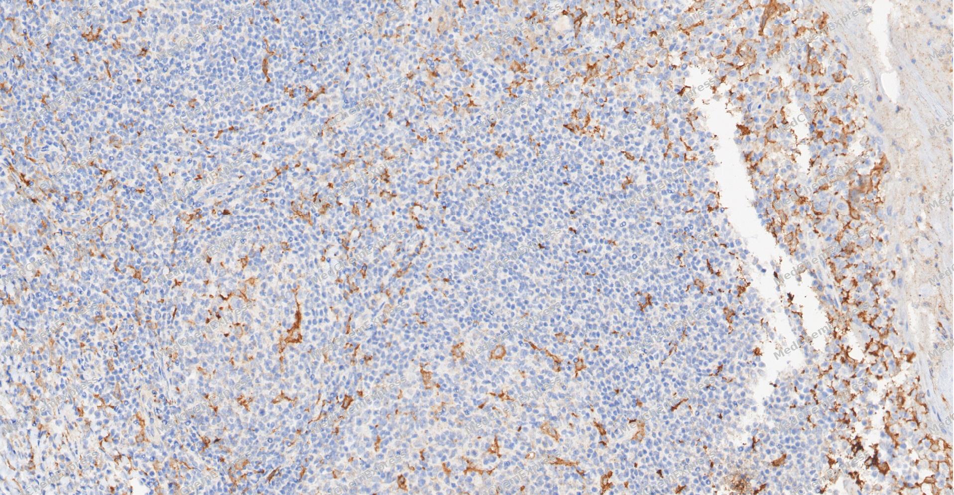 Immunohistochemical analysis of paraffin-embedded human Tonsil tissue using CHRNA7 antibody was performed. The section was pretreated using high-temperature and high-pressure mediated EDTA antigen retrieval buffer (pH 9.0), for 5 minutes. The tissues were incubated with primary antibody (HY-P83668, 1:500 dilution) at room temperature for 60 minutes. Detection was performed using an HRP conjugated compact polymer system. DAB was used as the chromogen. The tissues were counterstained with hematoxylin and mounted with neutral balsam mounting medium.
 -
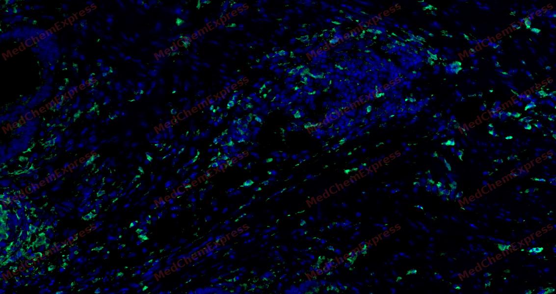 Tyramide signaling amplification based immunofluorescence was performed on paraffin-embedded human Colon cancer tissue using CHRNA7 antibody. Antigen retrieval was performed in EDTA buffer pH 9.0 (95 °C, 20 min) followed by cooling to RT. Then incubated with primary antibody (HY-P83668, 1:500 dilution) at room temperature for 60 minutes and HRP conjugated secondary antibody for 10 minutes. Fluorescence was then developed with Vari Fluor 532 TSA (200×)(HY-D1832). The tissues were counterstained with DAPI and mounted with Anti-fade mounting medium.
 -
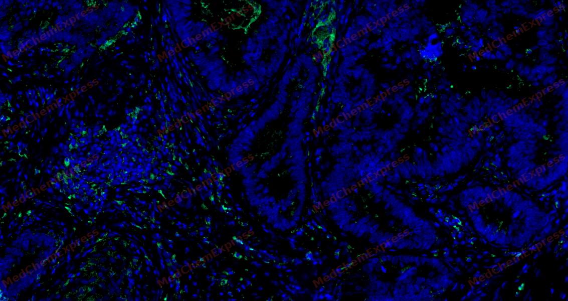 Tyramide signaling amplification based immunofluorescence was performed on paraffin-embedded human Colon cancer tissue using CHRNA7 antibody. Antigen retrieval was performed in EDTA buffer pH 9.0 (95 °C, 20 min) followed by cooling to RT. Then incubated with primary antibody (HY-P83668, 1:500 dilution) at room temperature for 60 minutes and HRP conjugated secondary antibody for 10 minutes. Fluorescence was then developed with Vari Fluor 532 TSA (200×)(HY-D1832). The tissues were counterstained with DAPI and mounted with Anti-fade mounting medium.
 -
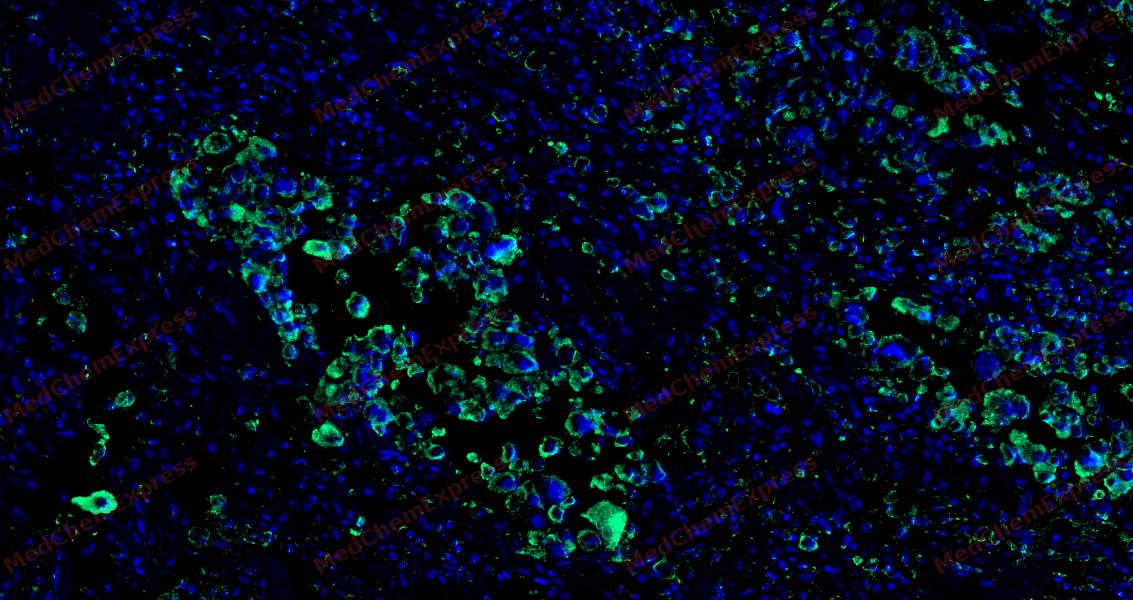 Tyramide signaling amplification based immunofluorescence was performed on paraffin-embedded human Lung Adenocarcinoma tissue using CHRNA7 antibody. Antigen retrieval was performed in EDTA buffer pH 9.0 (95 °C, 20 min) followed by cooling to RT. Then incubated with primary antibody (HY-P83668, 1:500 dilution) at room temperature for 60 minutes and HRP conjugated secondary antibody for 10 minutes. Fluorescence was then developed with Vari Fluor 532 TSA (200×)(HY-D1832). The tissues were counterstained with DAPI and mounted with Anti-fade mounting medium.
 -
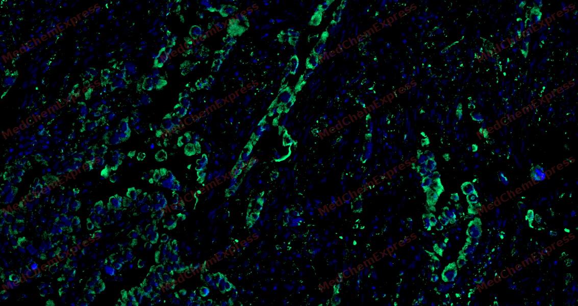 Tyramide signaling amplification based immunofluorescence was performed on paraffin-embedded human Lung Adenocarcinoma tissue using CHRNA7 antibody. Antigen retrieval was performed in EDTA buffer pH 9.0 (95 °C, 20 min) followed by cooling to RT. Then incubated with primary antibody (HY-P83668, 1:500 dilution) at room temperature for 60 minutes and HRP conjugated secondary antibody for 10 minutes. Fluorescence was then developed with Vari Fluor 532 TSA (200×)(HY-D1832). The tissues were counterstained with DAPI and mounted with Anti-fade mounting medium.
 -
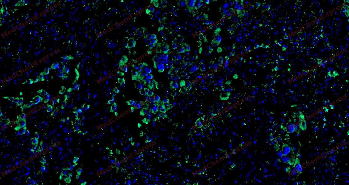 Tyramide signaling amplification based immunofluorescence was performed on paraffin-embedded human Lung Adenocarcinoma tissue using CHRNA7 antibody. Antigen retrieval was performed in EDTA buffer pH 9.0 (95 °C, 20 min) followed by cooling to RT. Then incubated with primary antibody (HY-P83668, 1:500 dilution) at room temperature for 60 minutes and HRP conjugated secondary antibody for 10 minutes. Fluorescence was then developed with Vari Fluor 532 TSA (200×)(HY-D1832). The tissues were counterstained with DAPI and mounted with Anti-fade mounting medium.
 -
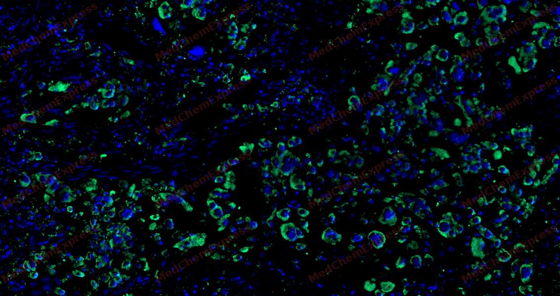 Tyramide signaling amplification based immunofluorescence was performed on paraffin-embedded human Lung Adenocarcinoma tissue using CHRNA7 antibody. Antigen retrieval was performed in EDTA buffer pH 9.0 (95 °C, 20 min) followed by cooling to RT. Then incubated with primary antibody (HY-P83668, 1:500 dilution) at room temperature for 60 minutes and HRP conjugated secondary antibody for 10 minutes. Fluorescence was then developed with Vari Fluor 532 TSA (200×)(HY-D1832). The tissues were counterstained with DAPI and mounted with Anti-fade mounting medium.
 -
 Flow cytometric analysis of 1X10⁶ Jurkat cells labeling CHRNA7 Antibody(HY-P83668, red). Cells were fixed with 4% paraformaldehyde and permeabilised with 90% methanol. Then stained with the primary antibody at 1/50 dilution for an hour at 4℃. AF488-conjugated Goat Anti-Mouse IgG H&L (HY-P8005) was used as the secondary antibody at 1/1,000 dilution for 30 minutes at 4℃. Mouse IgG Isotype Control (HY-P80757, blue) was used as the isotype control, cells without incubation with primary antibody were used as the unlabeled control (black).


Background
-
Function
CHRNA7 component of neuronal acetylcholine receptors (nAChRs) that function as pentameric, ligand-gated cation channels with high calcium permeability among other activities. nAChRs are excitatory neurotrasnmitter receptors formed by a collection of nAChR subunits known to mediate synaptic transmission in the nervous system and the neuromuscular junction. Each nAchR subunit confers differential attributes to channel properties, including activation, deactivation and desensitization kinetics, pH sensitivity, cation permeability, and binding to allosteric modulators. CHRNA7 forms homopentameric neuronal acetylcholine receptors abundantly expressed in the central nervous system, characterized by fast desensitization and high calcium permeability. Also forms heteropentamers with CHRNB2, mainly expressed in basal forebrain cholinergic neurons. Involved in the modulation of calcium-dependent signaling pathways and influences the release of neurotransmitters, including dopamine, glutamate and GABA. Also expressed in non-neuronal cells such as immune cells like lymphocytes, monocytes and macrophages. In T cells, activation induces metabotropic signaling that results in an increase of intracellular Ca2+ concentrations, independent of ionotropic receptor functions. In macrophages, required for acetylcholine-mediated inhibition of TNF and other inflammatory cytokine release. Once activated by acetylcholine, nicotine or other agonists, selectively inhibits production of pro-inflammatory cytokines while leaving anti-inflammatory cytokines undisturbed. Stimulates the cholinergic anti-inflammatory pathway, controlling inflammation by inhibiting NFKB nuclear translocation and activating the JAK2-STAT3 pathway, independently of ion channel activity. Also expressed in the urothelium where it modulates reflex bladder activity by increasing intracellular calcium through internal stores and decreasing basal ATP release[1][2][3][4][5][6][7][8][9][10].
-
Subcellular Localization
Postsynaptic cell membrane; Multi-pass membrane protein; Cell membrane; Multi-pass membrane protein
-
Expression
Tissue_specificity:Expression in neurons (PubMed: 8145738) . Expression in macrophages (protein level) (PubMed: 12508119) . -
Isoforms & Post-Translational Modification
P36544 has 3 isomers: P36544-1: 56449 Da (predicted); P36544-2: 59235 Da (predicted); P36544-3: 11485 Da (predicted).
Glycosylations at Asn-46, Asn-90 and Asn-133 are essential for TMEM35A/NACHO-mediated proper subunit assembly and trafficking to the cell membrane -
Subunit
Homopentamer (PubMed:33735609, PubMed:38382524). Can also form heteropentamers with CHRNB2, mainly found in basal forebrain cholinergic neurons (PubMed:33239400, PubMed:38161283). Interacts with RIC3; which is required for proper folding and assembly (PubMed:15504725, PubMed:16120769). Interacts with LYPD6 (PubMed:27344019). Interacts with CANX (PubMed:32783947)
-
SwissProt ID
-
Synonyms
7nAChR,CHRFAM7A,CHRNA7 (cholinergic receptor; nicotinic; alpha 7; exons 5-10) and FAM7A (family with sequence similarity 7A; exons A-E) fusion; D-10; CHRNA7; CHRNA7-DR1


Documentation
-
Data Sheet (235 KB)
-
SDS (252 KB)
- English - EN (252 KB)
- Français - FR (252 KB)
- Deutsch - DE (252 KB)
- Norwegian - NO (252 KB)
- Español - ES (252 KB)
- Swedish - SV (252 KB)
- Italian - IT (252 KB)
- Korean - KR (252 KB)
- Portuguese - PT (252 KB)
-
User Guide for Antibodies (1077 KB)

 Powered by Bioz
