



Raptor Antibody (YA1519)
(Synonyms: RPTOR; KOG1; KIAA1303; RAPTOR; Mip1)Based on 1 publication(s) in Google Scholar
Raptor Antibody (YA1519) is a Rabbit-derived and non-conjugated IgG monoclonal antibody, targeting to Raptor.
-
Host:
Rabbit
-
Isotype:
IgG
-
Application:
WB, IHC-P, ICC/IF, IP, FC
-
Reactivity :
Human, Mouse, Rat
-
Formulation:
Supplied in rabbit IgG in 50 mM Tris-Glycine(pH 7.4), 0.15 M NaCl, 40% Glycerol, 0.01% sodium azide and 0.05% BSA.
-
Conjugation:
Non-conjugated


To place orders, for customer services and technical support, please contact: MedChemExpress USA
Tel: 609-228-6898 E-mail: [email protected] [email protected]



Publications Citing Use of MedChemExpress (MCE) Raptor Antibody (YA1519)
More

Applications
| Application |
WB

WB: Western Blot
|
IHC-P
IHC-P: Immunohistochemistry-Paraffin
|
ICC/IF
ICC/IF: Immunocytochemistry/
Immunofluorescence |
IP
IP: Immunoprecipitation
|
FC
FC: Flow Cytometry
|
|---|---|---|---|---|---|
| Dilution Ratio | 1:500-1:1000 | 1:50-1:100 | 1:50-1:200 | 1:30 | 1:50-1:100 |


Product Details
Raptor Antibody (YA1519) is a Rabbit-derived and non-conjugated IgG monoclonal antibody, targeting to Raptor.
-
Host Rabbit

-
Clonality Recombinant,Monoclonal

-
Species ReactivityHuman, Mouse, Rat
-
Observed Molecular WeightObserved band size: 149 kDa
Note: Due to possible protein modifications or aggregation, the molecular weight should be confirmed by actual measurement, and the predicted value is for reference only.

-
Calculated Molecular Weight Predicted band size: 149 kDa

Entrez Gene: 57521 Human ; 74370 Mouse ; 287871 Rat
SwissProt: Q8N122 Human ; Q8K4Q0 Mouse ;
OMIM: 607130 Human
A synthesized peptide derived from human Raptor aa1300-1335.
Endogenous
Affinity Chromatography
Non-conjugated
Unmodified
IgG


Product Properties
-
Appearance
Solution
-
Formulation
Supplied in rabbit IgG in 50 mM Tris-Glycine(pH 7.4), 0.15 M NaCl, 40% Glycerol, 0.01% sodium azide and 0.05% BSA.
-
Concentration
Batch-dependent, Please check the COA for the concentration of each lot. Check Lot Concentration
-
Storage & Stability
Stored at -20°C for 1 year. Avoid repeated freeze / thaw cycles.
-
Shipping
Shipping with blue ice.


Publications (1)
-
Journal Impact Factor
-
Most Recent


Verification Images
-
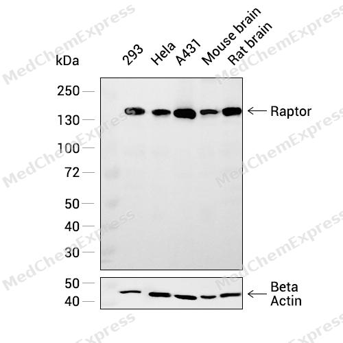 Western blot analysis was performed on extracts from 293 (lane 1, 20 μg), HeLa (lane 2, 20 μg), A431 (lane 3, 20 μg), Mouse brain (lane 4, 20 μg), and Rat brain (lane 5, 20 μg) using Raptor Rabbit mAb.Proteins were transferred to a PVDF membrane and blocked with 5% non - fat milk in TBST at 4°C overnight.The primary antibody (1:1000 dilution) and the loading control antibody (beta-Actin, HY-P83730, 1:5000 dilution) were incubated in 5% non-fat milk in TBST for 1 hour at 37°C.Goat Anti - Rabbit IgG - HRP Secondary Antibody (1:20000 dilution) was then applied for 40 minutes at 37°C.
 -
 Immunohistochemical analysis of paraffin-embedded rat heart muscle tissue using Raptor Antibody (HY-P81774,1/100). The section was pretreated using heat mediated antigen retrieval with sodium citrate buffer (pH 6.0) for 8 minutes. The tissues were blocked with quick block buffer for 0.5 hours at room temperature, washed with PBS and PBST, and then incubated with the primary antibody overnight at 4℃. The detection was performed using an HRP conjugated compact polymer system. DAB was used as the chromogen. Tissues were counterstained with hematoxylin and mounted with DPX.
 -
 Immunohistochemical analysis of paraffin-embedded rat endometrium tissue using Raptor Antibody (HY-P81774,1/100). The section was pretreated using heat mediated antigen retrieval with sodium citrate buffer (pH 6.0) for 8 minutes. The tissues were blocked with quick block buffer for 0.5 hours at room temperature, washed with PBS and PBST, and then incubated with the primary antibody overnight at 4℃. The detection was performed using an HRP conjugated compact polymer system. DAB was used as the chromogen. Tissues were counterstained with hematoxylin and mounted with DPX.
 -
 Immunohistochemical analysis of paraffin-embedded rat spleen tissue using Raptor Antibody (HY-P81774,1/100). The section was pretreated using heat mediated antigen retrieval with sodium citrate buffer (pH 6.0) for 8 minutes. The tissues were blocked with quick block buffer for 0.5 hours at room temperature, washed with PBS and PBST, and then incubated with the primary antibody overnight at 4℃. The detection was performed using an HRP conjugated compact polymer system. DAB was used as the chromogen. Tissues were counterstained with hematoxylin and mounted with DPX.
 -
 Immunohistochemical analysis of paraffin-embedded rat thyroid gland tissue using Raptor Antibody (HY-P81774,1/100). The section was pretreated using heat mediated antigen retrieval with sodium citrate buffer (pH 6.0) for 8 minutes. The tissues were blocked with quick block buffer for 0.5 hours at room temperature, washed with PBS and PBST, and then incubated with the primary antibody overnight at 4℃. The detection was performed using an HRP conjugated compact polymer system. DAB was used as the chromogen. Tissues were counterstained with hematoxylin and mounted with DPX.
 -
 Immunohistochemical analysis of paraffin-embedded rat tonsil tissue using Raptor Antibody (HY-P81774,1/100). The section was pretreated using heat mediated antigen retrieval with sodium citrate buffer (pH 6.0) for 8 minutes. The tissues were blocked with quick block buffer for 0.5 hours at room temperature, washed with PBS and PBST, and then incubated with the primary antibody overnight at 4℃. The detection was performed using an HRP conjugated compact polymer system. DAB was used as the chromogen. Tissues were counterstained with hematoxylin and mounted with DPX.
 -
 Immunohistochemical analysis of paraffin-embedded rat cerebral cortex tissue using Raptor Antibody (HY-P81774,1/100). The section was pretreated using heat mediated antigen retrieval with sodium citrate buffer (pH 6.0) for 8 minutes. The tissues were blocked with quick block buffer for 0.5 hours at room temperature, washed with PBS and PBST, and then incubated with the primary antibody overnight at 4℃. The detection was performed using an HRP conjugated compact polymer system. DAB was used as the chromogen. Tissues were counterstained with hematoxylin and mounted with DPX.
 -
 Flow cytometric analysis of 1X10⁶ HeLa cells labeling Raptor Antibody (HY-P81774, red). Cells were fixed with 4% paraformaldehyde and permeabilised with 90% methanol. Then stained with the primary antibody at 1/100 dilution for an hour at 4℃. AF488-conjugated Goat Anti-Rabbit IgG H&L (HY-P8002) was used as the secondary antibody at 1/1,000 dilution for 30 minutes at 4℃. Rabbit IgG Isotype Control (HY-P80879, blue) was used as the isotype control, cells without incubation with primary antibody were used as the unlabeled control (black).
 -
 Immunocytochemistry analysis of PC-3 cells labeling Raptor with Raptor Antibody (HY-P81774) at 1/100 dilution. Cells were fixed in 4% paraformaldehyde for 15 minutes at room temperature, permeabilized with 0.1% Triton X-100 in PBS for 15 minutes at room temperature, then blocked with quick block buffer for 10 minutes at room temperature. Cells were then incubated with Raptor Antibody (HY-P81774) at 1/100 dilution in quick block buffer overnight at 4 ℃. AF488-conjugated Goat Anti-Rabbit IgG H&L(HY-P8002, Green) was used as the secondary antibody at 1/1,000 dilution. PBS instead of the primary antibody was used as the secondary antibody only control. The Nuclear counterstain was DAPI (Blue).
 -
 Immunocytochemistry analysis of PC-3 cells labeling Raptor with Raptor Antibody (HY-P81774) at 1/200 dilution. Cells were fixed in 4% paraformaldehyde for 15 minutes at room temperature, permeabilized with 0.1% Triton X-100 in PBS for 15 minutes at room temperature, then blocked with quick block buffer for 10 minutes at room temperature. Cells were then incubated with Raptor Antibody (HY-P81774) at 1/200 dilution in quick block buffer overnight at 4 ℃. AF488-conjugated Goat Anti-Rabbit IgG H&L(HY-P8002, Green) was used as the secondary antibody at 1/1,000 dilution. PBS instead of the primary antibody was used as the secondary antibody only control. The Nuclear counterstain was DAPI (Blue).


Background
-
Function
Raptor component of the mechanistic target of rapamycin complex 1 (mTORC1), an evolutionarily conserved central nutrient sensor that stimulates anabolic reactions and macromolecule biosynthesis to promote cellular biomass generation and growth. In response to nutrients, growth factors or amino acids, mTORC1 is recruited to the lysosome membrane and promotes protein, lipid and nucleotide synthesis by phosphorylating several substrates, such as ribosomal protein S6 kinase (RPS6KB1 and RPS6KB2) and EIF4EBP1 (4E-BP1). In the same time, it inhibits catabolic pathways by phosphorylating the autophagy initiation components ULK1 and ATG13, as well as transcription factor TFEB, a master regulators of lysosomal biogenesis and autophagy. The mTORC1 complex is inhibited in response to starvation and amino acid depletion. Within the mTORC1 complex, RPTOR acts both as a molecular adapter, which (1) mediates recruitment of mTORC1 to lysosomal membranes via interaction with small GTPases Rag (RagA/RRAGA, RagB/RRAGB, RagC/RRAGC and/or RagD/RRAGD), and a (2) substrate-specific adapter, which promotes substrate specificity by binding to TOS motif-containing proteins and direct them towards the active site of the MTOR kinase domain for phosphorylation. mTORC1 complex regulates many cellular processes, such as odontoblast and osteoclast differentiation or neuronal transmission. mTORC1 complex in excitatory neuronal transmission is required for the prosocial behavior induced by the psychoactive substance lysergic acid diethylamide (LSD)[1][2][3][4][5][6][7].
-
Subcellular Localization
Lysosome membrane; Cytoplasm; Cytoplasmic granule
-
Expression
Tissue_specificity:It is highly expressed in skeletal muscle, and less expressed in the brain, lungs, small intestine, kidneys and placenta; it is widely expressed, with the highest expression levels in the nasal mucosa and pituitary gland, and the lowest expression levels in the spleen. -
Isoforms & Post-Translational Modification
Q8N122 has 3 isomers: Q8N122-1: 149038 Da (predicted); Q8N122-2: 43256 Da (predicted); Q8N122-3: 131515 Da (predicted).
Insulin-stimulated phosphorylation at Ser-863 by MTOR and MAPK8 regulates mTORC1 activity (PubMed:19864431). Phosphorylated at Ser-863 by NLK in response to stress, disrupting the interaction with small GTPases Rag (RagA/RRAGA, RagB/RRAGB, RagC/RRAGC and/or RagD/RRAGD), thereby preventing lysosome recruitment and activation of the mTORC1 complex (PubMed:26588989). Osmotic stress also induces phosphorylation at Ser-696, Thr-706 and Ser-863 by MAPK8 (PubMed:19864431, PubMed:22493283). Ser-863 phosphorylation is required for phosphorylation at Ser-855 and Ser-859 (PubMed:19864431). In response to nutrient limitation, phosphorylated at Ser-722 and Ser-792 by AMPK; phosphorylation promotes interaction with 14-3-3 proteins, leading to negative regulation of the mTORC1 complex (PubMed:18439900). Phosphorylation at Ser-722 and Ser-792 by AMPK in response to glucose starvation inhibits O-GlcNAcylation by OGT and subsequent activation of mTORC1 (PubMed:37541260). In response to growth factors, phosphorylated at Ser-719, Ser-721 and Ser-722 by RPS6KA1, which stimulates mTORC1 activity (PubMed:18439900, PubMed:18722121). Phosphorylation at Ser-791 by PKA downstream of cAMP inhibits the mTORC1 complex (PubMed:31112131). Phosphorylated at Ser-877 by TBK1, leading to negative regulation of the mTORC1 complex (PubMed:31530866);O-GlcNAcylated by OGT upon glucose sufficiency, promoting interaction with small GTPases Rag (RagA/RRAGA, RagB/RRAGB, RagC/RRAGC and/or RagD/RRAGD) and subsequent recruitment of mTORC1 to lysosomal membranes, leading to activation of the mTORC1 complex (PubMed:37541260). Phosphorylation at Ser-722 and Ser-792 by AMPK in response to glucose starvation inhibits O-GlcNAcylation (PubMed:37541260);Acetylation at Lys-1097 by EP300/p300 in response to leucine metabolite acetyl-coA promotes its activity, leading to activation of the mTORC1 complex (PubMed:30197302, PubMed:32561715). Acetylation is decreased in response to fasting (By similarity);Ubiquitinated, leading to its degradation by the proteasome (PubMed:34290272, PubMed:35927303). Deubiquitinated by OTUB1 via a non-catalytic mechanism (PubMed:35927303). Ubiquitinated by an E3 ubiquitin ligase complex containing VHL (PubMed:34290272) -
Subunit
Part of the mechanistic target of rapamycin complex 1 (mTORC1) which contains MTOR, MLST8 and RPTOR (PubMed:12408816, PubMed:24403073, PubMed:25940091, PubMed:27909983, PubMed:29236692, PubMed:31601708, PubMed:31601764, PubMed:36697823). mTORC1 associates with AKT1S1/PRAS40, which inhibits its activity (PubMed:17386266, PubMed:31601764). mTORC1 associates with DEPTOR, which regulates its activity (PubMed:34519268, PubMed:34519269). mTORC1 binds to and is inhibited by FKBP12-rapamycin (PubMed:12408816, PubMed:15066126). Forms a complex with MTOR under both leucine-rich and -poor conditions (PubMed:12408816, PubMed:25940091, PubMed:31601708, PubMed:31601764). Interacts with (via TOS motifs) EIF4EBP1 and RPS6KB1; interaction is independent of its association with MTOR (PubMed:12150925, PubMed:12150926, PubMed:12747827, PubMed:24403073). Binds preferentially to poorly or non-phosphorylated forms of EIF4EBP1, and this binding is critical to the ability of MTOR to catalyze phosphorylation (PubMed:12747827, PubMed:24403073). Interacts with ULK1 in a nutrient-dependent manner; the interaction is reduced during starvation (PubMed:19211835). Interacts with GTP-bound form of RagA/RRAGA or RagB/RRAGB and GDP-bound form of RagC/RRAGC or RagD/RRAGD, promoting recruitment of mTORC1 to the lysosomes (PubMed:31601708, PubMed:31601764). Interacts (when phosphorylated by AMPK) with 14-3-3 protein, leading to inhibition of its activity (PubMed:18439900). Interacts with SPAG5; SPAG5 competes with MTOR for RPTOR-binding, resulting in decreased mTORC1 formation (PubMed:23953116). Interacts with WAC; WAC positively regulates MTOR activity by promoting the assembly of the TTT complex composed of TELO2, TTI1 and TTI2 and the RUVBL complex composed of RUVBL1 and RUVBL2 into the TTT-RUVBL complex which leads to the dimerization of the mTORC1 complex and its subsequent activation (PubMed:26812014). Interacts with G3BP1 (PubMed:23953116). The complex formed with G3BP1 and SPAG5 is increased by oxidative stress (PubMed:23953116). Interacts with HTR6 (PubMed:23027611). Interacts with PIH1D1 (PubMed:24036451). Interacts with LARP1 (PubMed:25940091). Interacts with BRAT1 (PubMed:25657994). Interacts with SIK3 (PubMed:30232230). Interacts with SLC38A7; this interaction mediates the recruitment of mTORC1 to the lysosome and its subsequent activation (PubMed:35561222)
-
SwissProt ID
-
Synonyms
RPTOR; KOG1; KIAA1303; RAPTOR; Mip1
-
Research Field
Cell Biology
Documentation
-
Data Sheet (234 KB)
-
SDS (251 KB)
- English - EN (251 KB)
- Français - FR (251 KB)
- Deutsch - DE (251 KB)
- Norwegian - NO (251 KB)
- Español - ES (251 KB)
- Swedish - SV (251 KB)
- Italian - IT (251 KB)
- Korean - KR (251 KB)
- Portuguese - PT (251 KB)
-
User Guide for Antibodies (1077 KB)

 Powered by Bioz
