



CADD522
Based on 5 publication(s) in Google Scholar
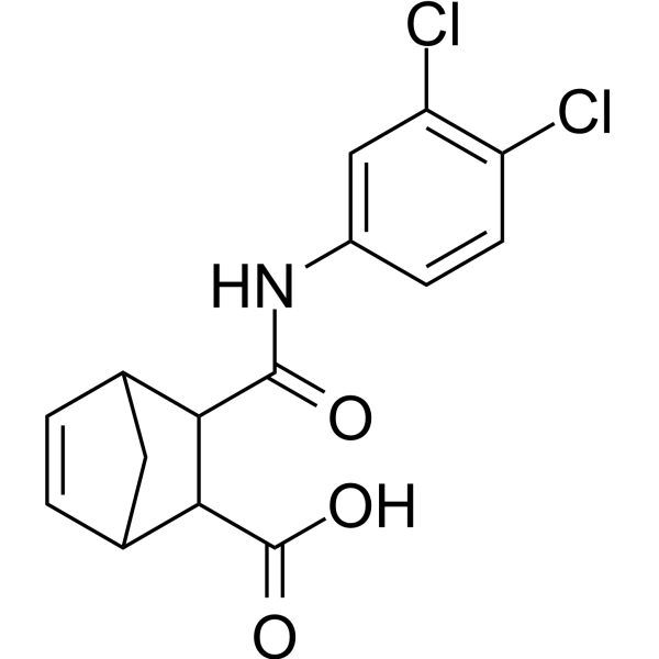
CADD522 is a RUNX2-DNA binding inhibitor (downregulates RUNX2-mediated transcription of downstream target genes), with an IC50 of 10 nM. CADD522 inhibits primary tumor growth and experimental metastasis of tumor cells in the lungs of immune-compromised mice. CADD522 can be used in study of cancer.
For research use only. We do not sell to patients.
- Purity : 98.17%
- CAS No.: 199735-88-1
- Formula: C15H13Cl2NO3
- Molecular Weight:326.17
-
Storage:Powder -20°C, 3 years , 4°C, 2 years ; In solvent -80°C, 2 years , -20°C, 1 year

To place orders, for customer services and technical support, please contact: MedChemExpress USA
Tel: 609-228-6898 E-mail: [email protected] [email protected]


-
 Biological Activity
-
Chemical Information
-
Solvent & Solubility
- Protocols
- Purity & Documentation
- References
-
 Help & FAQs


-
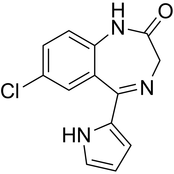
HY-108470
Ro5-3335RUNX1-CBFB Inhibitor -
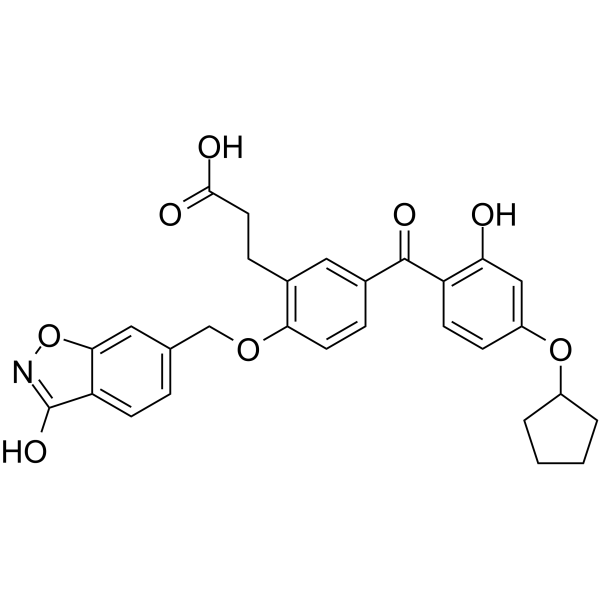
HY-12270
T-5224c-Fos/AP-1 Inhibitor -

HY-10219
RapamycinmTOR Inhibitor -
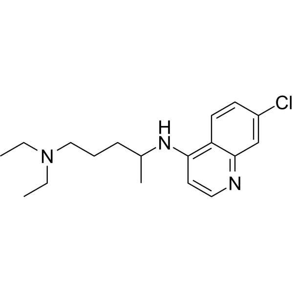
HY-17589A
ChloroquineAntimalarial Agent -
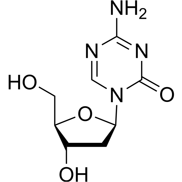
HY-A0004
DecitabineDNMT Inhibitor -
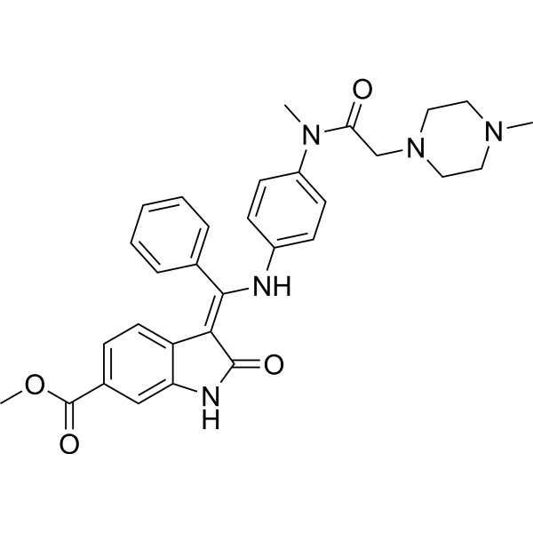
HY-50904
NintedanibPDGFR/VEGFR/FGFR Inhibitor -

HY-100558
Bafilomycin A1V-ATPase Inhibitor -
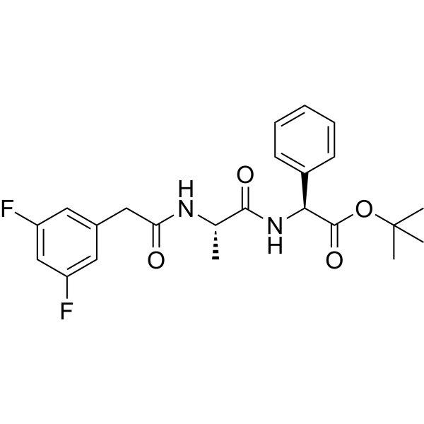
HY-13027
DAPTγ-secretase Inhibitor -
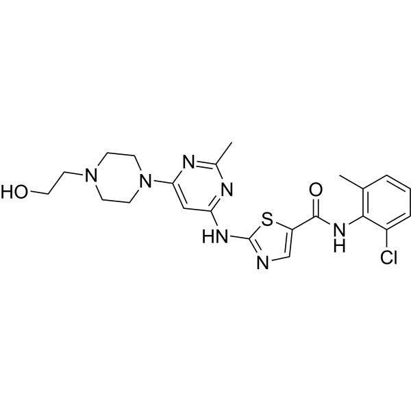
HY-10181
DasatinibSrc/Bcr-Abl Inhibitor -
HY-15531
VenetoclaxBCL-2 Inhibitor -
HY-13012
RepSoxTGFβR-1/ALK5 Inhibitor -
HY-13653
(-)-Epigallocatechin GallateAnticancer Agent
Publications Citing Use of MedChemExpress (MCE) CADD522
More Customer Validation & Images

-
Histological Imaging/Staining
-
WB
-
In Vivo Efficacy Study
-
Histological Imaging/Staining
-
Bio/Physico-chemical Assay


Biological Activity
Description
IC50 & Target
[1]|
RUNX2 10 nM () |
In Vitro
CADD522 (0-100 μM; 24-72 h) exhibits a strong inhibitory effect on BC cell growth and survival[1].
CADD522 (50 μM; 72 h) shows anti-proliferative effect by inducing cell cycle arrest (G1 phase)[1].
CADD522 (50 μM; 8 days) inhibits tumorsphere formation and (50 μM; 24 h) in vitro invasion of BC cells (without cellular toxicity)[1].
CADD522 (2, 10, 25, 50, 100 μM; 48 h) inhibits RUNX2 transcriptional activity by inhibiting RUNX2-DNA binding in T47D-RUNX2 and T47D-Empty cells[1].
CADD522 (50 μM; 72 h) upregulates RUNX2 levels through increased RUNX2 stability in cells[1].
CADD522 (50 μM; 6 or 24 h) increases ROS generation of mitochondrial in MCF7 and MDA-468 cells[2].
CADD522 (0-2000 nM, 30 min) inhibits mitochondrial ATP synthase activity in MDA-231 and MDA-468 cells[2].
MedChemExpress (MCE) has not independently confirmed the accuracy of these methods. They are for reference only.
-
Cell Line:MDA-MB-468, MCF7, MCF10A, IEC-6, GES-1 and C2C12 cells
-
Concentration:0-100 μM
-
Incubation Time:24-72 h
-
Result:Displayed a dose- and time-dependent cell growth inhibition over 72 h.
Exhibited low cytotoxicity for normal cell growth.
-
Cell Line:MCF7, MDA-468 and MDA-231 cells
-
Concentration:50 μM
-
Incubation Time:72 h
-
Result:Induced MDA-231 cells accumulated at the G1 and G2/M phase whereas MCF7 and MDA-468 cells were at the G1 phase.
-
Cell Line:MCF7, MCF7-tet-off cells
-
Concentration:50 μM
-
Incubation Time:8 days
-
Result:Dramatically decreased the size as well as the number of tumorspheres, and severely disrupted tumorspheres at day 4.
Showed a relatively selective effect on BC cells (did not have a significant influence on mammosphere formation of the MCF10A non-malignant mammary epithelial cells).
-
Cell Line:MCF7-tet-off (+Doxy), MCF7-tet-off (-Doxy) cells
-
Concentration:50 μM
-
Incubation Time:24 h
-
Result:Almost abrogated the invasiveness of both MCF7-tet-off (+Doxy) and MCF7-tet-off (-Doxy) cells without cellular toxicity.
-
Cell Line:T47D-RUNX2 and T47D-Empty cells
-
Concentration:2, 10, 25, 50, 100 μM
-
Incubation Time:48 h
-
Result:Resulted in a dramatic decrease of the promoter-luciferase (Luc) activities of RUNX2 downstream target genes such as MMP13 and VEGF (metastasis markers) and OC (osteogenesis marker).
-
Cell Line:T47D and MCF7 cells (ectopic expressing RUNX2)
-
Concentration:50 μM
-
Incubation Time:72 h
-
Result:Significantly inhibited the mRNA level (RUNX2-mediated) of Glut-1 and LDHA.
-
Cell Line:T47D-RUNX2 and MCF7-RUNX2 cells
-
Concentration:50 μM
-
Incubation Time:72 h
-
Result:Enhanced both mRNA and protein expression of RUNX2.
-
Cell Line:MDA-468 and MDA-231 cells
-
Concentration:50 μM
-
Incubation Time:2, 4, 6 h
-
Result:Increased RUNX2 stability by delaying protein degradation.
-
Cell Line:MCF7 and MDA-468 cells
-
Concentration:50 μM
-
Incubation Time:6 or 24 h
-
Result:Increased the level of mitochondrial ROS, which was more evident in serum-free than serum-containing condition.
-
Cell Line:MDA-231 and MDA-468 cells
-
Concentration:50, 250, 2000 nM (for MDA-231); 500, 2000 nM (for MDA-468)
-
Incubation Time:30 min
-
Result:Inhibited the activity of A TP synthase.
In Vivo
CADD522 (10 mg/kg; i.p.; twice a week for 11 days) suppresses tumor metastasis and inhibits expression of Ki-67 in mice[1].
MedChemExpress (MCE) has not independently confirmed the accuracy of these methods. They are for reference only.
-
Animal Model:Female mice (6-week-old; MMTV-PyMT transgenic model)[1].
-
Dosage:1, 5 and 20 mg/kg
-
Administration:Intraperitoneal injection; twice a week for 45 days.
-
Result:Delayed the onset of the tumors, delayed tumor development and reduced tumor burden in transgenic MMTV-PyMT mice.
Reduced the tumor weight in mice.
-
Animal Model:Female NOD scid gamma (NSG) mice and nude mice (TNBC-PDX Br-001 model)[1].
-
Dosage:10 mg/kg
-
Administration:Intraperitoneal injection; twice a week for 11 days.
-
Result:Significant decreased tumor volume and markedly inhibited expression of Ki-67.
Inhibited experimental metastasis of BC cells in vivo.(did not significantly decrease body weight or influence the general health of animals).


Chemical Information
-
CAS No. 199735-88-1
-
Appearance Solid
-
Molecular Weight 326.17
-
Formula C15H13Cl2NO3
-
Color White to off-white
-
SMILES
ClC1=C(Cl)C=CC(NC(C2C(C3)C=CC3C2C(O)=O)=O)=C1
-
Shipping
Room temperature in continental US; may vary elsewhere.
-
Storage
Powder -20°C 3 years 4°C 2 years In solvent -80°C 2 years -20°C 1 year


Publications (5)
-
Journal Impact Factor
-
Most Recent
-
Nat Commun
2022 Nov 4;13(1):6648. PMID: 36333322 -
Adv Sci (Weinh)
RUNX2 Activation in Fibro/Adipogenic Progenitors Promotes Muscle Fibrosis in Muscular Dystrophy. [Abstract]2025 Dec 22:e10850. PMID: 41431178
CADD522 purchased from MedChemExpress. Usage Cited in: Adv Sci (Weinh). 2025 Dec 22:e10850. [Abstract]
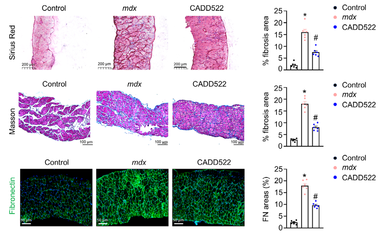
CADD522 (10 mg/kg; 6 weeks) significantly alleviated muscle fibrosis, as indicated by reduced areas positive for Picrosirius red, Masson's trichrome, and fibronectin in the DIA muscles of mdx mice.
CADD522 purchased from MedChemExpress. Usage Cited in: Adv Sci (Weinh). 2025 Dec 22:e10850. [Abstract]
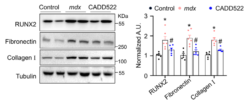
CADD522 (10 mg/kg; i.p.; twice a week for 6 weeks) reduced the mRNA and protein levels of RUNX2, fibronectin, and collagen I in the DIA muscles of mdx mice.
CADD522 purchased from MedChemExpress. Usage Cited in: Adv Sci (Weinh). 2025 Dec 22:e10850. [Abstract]
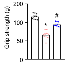
CADD522 (10 mg/kg; i.p.; twice a week for 6 weeks) significantly improved grip strength of mdx mice.
CADD522 purchased from MedChemExpress. Usage Cited in: Adv Sci (Weinh). 2025 Dec 22:e10850. [Abstract]
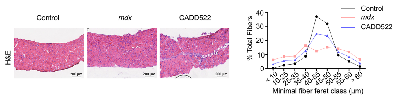
CADD522 (10 mg/kg; i.p.; twice a week for 6 weeks) effectively mitigated fibrosis and increased myofiber diameter in the DIA muscles of mdx mice, as demonstrated by haematoxylin and eosin staining.
CADD522 purchased from MedChemExpress. Usage Cited in: Adv Sci (Weinh). 2025 Dec 22:e10850. [Abstract]
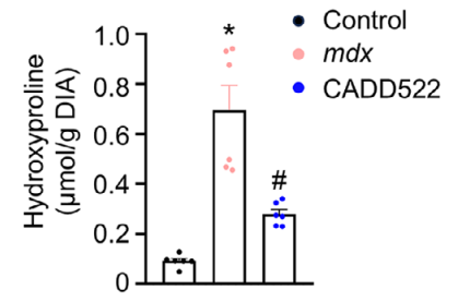
CADD522 (10 mg/kg; i.p.; twice a week for 6 weeks) significantly alleviated muscle fibrosis of mdx mice, as indicated by decreased hydroxyproline levels in the DIA muscles.
-
Cell Biosci
ALKBH5-mediated m6A demethylation of Runx2 mRNA promotes extracellular matrix degradation and intervertebral disc degeneration. [Abstract]2024 Jun 14;14(1):79. PMID: 38877576 -
Mol Cell Biol
Unveiling the Role of Sik1 in Osteoblast Differentiation: Implications for Osteoarthritis. [Abstract]2024;44(10):411-428. PMID: 39169784 -


Solvent & Solubility
In Vitro:
DMSO : ≥ 100 mg/mL (306.59 mM; Hygroscopic DMSO has a significant impact on the solubility of product, please use newly opened DMSO)
* "≥" means soluble, but saturation unknown.
Please refer to the solubility information to select the appropriate solvent. Once prepared, please aliquot and store the solution to prevent product inactivation from repeated freeze-thaw cycles.
Storage method and period of stock solution: -80°C, 2 years; -20°C, 1 year. When stored at -80°C, please use it within 2 years. When stored at -20°C, please use it within 1 year.
Please refer to the solubility information to select the appropriate solvent. Once prepared, please aliquot and store the solution to prevent product inactivation from repeated freeze-thaw cycles.
Storage method and period of stock solution: -80°C, 2 years; -20°C, 1 year. When stored at -80°C, please use it within 2 years. When stored at -20°C, please use it within 1 year.
Concentration (start) × Volume (start) = Concentration (final) × Volume (final)
In Vivo:
Select the appropriate dissolution method based on your experimental animal and administration route.
- For the following dissolution methods, please ensure to first prepare a clear stock solution using an In Vitro approach and then sequentially add co-solvents:
- To ensure reliable experimental results, the clarified stock solution can be appropriately stored based on storage conditions. As for the working solution for In Vivo experiments, it is recommended to prepare freshly and use it on the same day.
- The percentages shown for the solvents indicate their volumetric ratio in the final prepared solution. If precipitation or phase separation occurs during preparation, heat and/or sonication can be used to aid dissolution.
Add each solvent one by one: 10% DMSO 40% PEG300 5% Tween-80 45% Saline
Solubility: ≥ 2.17 mg/mL (6.65 mM); Clear solution
This protocol yields a clear solution of ≥ 2.17 mg/mL (saturation unknown).
Taking 1 mL working solution as an example, add 100 μL DMSO stock solution (21.7 mg/mL) to 400 μL PEG300, and mix evenly; then add 50 μL Tween-80 and mix evenly; then add 450 μL Saline to adjust the volume to 1 mL.
Preparation of Saline: Dissolve 0.9 g sodium chloride in ddH₂O and dilute to 100 mL to obtain a clear Saline solution.
In Vivo Dissolution Calculator
Please enter the basic information of animal experiments:
-
-
-
-
Recommended: Prepare an additional quantity of animals to account for potential losses during experiments.
Please enter your animal formula composition:
-
%DMSO +
Recommended: Keep the proportion of DMSO in working solution below 2% if your animal is weak.
-
%+
-
+%Tween-80 + +
-
%Saline +
The co-solvents required include: DMSO, . All of co-solvents are available by MedChemExpress (MCE). , Tween 80. All of co-solvents are available by MedChemExpress (MCE).
Working solution concentration: 0.22 mg/mL
Method for preparing stock solution: mg drug dissolved in μL DMSO. Stock solution concentration: mg/mL.
1. Take μL DMSO stock solution;
2. Add μL .
μL , mix evenly;
3. Then add μL Tween 80, mix evenly;
4. Then add μL
Please ensure that the stock solution in the first step is dissolved to a clear state, and add co-solvents in sequence. You can use ultrasonic heating (ultrasonic cleaner, recommended frequency 20-40 kHz), vortexing, etc. to assist dissolution.


Purity & Documentation
-
Data Sheet (289 KB)
-
SDS (396 KB)
- English - EN (396 KB)
- Français - FR (396 KB)
- Deutsch - DE (396 KB)
- Norwegian - NO (396 KB)
- Español - ES (396 KB)
- Swedish - SV (396 KB)
- Italian - IT (396 KB)
- Korean - KR (396 KB)
- Portuguese - PT (396 KB)
-
Handling Instructions (2659 KB)
References
[1]. Kim MS, et al. Characterization of CADD522, a small molecule that inhibits RUNX2-DNA binding and exhibits antitumor activity. Oncotarget. 2017 Aug 10;8(41):70916-70940. [Content Brief]
[2]. Kim MS, et al. Targeting breast cancer metabolism with a novel inhibitor of mitochondrial ATP synthesis. Oncotarget. 2020 Oct 27;11(43):3863-3885. [Content Brief]


Complete Stock Solution Preparation Table
Please refer to the solubility information to select the appropriate solvent. Once prepared, please aliquot and store the solution to prevent product inactivation from repeated freeze-thaw cycles.
Storage method and period of stock solution: -80°C, 2 years; -20°C, 1 year. When stored at -80°C, please use it within 2 years. When stored at -20°C, please use it within 1 year.
| Optional Solvent | Concentration Solvent Mass | 1 mg | 5 mg | 10 mg | 25 mg |
|---|---|---|---|---|---|
| DMSO | 1 mM | 3.0659 mL | 15.3294 mL | 30.6589 mL | 76.6471 mL |
| 5 mM | 0.6132 mL | 3.0659 mL | 6.1318 mL | 15.3294 mL | |
| 10 mM | 0.3066 mL | 1.5329 mL | 3.0659 mL | 7.6647 mL | |
| 15 mM | 0.2044 mL | 1.0220 mL | 2.0439 mL | 5.1098 mL | |
| 20 mM | 0.1533 mL | 0.7665 mL | 1.5329 mL | 3.8324 mL | |
| 25 mM | 0.1226 mL | 0.6132 mL | 1.2264 mL | 3.0659 mL | |
| 30 mM | 0.1022 mL | 0.5110 mL | 1.0220 mL | 2.5549 mL | |
| 40 mM | 0.0766 mL | 0.3832 mL | 0.7665 mL | 1.9162 mL | |
| 50 mM | 0.0613 mL | 0.3066 mL | 0.6132 mL | 1.5329 mL | |
| 60 mM | 0.0511 mL | 0.2555 mL | 0.5110 mL | 1.2775 mL | |
| 80 mM | 0.0383 mL | 0.1916 mL | 0.3832 mL | 0.9581 mL | |
| 100 mM | 0.0307 mL | 0.1533 mL | 0.3066 mL | 0.7665 mL |


CADD522 Related Classifications
HY-107999 Related Classifications
 Powered by Bioz