



Levobunolol
Based on 1 publication(s) in Google Scholar
Levobunolol (l-Bunolol) is a non-selective β-adrenergic antagonist and vasodilator. By blocking calcium ion influx and reducing the sensitivity of vascular smooth muscle to calcium, Levobunolol effectively dilates the ciliary arteries and increases ocular blood flow, so it is widely used in research on glaucoma and ocular hypertension. Levobunolol inhibits the β-receptor signaling pathway and the expression of related proliferation markers (such as CK3, CK14, CK19, Ki67) in corneal cells. In rabbit models, Levobunolol not only does not inhibit corneal epithelial regeneration, but also accelerates the healing of mechanical injury without adverse effects. Levobunolol also inhibits histamine-induced vasoconstriction and intracellular calcium elevation, exhibiting unique vascular regulatory activity. Levobunolol protects ocular blood flow and promotes corneal repair.
For research use only. We do not sell to patients.
- CAS No.: 47141-42-4
- Formula: C17H25NO3
- Molecular Weight:291.39
-
Storage:
Please store the product under the recommended conditions in the Certificate of Analysis.

To place orders, for customer services and technical support, please contact: MedChemExpress USA
Tel: 609-228-6898 E-mail: [email protected] [email protected]


-
 Biological Activity
-
Chemical Information
- Protocols
- Purity & Documentation
- References
-
 Help & FAQs


Publications Citing Use of MedChemExpress (MCE) Levobunolol
MoreAll Adrenergic Receptor Isoforms
MoreAll Calcium Channel Isoforms
More

Biological Activity
Description
IC50 & Target
|
β adrenergic receptor |
In Vitro
Levobunolol (50 μM; 24 h) significantly inhibits the migration and proliferation of corneal epithelial stem/progenitor cells in TKE2 mice[1].
Levobunolol (50 μM; 7 d) reduces the clone size of corneal epithelial stem/progenitor cells in TKE2 mice without affecting the number of clones[1].
Levobunolol (10 μM-0.3 mM; 10-30 min) induces concentration-dependent relaxation in isolated rabbit ciliary arterial rings precontracted by various stimuli, with the strongest inhibitory potency against histamine-induced contraction (EC50=20.7 μM). This relaxation effect is independent of endothelial nitric oxide synthase activity and vascular endothelium[3].
Levobunolol (10 μM) does not bind to recombinant human histamine H1 receptors, but exhibits partial inhibitory effects on the binding of [3H]tiotidine to histamine H2 receptors[3].
Levobunolol (3 μM) significantly inhibits histamine-induced elevation of [Ca2+]i in human aortic smooth muscle cells cultured in calcium-containing medium, but exerts no effect on this response in calcium-free medium[3].
MedChemExpress (MCE) has not independently confirmed the accuracy of these methods. They are for reference only. Further protocols information, click here.
In Vivo
Levobunolol (0.25%; topical; every 12 hours) accelerates rabbit corneal epithelial wound healing, with a mean wound radius of 1.58 mm at 50% closure, a healing rate of 5.00 μm/h, and projected closure before 60 hours[2].
MedChemExpress (MCE) has not independently confirmed the accuracy of these methods. They are for reference only.


Chemical Information
-
CAS No. 47141-42-4
-
Molecular Weight 291.39
-
Formula C17H25NO3
-
SMILES
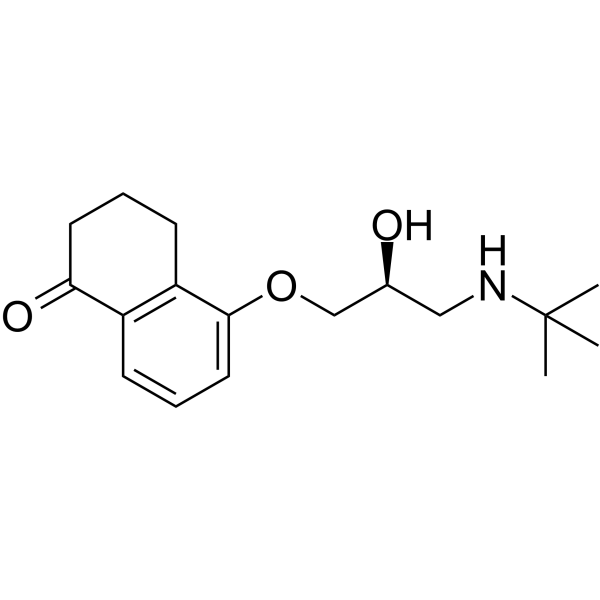
O=C1CCCC2=C1C=CC=C2OC[C@@H](O)CNC(C)(C)C
-
Synonyms
l-Bunolol
-
Shipping
Room temperature in continental US; may vary elsewhere.
-
Storage
Please store the product under the recommended conditions in the Certificate of Analysis.


Publications (1)
-
Journal Impact Factor
-
Most Recent
-
BMC Ophthalmol
β-blocker eye drops affect ocular surface through β2 adrenoceptor of corneal limbal stem cells. [Abstract]2021 Dec 5;21(1):419. PMID: 34863129

Protocols
-
RNA extraction experimental
By lysing cells, releasing RNA, and removing impurities such as proteins and DNA, high-purity RNA products are finally obtained. The commonly used traditional method is the guanidine isothiocyanate/phenol/chloroform method (Trizol), which is suitable for a variety of animal materials including animal tissues, microorganisms, cultured cells, etc., and most plant materials.
-
Ki-67 Immunostaining Proliferation Assay
Ki-67 immunostaining measures the growth fraction of a cell population by detecting Ki-67, a nuclear antigen present in proliferating cells and absent in quiescent G0 cells. The readout is the percentage of Ki-67-positive nuclei among total counted cells, commonly called the Ki-67 labeling index or proliferation index.
-
Calcium Spark Assay
Calcium sparks are localized, transient increases in intracellular calcium concentration ([Ca2+]i) that occur in cardiac myocytes and represent elementary events underlying excitation-contraction coupling. These events are generated by the coordinated opening of clusters of ryanodine receptors (RyRs) on the sarcoplasmic reticulum membrane, leading to a brief release of Ca2+ into the cytosol. The detection and analysis of calcium sparks provide insights into the mechanisms of calcium handling and signaling in cardiac cells. Imaging techniques using fluorescent calcium indicators such as Fluo-3 are employed to visualize these subcellular calcium transients with high spatial and temporal resolution. The protocol is based on established methodologies described in primary literature for both experimental measurement and automated analysis of calcium sparks.
-
Research Protocol for Cardiovascular Diseases
Cardiovascular disease can be modeled as maladaptive cardiac remodeling, where ischemic injury or pressure overload activates inflammatory signaling, fibroblast activation, extracellular-matrix deposition, cardiomyocyte hypertrophy, vascular remodeling, and progressive ventricular dysfunction. The TGF-β/SMAD axis is a central profibrotic pathway after myocardial injury and pressure overload, while innate immune and cytokine pathways regulate leukocyte recruitment, scar formation, and adverse remodeling. Key unresolved questions include which inflammatory signals are reparative versus harmful, when fibrosis is protective versus maladaptive, and whether pathway inhibition improves function without weakening necessary infarct healing or compensatory remodeling.
-
Ca2+ Staining Technique
Ca2+ staining is an experimental technique that utilizes specific fluorescent probes (such as Fluo-4 AM, Fura-2, etc.) to qualitatively or quantitatively detect dynamic changes in intracellular Ca2+ concentrations; this is achieved by monitoring the changes in fluorescent signals generated when these probes bind to free intracellular calcium ions. The underlying principle relies primarily on the presence of chelating groups within the probe's molecular structure that possess high affinity for calcium ions.
-
Cell Viability Determination by MTT Colorimetric Assay
The following protocol uses the MTT colorimetric assay as a classic literature-established method for assessing cell viability/metabolic activity in cultured mammalian cells. MTT[3-(4,5-dimethylthiazol-2-yl)-2,5-diphenyltetrazolium bromide] is reduced by metabolically active cells to a colored formazan product; the amount of formazan is quantified spectrophotometrically and provides an indirect measure of metabolically active viable cells. Importantly, MTT reduction reflects cellular oxidoreductase/metabolic activity rather than an absolute direct count of living cells, so changes in cellular metabolism can alter the signal independently of cell number.


Purity & Documentation
References

Calculators
Concentration (start) × Volume (start) = Concentration (final) × Volume (final)


Levobunolol Related Classifications
HY-B1035A Related Classifications
 Powered by Bioz