



LL-37, human TFA
Based on 6 publication(s) in Google Scholar
LL-37, human TFA is a 37-residue, amphipathic, cathelicidin-derived antimicrobial peptide, which exhibits a broad spectrum of antimicrobial activity. LL-37, human TFA could help protect the cornea from infection and modulates wound healing.
Nos produits utilisent uniquement pour la recherche. Nous ne vendons pas aux patients.
- Pureté : 99.96%
- Formule: C205H340N60O53.xC2HF3O2
- Masse moléculaire:4493.26 (free base)
-
Stockage:
Sealed storage, away from moisture.
Powder -80°C, 2 years , -20°C, 1 year* In solvent : -80°C, 6 months; -20°C, 1 month (sealed storage, away from moisture)


To place orders, for customer services and technical support, please contact: MedChemExpress USA
Tel: 609-228-6898 E-mail: [email protected] [email protected]


-
 Activité biologique
-
Chemical Information
-
Solvant et solubilité
- Protocole
- Pureté et documentation
- Références
-
 Help & FAQs



Publications Citing Use of MedChemExpress (MCE) LL-37, human TFA
More Customer Validation & Images

-
2D/3D Cell Culture and Differentiation
-
2D/3D Cell Culture and Differentiation
-
Cell Imaging/Staining
-
Cell Migration/Invasion Assay
-
ELISA


Activité biologique
Description
In Vitro
LL-37, human TFA (1-20 μg/mL; 24 h) affects HCECs migration[2]. LL-37, human TFA (0.0001-5 μg/mL; 6-24 h) affects cytokine secretion in HCECs[2]. LL-37, human TFA (1-100 μg/mL; 24 h) shows dose-dependently cytotoxic to HCECs at concentrations over 10 μg/mL[2].
MedChemExpress (MCE) has not independently confirmed the accuracy of these methods. They are for reference only.
-
Cell Line:Human corneal epithelial cell (HCEC)
-
Concentration:1, 2.5, 5, 10 and 20 μg/mL
-
Incubation Time:24 hours
-
Result:Dose-dependently stimulated HCECs migration but showed no effect on cells proliferation.
-
Cell Line:Human corneal epithelial cell (HCEC)
-
Concentration:0.0001, 0.001, 0.01, 0.1, 0.5, 1, and 5 μg/mL
-
Incubation Time:6 and 24 hours
-
Result:Dose-dependently increased IL-8, IL-6, IL-1β and TNF-α secretion at 6 and 24 hours in HCECs.
In Vivo
MedChemExpress (MCE) has not independently confirmed the accuracy of these methods. They are for reference only.
-
Animal Model:6-8 week-old C57BL/6 mice with MRSA-induced pneumonia[3]
-
Dosage:0.4, 0.8, 1.2, 1.6 and 2.0 mg/kg
-
Administration:Intratracheal injection; 0.4-2.0 mg/kg once
-
Result:Decreased IL-6 and TNF-α release to attenuated MRSA-induced pneumonia of testing mice.


Chemical Information
-
Appearance Solid
-
Masse moléculaire 4493.26 (free base)
-
Formule C205H340N60O53.xC2HF3O2
-
Color White to off-white
-
Sequence
Leu-Leu-Gly-Asp-Phe-Phe-Arg-Lys-Ser-Lys-Glu-Lys-Ile-Gly-Lys-Glu-Phe-Lys-Arg-Ile-Val-Gln-Arg-Ile-Lys-Asp-Phe-Leu-Arg-Asn-Leu-Val-Pro-Arg-Thr-Glu-Ser
-
Sequence Shortening
LLGDFFRKSKEKIGKEFKRIVQRIKDFLRNLVPRTES
-
Livraison
Room temperature in continental US; may vary elsewhere.
-
Stockage
Sealed storage, away from moisture
Powder -80°C 2 years -20°C 1 year * In solvent : -80°C, 6 months; -20°C, 1 month (sealed storage, away from moisture)


Publications (6)
-
Journal Impact Factor
-
Most Recent
-
Commun Biol
Vibrio cholerae senses human enteric α-defensin 5 through a CarSR two-component system to promote bacterial pathogenicity. [Abstract]2022 Jun 8;5(1):559. PMID: 35676416 -
Int Immunopharmacol
Oroxylin A suppress LL-37 generated rosacea-like skin inflammation through the modulation of SIRT3-SOD2-NF-κB signaling pathway. [Abstract]2024 Mar 10:129:111636. PMID: 38364746
LL-37, human TFA purchased from MedChemExpress. Usage Cited in: Int Immunopharmacol. 2024 Mar 10:129:111636. [Abstract]
LL-37, human TFA (320 μM; 40 μL; every 12 h for 2 d) generated the redness and telangiectasis in the skin of BALB/c mice, with increased range of redness and the redness score.
LL-37, human TFA purchased from MedChemExpress. Usage Cited in: Int Immunopharmacol. 2024 Mar 10:129:111636. [Abstract]
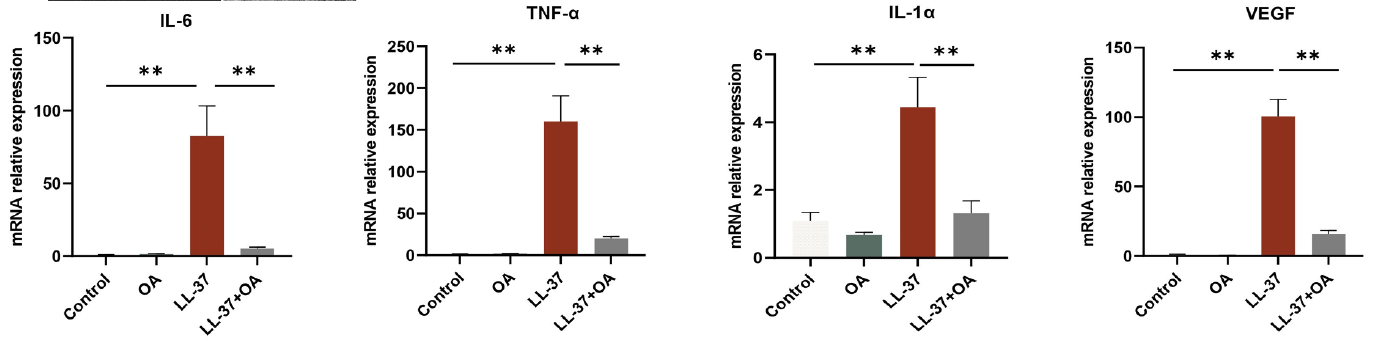
LL-37, human TFA (320 μM; 40 μL; every 12 h for 2 d) elevated the levels of IL-6, TNF-α, IL-1α, and VEGF in BALB/c mice tissues.
LL-37, human TFA purchased from MedChemExpress. Usage Cited in: Int Immunopharmacol. 2024 Mar 10:129:111636. [Abstract]
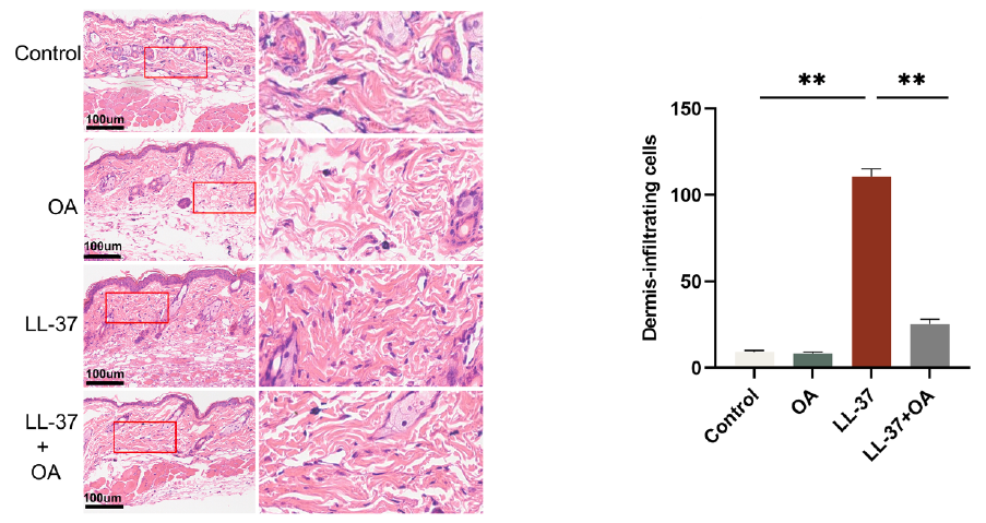
The present study employed OCT to examine the structural characteristics of the skin of BALB/c mice treated with LL-37, human TFA (320 μM; 40 μL; every 12 h for 2 d).
LL-37, human TFA purchased from MedChemExpress. Usage Cited in: Int Immunopharmacol. 2024 Mar 10:129:111636. [Abstract]
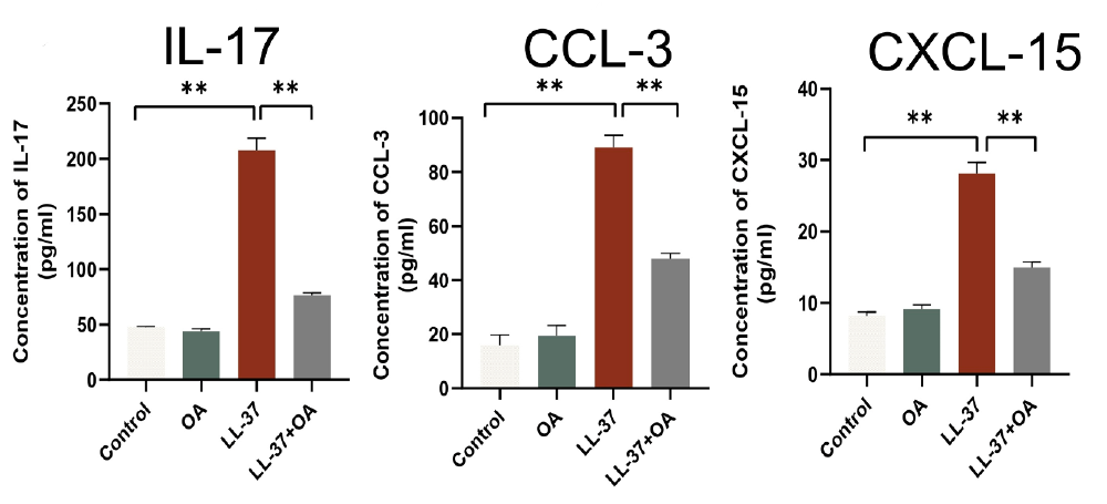
ELISA analysis of IL-17, CCL-3, CXCL-15 level in BALB/c mice treated with LL-37, human TFA (320 μM; 40 μL; every 12 h for 2 d).
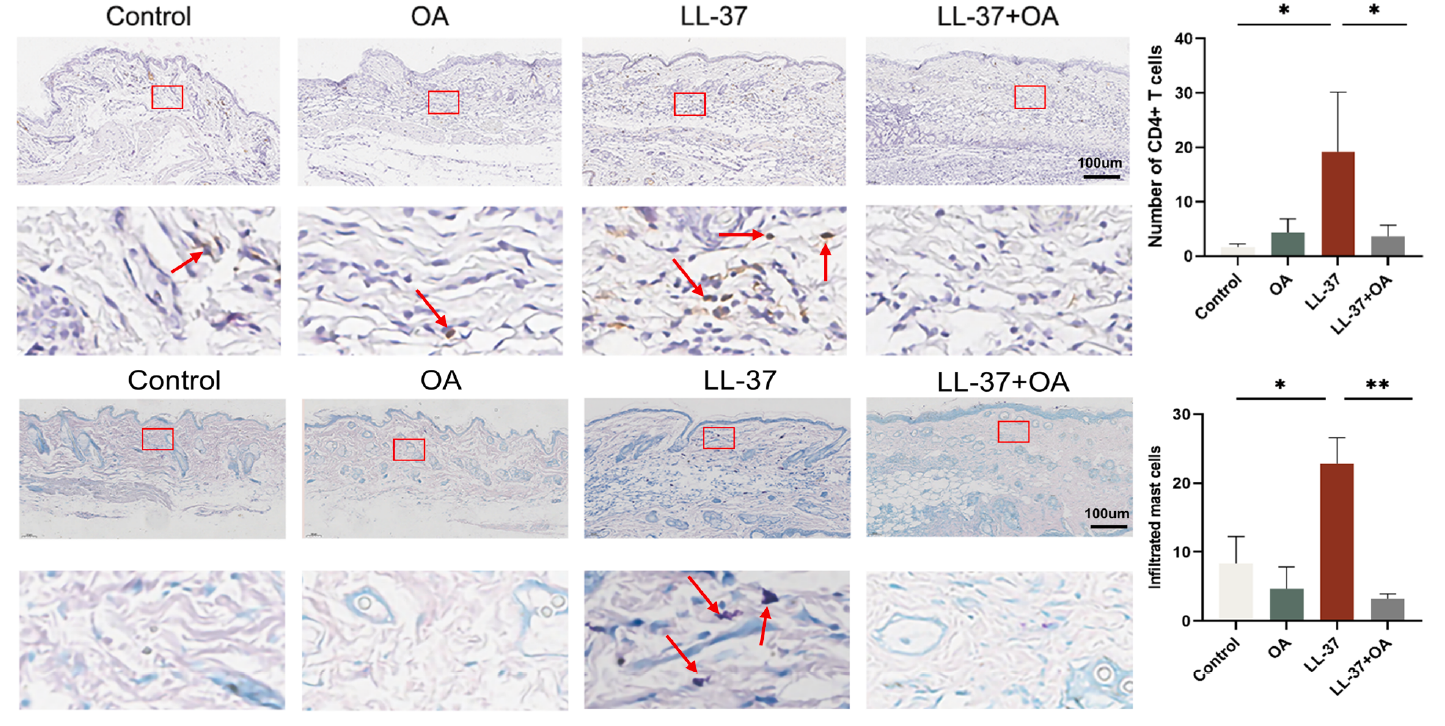
LL-37, human TFA purchased from MedChemExpress. Usage Cited in: Int Immunopharmacol. 2024 Mar 10:129:111636. [Abstract]
LL-37, human TFA (320 μM; 40 μL; every 12 h for 2 d) caused a statistically significant increase in the presence of CD4+T cells and MCs within the dermis of BALB/c mice.
-
ACS Omega
Potential Antiphotoaging Effect of Human Cathelicidin LL-37 Fragments and KR-12 Analogs on UVB-Induced HaCaT Cells and UVA-Induced HDF Cells. [Abstract]2025 Aug 12;10(33):36994-37003. PMID: 40893218
LL-37, human TFA purchased from MedChemExpress. Usage Cited in: ACS Omega. 2025 Aug 12;10(33):36994-37003. [Abstract]
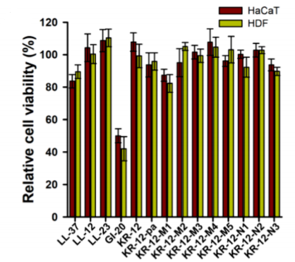
LL-37 (50 μM, 24 h) showed no cytotoxicity to HaCaT and HDF cells.
LL-37, human TFA purchased from MedChemExpress. Usage Cited in: ACS Omega. 2025 Aug 12;10(33):36994-37003. [Abstract]
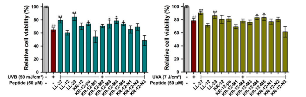
LL-37 (50 μM, 24 h) demonstrated effective photodamage repair, increasing the survival of UVB-irradiated HaCaT cells by 14.6% and UVA-irradiated HDF cells by 11.8%.
LL-37, human TFA purchased from MedChemExpress. Usage Cited in: ACS Omega. 2025 Aug 12;10(33):36994-37003. [Abstract]
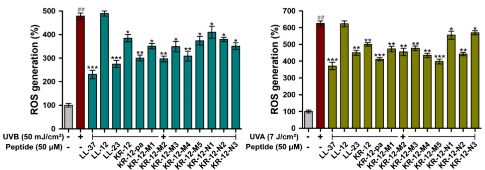
LL-37 (50 μM, 24 h) reduced ROS levels in UVB-irradiated HaCaT cells and UVA-irradiated HDF cells.
LL-37, human TFA purchased from MedChemExpress. Usage Cited in: ACS Omega. 2025 Aug 12;10(33):36994-37003. [Abstract]
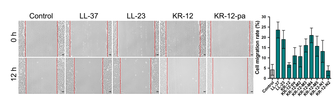
LL-37 (50 μM, 12 h) increased the migration rate of HaCaT cells.
LL-37, human TFA purchased from MedChemExpress. Usage Cited in: ACS Omega. 2025 Aug 12;10(33):36994-37003. [Abstract]
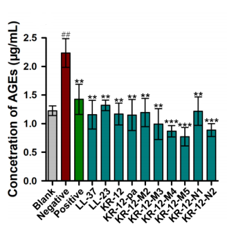
LL-37 (50 μM, 24 h) mitigated MGO-induced AGE formation in HaCaT cells.
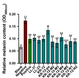
LL-37, human TFA purchased from MedChemExpress. Usage Cited in: ACS Omega. 2025 Aug 12;10(33):36994-37003. [Abstract]
LL-37 (50 μM, 24 h) suppressed α-MSH-induced melanin production in HaCaT cells.
-
FASEB J
Imperatorin Mitigates Rosacea-Like Inflammation by Modulating the Crosstalk Between JNK1 and STAT1. [Abstract]2026 Mar 15;40(5):e71641. PMID: 41773812 -
FASEB J
Integrated Multi-Omics and Experimental Validation Unveil the GZMK/NF-κB Axis Driving Inflammation and Fibroblast Proliferation in Rosacea: A Novel Drug Target Selection Strategy. [Abstract]2025 Oct 31;39(20):e71164. PMID: 41139274 -


Solvant et solubilité
In Vitro:
H2O : 100 mg/mL (Need ultrasonic)
DMSO : 50 mg/mL (Need ultrasonic; Hygroscopic DMSO has a significant impact on the solubility of product, please use newly opened DMSO)
In Vivo:
For the following dissolution methods, please prepare the working solution directly:
It is recommended to prepare fresh solutions and use them promptly within a short period of time.
The percentages shown for the solvents indicate their volumetric ratio in the final prepared solution. If precipitation or phase separation occurs during preparation, heat and/or sonication can be used to aid dissolution.
Add each solvent one by one: PBS
Solubility: 16.67 mg/mL; Clear solution; Need ultrasonic


Pureté et documentation
-
Fiche technique (287 KB)
-
SDS (252 KB)
- English - EN (252 KB)
- Français - FR (252 KB)
- Deutsch - DE (252 KB)
- Norwegian - NO (252 KB)
- Español - ES (252 KB)
- Swedish - SV (252 KB)
- Italian - IT (252 KB)
- Korean - KR (252 KB)
- Portuguese - PT (252 KB)
-
Instruction de manipulation (2659 KB)
Références
[1]. Dürr UH, et al. LL-37, the only human member of the cathelicidin family of antimicrobial peptides. Biochim Biophys Acta. 2006 Sep;1758(9):1408-25. [Content Brief]
[2]. Huang LC, et al. Multifunctional roles of human cathelicidin (LL-37) at the ocular surface. Invest Ophthalmol Vis Sci. 2006 Jun;47(6):2369-80. [Content Brief]
[3]. Hou M, et al. Antimicrobial peptide LL-37 and IDR-1 ameliorate MRSA pneumonia in vivo. Cell Physiol Biochem. 2013;32(3):614-23. [Content Brief]

Calculators
Concentration (start) × Volume (start) = Concentration (final) × Volume (final)


LL-37, human TFA Related Classifications
HY-P1222A Related Classifications
 Powered by Bioz