



KGF/FGF-7 Protein, Human
Based on 5 publication(s) in Google Scholar
KGF/FGF-7 Protein is an epithelial cell-specific mitogen secreted by normal stromal fibroblasts. KGF/FGF-7 Protein activates plasminogen activator (PA) activity to promote extracellular matrix degradation. KGF/FGF-7 Protein, Human has activities such as promoting the proliferation and differentiation of epithelial cells (such as keratinocytes, thymic epithelial cells), repairing tissue damage (such as intestinal ischemia/reperfusion injury, bone defects), and regulating immune function (such as improving thymus function in aged mice). KGF/FGF-7 Protein, Human is a recombinant KGF/FGF-7 protein expressed by E. coli without a tag.
- Species: Human
- Source: E. coli
-
Storage:Stored at -20°C for 2 years from date of receipt. After reconstitution, it is stable at 4°C for 1 week or -20°C for longer (with carrier protein). It is recommended to freeze aliquots at -20°C or -80°C for extended storage.


To place orders, for customer services and technical support, please contact: MedChemExpress USA
Tel: 609-228-6898 E-mail: [email protected] [email protected]


-
 Biological Activity
-
Technical Parameters
-
Product Properties
- Documentation
- References
- Help & FAQs


-

HY-P7048
KGF-2/FGF-10 Protein, Human (169a.a) -

HY-10182
Laduviglusib -
HY-10432
A 83-01 -

HY-B0215
Acetylcysteine -
HY-P70311
Activin A Protein, Human/Mouse/Rat (HEK293) -
HY-P7109
EGF Protein, Human -
HY-B0150
Nicotinamide -

HY-10071
Y-27632 -
HY-P70558
Noggin Protein, Human (HEK293) -

HY-10583
Y-27632 dihydrochloride -

HY-15371
Forskolin -
HY-10295
SB 202190


Biological Activity
Description
KGF/FGF-7 Protein is an epithelial cell-specific mitogen secreted by normal stromal fibroblasts. KGF/FGF-7 Protein activates plasminogen activator (PA) activity to promote extracellular matrix degradation. KGF/FGF-7 Protein, Human has activities such as promoting the proliferation and differentiation of epithelial cells (such as keratinocytes, thymic epithelial cells), repairing tissue damage (such as intestinal ischemia/reperfusion injury, bone defects), and regulating immune function (such as improving thymus function in aged mice). KGF/FGF-7 Protein, Human is a recombinant KGF/FGF-7 protein expressed by E. coli without a tag.
Background
KGF/FGF-7 Protein belongs to the fibroblast growth factor family[1]. KGF/FGF-7 Protein is an epithelial cell-specific mitogen secreted by normal stromal fibroblasts[1]. KGF/FGF-7 Protein stimulates epithelial cell proliferation and migration and regulates differentiation processes by binding to specific receptors on the surface of epithelial cells. For example, under high calcium conditions, it promotes keratinocytes to express early (K1) and late (filagrin) differentiation markers, and activates plasminogen activator (PA) activity to promote extracellular matrix degradation[1][3]. KGF/FGF-7 Protein is expressed in mesenchymal cells of various tissues such as skin, gastrointestinal tract, lung, and thymus, and acts on epithelial cells through paracrine effects[1][2][5][6][7]. KGF/FGF-7 Protein, Human has activities such as promoting the proliferation and differentiation of epithelial cells (such as keratinocytes and thymic epithelial cells), repairing tissue damage (such as intestinal ischemia/reperfusion injury and bone defects), and regulating immune function (such as improving thymus function in aged mice). For example, it stimulates keratinocyte migration and PA activity in wound repair, and promotes the expression of osteogenic markers and new bone formation in bone defect models[1][3][4][5][6][7].
In Vitro
KGF/FGF-7 Protein, Human (10 ng/mL; 4 days) promotes human keratinocyte proliferation with an effect comparable to that of EGF[1].
KGF/FGF-7 Protein, Human (20 ng/mL; 6-48 h) induces increased SCF mRNA and protein secretion in human HaCaT keratinocytes[2].
KGF/FGF-7 Protein, Human (1 nM; 16 h) significantly stimulates human keratinocyte migration and urokinase-type plasminogen activator (uPA) activity[3].
KGF/FGF-7 Protein, Human (25-200 ng/mL; 48 h for migration test) stimulates migration of rat bone marrow stromal cells (BMSCs) and increases the expression of SDF-1 and CXCR4[4].
In Vivo
KGF/FGF-7 Protein, Human (5 mg/kg; s.c.; 3 days; before transplantation) reduces GVHD mortality and improves liver, lung and other organ damage in the B10.BR mouse allogeneic bone marrow transplantation model[5].
KGF/FGF-7 Protein, Human (5 mg/kg/d; i.p.; 5 days; before surgery) increases intestinal wet weight, reduces epithelial apoptosis, and restores tight junction protein distribution in the intestinal ischemia/reperfusion model of C57BL/6J mice[6].
KGF/FGF-7 Protein, Human (5 mg/kg/d; s.c.; 3 days) induces thymic regeneration, increases thymocyte number and naive T cell output in C57BL/6J mice[7].
Verified Bioactivity
Measured in a cell proliferation assay using 4MBr-5 rhesus monkey epithelial cells.The ED50 for this effect is ≤110.3 ng/mL, corresponding to a specific activity is ≥9.07×10³ U/mg.
MCE Validation Data
-
Purity - SDS-PAGE
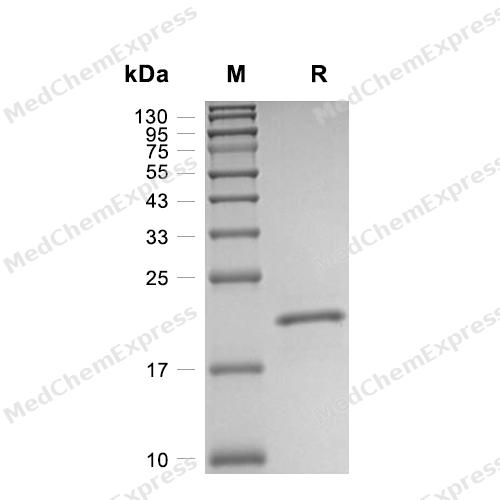
Purity - SDS-PAGE
-
Bioactivity - Cell-Based Assay
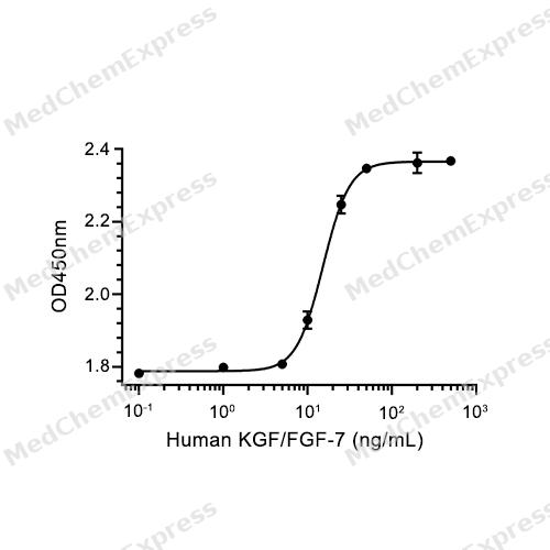
Bioactivity - Cell-Based Assay


Publications (5)
-
Journal Impact Factor
-
Most Recent
-
Nat Commun
2025 Dec 8. PMID: 41361241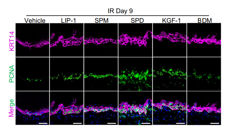
KGF/FGF-7 Protein, Human purchased from MedChemExpress. Usage Cited in: Nat Commun. 2025 Dec 8. [Abstract]
Quantification of PCNA+ basal cells before irradiation and for 9 days post-irradiation. Vehicle, LIP-1 (5 mg/kg), KGF-1 (6.25 mg/kg), Spermidine (10 mg/kg), Spermine (10 mg/kg) (all administered via intraperitoneal injection), and 0.15% Benzydamine mouthwash (topical application), starting 7 days prior to irradiation and continuing until 9 days post-irradiation.
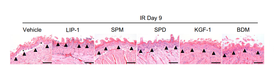
KGF/FGF-7 Protein, Human purchased from MedChemExpress. Usage Cited in: Nat Commun. 2025 Dec 8. [Abstract]
H&E staining and epithelial thickness of mouse mucosa before irradiation and for 9 days post-irradiation. Vehicle, LIP-1 (5 mg/kg), KGF-1 (6.25 mg/kg), Spermidine (10 mg/kg), Spermine (10 mg/kg) (all administered via intraperitoneal injection), and 0.15% Benzydamine mouthwash (topical application), starting 7 days prior to irradiation and continuing until 9 days post-irradiation.
-
Stem Cell Res Ther
Modulation of senescent Lepr+ skeletal stem cells via suppression of leptin-induced STAT3‒FGF7 axis activation alleviates abnormal subchondral bone remodeling and osteoarthritis progression. [Abstract]2025 May 5;16(1):227. PMID: 40325465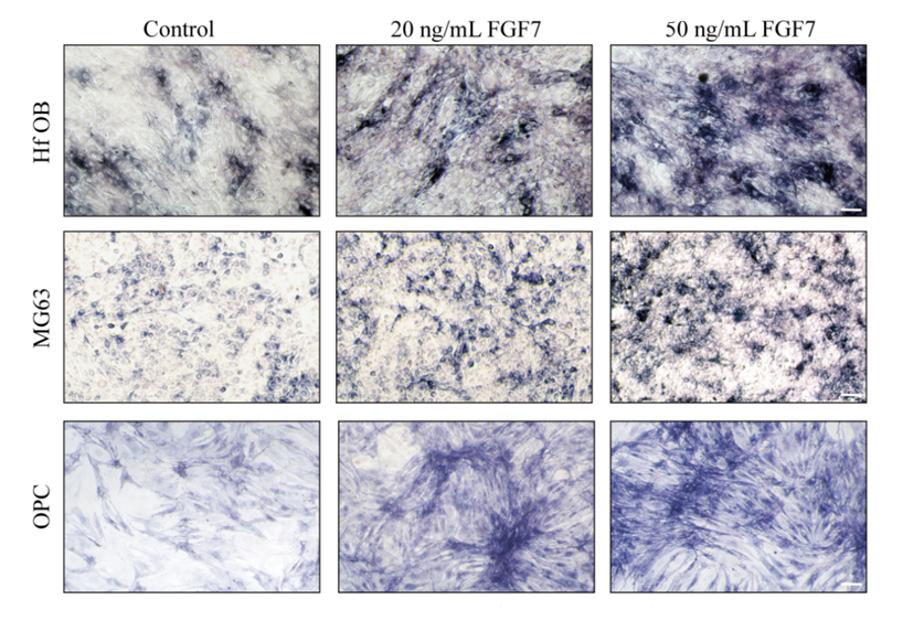
KGF/FGF-7 Protein, Human purchased from MedChemExpress. Usage Cited in: Stem Cell Res Ther. 2025 May 5;16(1):227. [Abstract]
The influence of varying FGF7 (20-50 ng/mL) concentrations on the osteogenic promotion of MG63, hFOB, and OPC cells under conditions of osteogenic induction.
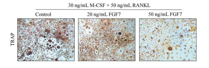
KGF/FGF-7 Protein, Human purchased from MedChemExpress. Usage Cited in: Stem Cell Res Ther. 2025 May 5;16(1):227. [Abstract]
Effect of different concentrations of FGF7 (20-50 ng/mL) on bone marrow-derived osteoclastic progenitors under osteoclastogenesis induction.
-
-
-


Technical Parameters
-
Species Human
-
Source E. coli
-
Tag Tag Free
-
Accession
P21781-1 (C32-T194)
-
Molecular Construction
-
N-term
-
FGF-7 (C32-T194)
Accession # P21781 -
C-term
-
-
Protein Length
Full Length of Isoform-1
-
Synonyms
FGF7; HBGF-7; Fibroblast Growth Factor 7; FGF-7; KGF; Fibroblast Growth Factor 7 (Keratinocyte Growth Factor); Keratinocyte Growth Factor; Fibroblast Growth Factor; Heparin-Binding Growth Factor 7; FGF
-
AA Sequence
CNDMTPEQMATNVNCSSPERHTRSYDYMEGGDIRVRRLFCRTQWYLRIDKRGKVKGTQEMKNNYNIMEIRTVAVGIVAIKGVESEFYLAMNKEGKLYAKKECNEDCNFKELILENHYNTYASAKWTHNGGEMFVALNQKGIPVRGKKTKKEQKTAHFLPMAIT
-
Predicted Molecular Mass
18.9 kDa
-
Molecular Weight
Approximately 17-19 kDa, based on SDS-PAGE under reducing conditions.
-
Purity
≥ 95%, as determined by reducing SDS-PAGE.


Product Properties
Lyophilized powder
1.Lyophilized from a 0.22 μm filtered solution of 20 mM Tris, 1 mM EDTA, 5% trehalose, 0.02% Tween 80, pH 8.0.
2.Lyophilized from a 0.22 μm filtered solution of PBS.
3.Lyophilized from a 0.22 μm filtered solution of 50 mM Tris-HCl, 300 mM NaCl, pH 7.4.
4.Lyophilized from a 0.22 μm filtered solution of 50 mM Tris-HCl, 300 mM NaCl, pH 8.0, 5% trehalose, 5% mannitol, 0.01% Tween 80.
5.Lyophilized from a 0.22 μm filtered solution of 20 mM PB, 500 mM NaCl, 8% trehalose, pH 8.0.
Please refer to the lot-specific COA for specific buffer information.
Note: For SPR assay, please replace the buffer. Primary amine components (e.g., Tris, imidazole) can affect protein-coupled chips.
<1 EU/μg, determined by LAL method.
It is not recommended to reconstitute to a concentration less than 100 μg/mL in ddH2O. For long term storage it is recommended to add a carrier protein (0.1% BSA, 5% HSA, 10% FBS or 5% Trehalose).
Stored at -20°C for 2 years from date of receipt. After reconstitution, it is stable at 4°C for 1 week or -20°C for longer (with carrier protein). It is recommended to freeze aliquots at -20°C or -80°C for extended storage.
Room temperature in continental US; may vary elsewhere.


Documentation
-
Data Sheet (264 KB)
-
SDS (252 KB)
- English - EN (252 KB)
- Français - FR (252 KB)
- Deutsch - DE (252 KB)
- Norwegian - NO (252 KB)
- Español - ES (252 KB)
- Swedish - SV (252 KB)
- Italian - IT (252 KB)
- Korean - KR (252 KB)
- Portuguese - PT (252 KB)
-
Handling Instructions (2659 KB)


References
[1]. Marchese C, et al. Human keratinocyte growth factor activity on proliferation and differentiation of human keratinocytes: differentiation response distinguishes KGF from EGF family. J Cell Physiol. 1990 Aug;144(2):326-32. [Content Brief]
[2]. Belleudi F, et al. KGF Promotes Paracrine Activation of the SCF/c-KIT Axis from Human Keratinocytes to Melanoma Cells. Transl Oncol. 2010 Apr;3(2):80-90. [Content Brief]
[3]. Tsuboi R, et al. Keratinocyte growth factor (FGF-7) stimulates migration and plasminogen activator activity of normal human keratinocytes. J Invest Dermatol. 1993 Jul;101(1):49-53. [Content Brief]
[4]. Poudel SB, et al. Local delivery of recombinant human FGF7 enhances bone formation in rat mandible defects. J Bone Miner Metab. 2017 Sep;35(5):485-496. [Content Brief]
[5]. Panoskaltsis-Mortari A, et al. Keratinocyte growth factor administered before conditioning ameliorates graft-versus-host disease after allogeneic bone marrow transplantation in mice. Blood. 1998 Nov 15;92(10):3960-7. [Content Brief]
[6]. Cai Y, et al. Keratinocyte growth factor improves epithelial structure and function in a mouse model of intestinal ischemia/reperfusion. PLoS One. 2012;7(9):e44772. [Content Brief]
[7]. Min D, et al. Sustained thymopoiesis and improvement in functional immunity induced by exogenous KGF administration in murine models of aging. Blood. 2007 Mar 15;109(6):2529-37. [Content Brief]

Calculators
Concentration (start) × Volume (start) = Concentration (final) × Volume (final)


 Powered by Bioz

