



von Hippel-Lindau (VHL)
Von Hippel-Lindau (VHL) protein functions as a tumor suppressor and substrate recognition subunit of the Cullin2-RING E3 ligase complex (CRL2^VHL), mediating oxygen-sensing by targeting hypoxia-inducible factor alpha (HIF-α) for ubiquitination and proteasomal degradation[1][2]. Loss or mutation of VHL disrupts HIF-α regulation, resulting in differential activation of HIF isoforms, where HIF-1α can suppress and HIF-2α can promote tumorigenesis[3][4][5]. Mechanistically, VHL-mediated ubiquitination specifically regulates HIF-2α-driven transcriptional programs that control cell proliferation, angiogenesis, and erythropoiesis, as demonstrated in VHL-R200W polycythemia models and renal cell carcinoma (RCC) studies[6][7][8]. Compared with related isoforms, HIF-2α exhibits tissue-specific oncogenic activity, particularly in proximal tubular epithelial cells, whereas HIF-1α exhibits tumor-suppressive effects[4][9][10]. Experimental models, including zebrafish and conditional mouse knockouts, recapitulate VHL loss phenotypes such as hemangioblastomas, impaired visual function, and splenic erythropoiesis, facilitating pharmacological intervention studies[11][12][13]. VHL-targeted small molecules, such as VH298 and belzutifan, stabilize VHL protein or inhibit HIF-2α activity, thereby modulating downstream hypoxia responses and providing a framework for therapeutic strategies in VHL-associated RCC and other neoplasms[1][14]. Overall, precise regulation of VHL and HIF isoforms underpins disease pathogenesis, isoform-specific biology, and translational applications in inhibitor or agonist development[2][4][9].
References:
- [1]. Rabezanahary H, et al. Live virus neutralizing antibodies against pre and post Omicron strains in food and retail workers in Québec, Canada. Heliyon. 2024 May 21;10(10):e31026. [Content Brief]
- [2]. Takamori H, et al. Development of drugs targeting hypoxia-inducible factor against tumor cells with VHL mutation: Story of 127 years. Cancer Sci. 2023 Apr;114(4):1208-1217. [Content Brief]
- [3]. Schödel J, et al. Hypoxia, Hypoxia-inducible Transcription Factors, and Renal Cancer. Eur Urol. 2016 Apr;69(4):646-657. [Content Brief]
- [4]. Lima JDCC, et al. HIFα isoform specific activities drive cell-type specificity of VHL-associated oncogenesis. Nat Commun. 2025 Oct 16;16(1):9185. [Content Brief]
- [5]. Mazumder S, et al. Downstream Targets of VHL/HIF-α Signaling in Renal Clear Cell Carcinoma Progression: Mechanisms and Therapeutic Relevance. Cancers (Basel). 2023 Feb 19;15(4):1316. [Content Brief]
- [6]. Hickey MM, et al. von Hippel-Lindau mutation in mice recapitulates Chuvash polycythemia via hypoxia-inducible factor-2alpha signaling and splenic erythropoiesis. J Clin Invest. 2007 Dec;117(12):3879-89. [Content Brief]
- [7]. Ding Z, et al. Genetic and pharmacological strategies to refunctionalize the von Hippel Lindau R167Q mutant protein. Cancer Res. 2014 Jun 1;74(11):3127-36. [Content Brief]
- [8]. Cautain B, et al. Identification of the Lipodepsipeptide MDN-0066, a Novel Inhibitor of VHL/HIF Pathway Produced by a New Pseudomonas Species. PLoS One. 2015 May 27;10(5):e0125221. [Content Brief]
- [9]. Lee SJ, et al. Von Hippel-Lindau tumor suppressor gene loss in renal cell carcinoma promotes oncogenic epidermal growth factor receptor signaling via Akt-1 and MEK-1. Eur Urol. 2008 Oct;54(4):845-53. [Content Brief]
- [10]. Hermans A, et al. A 3D-Printed and Freely Available Device to Measure the Zebrafish Optokinetic Response Before and After Injury. Zebrafish. 2024 Apr;21(2):144-148. [Content Brief]
- [11]. Abimbola A, et al. Role of Targeted Therapy in the Management of von Hippel-Lindau Disease-Associated Renal Cell Carcinoma: A Single-Proportion Meta-Analysis. Cureus. 2025 Nov 23;17(11):e97606. [Content Brief]
- [12]. Kaelin WG Jr. Von Hippel-Lindau disease: insights into oxygen sensing, et al. Von Hippel-Lindau disease: insights into oxygen sensing, protein degradation, and cancer. J Clin Invest. 2022 Sep 15;132(18):e162480. [Content Brief]
- [13]. Soares P, et al. Group-Based Optimization of Potent and Cell-Active Inhibitors of the von Hippel-Lindau (VHL) E3 Ubiquitin Ligase: Structure-Activity Relationships Leading to the Chemical Probe (2S,4R)-1-((S)-2-(1-Cyanocyclopropanecarboxamido)-3,3-dimethy [Content Brief]
- [14]. Steenhard BM, et al. Deletion of von Hippel-Lindau in glomerular podocytes results in glomerular basement membrane thickening, ectopic subepithelial deposition of collagen {alpha}1{alpha}2{alpha}1(IV), expression of neuroglobin, and proteinuria. Am J Path [Content Brief]
-
von Hippel-Lindau (VHL) Related Products (152)
Related Products (152)

-
dTAGV-1 TFA
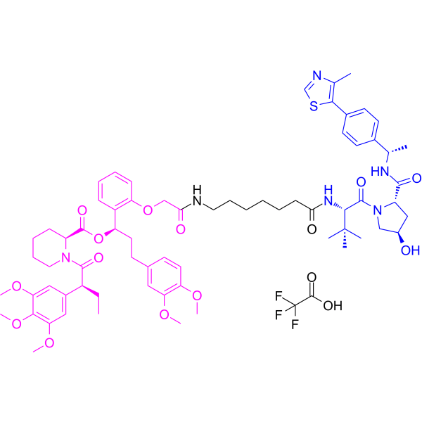 0 ImagesdTAGV-1 TFA is a selective FKBP12F36V PORTAC degrader. dTAGV-1 TFA induces rapid degradation of FKBP12F36V-tagged oncogenic fusion proteins, triggering collapse of downstream cellular signaling pathways, reduced proliferative capacity of cancer cells, and decreased levels of target proteins. dTAGV-1 TFA is applicable to functional validation studies of cancer and undegradable oncoproteins.
-
DT2216
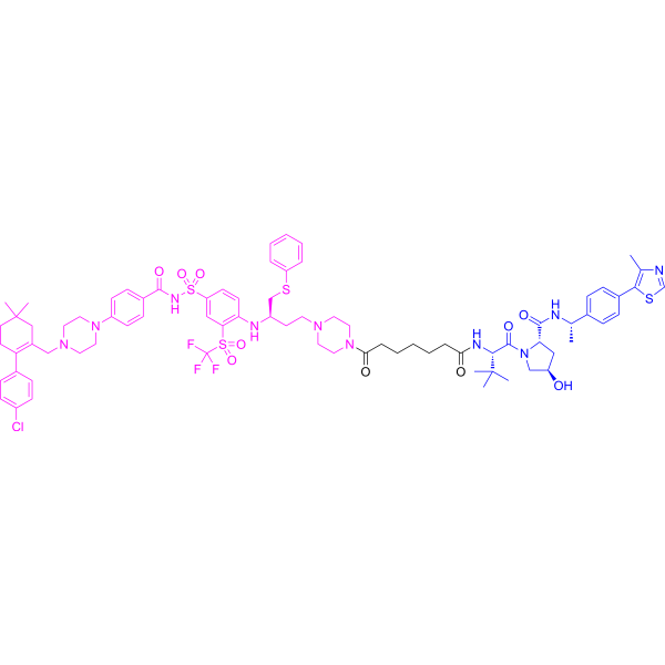 0 ImagesDT2216 is a potent and selective BCL-XL (Bcl-2 family member) degrader based on PROTAC technology. DT2216 causes effective degradation of BCL-XL protein by recruiting Von Hippel-Lindau (VHL) E3 ubiquitin ligase. DT2216 inhibits various BCL-XL-dependent leukemia and cancer cells but considerably less toxic to platelets.
-
AU-15330
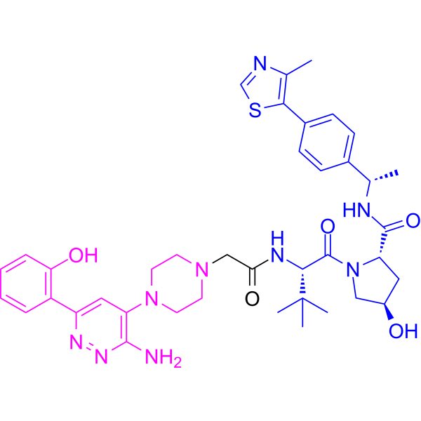 0 ImagesAU-15330 is a proteolysis-targeting chimera (PROTAC) degrader of the SWI/SNF ATPase subunits, SMARCA2 and SMARCA4. AU-15330 induces potent inhibition of tumour growth in xenograft models of prostate cancer and synergizes with the AR antagonist enzalutamide. AU-15330 induces disease remission in castration-resistant prostate cancer (CRPC) models without toxicity.
-
dTAGV-1 hydrochloride
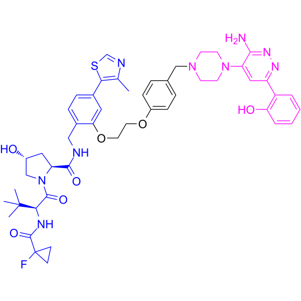
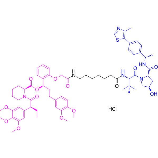 0 ImagesdTAGV-1 hydrochloride is a selective FKBP12F36V PORTAC degrader. dTAGV-1 hydrochloride induces rapid degradation of FKBP12F36V-tagged oncogenic fusion proteins, triggering collapse of downstream cellular signaling pathways, reduced proliferative capacity of cancer cells, and decreased levels of target proteins. dTAGV-1 hydrochloride is applicable to functional validation studies of cancer and undegradable oncoproteins.
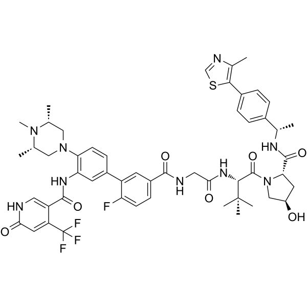
-
LC-2
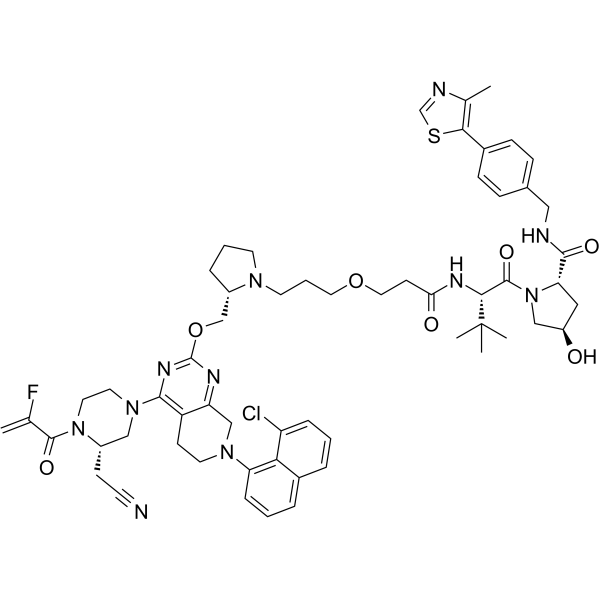 0 ImagesLC-2 is an orally active PROTAC-class degrader targeting KRASG12C and a MAPK inhibitor. LC-2 shows higher selectivity for KRASG12C than wild-type KRAS and other KRAS mutants. LC-2 covalently binds to KRASG12C via the MRTX849 (HY-130149) warhead, recruits the VHL E3 ligase to form a ternary complex, and induces ubiquitination and degradation of KRASG12C. Meanwhile, LC-2 inhibits the MAPK signaling pathway, reduces the phosphorylation levels of CRAF and AKT, thereby inducing apoptosis and suppressing cancer cell and tumor growth. LC-2 can be used for the research of KRASG12C-positive cancers, including non-small cell lung cancer and colorectal cancer.
-
PROTAC RIPK degrader-2
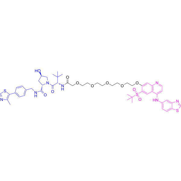 0 ImagesPROTAC RIPK degrader-2 is a non-peptidic PROTAC degrader targeting RIPK2, with a DC50 of 1.5 nM. PROTAC RIPK degrader-2 recruits VHL or cereblon E3 ubiquitin ligases, induces ubiquitination and proteasomal degradation of RIPK2, acts via substoichiometric catalysis, and inhibits downstream pro-inflammatory signaling pathways. PROTAC RIPK degrader-2 inhibits the NOD2/RIPK2 pathway and suppresses MDP (HY-127090)-stimulated TNFα release in human whole blood. PROTAC RIPK degrader-2 can be used in studies related to acute monocytic leukemia and glucose metabolism.
-
Setidegrasib
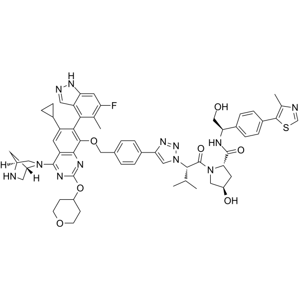 0 ImagesSynonyms: ASP-3082; KRAS G12D inhibitor 17Setidegrasib (ASP-3082) is a PROTAC KRAS degrader (DC50: 37 nM). Setidegrasib induces the degradation of G12D-mutation KRAS protein. Setidegrasib suppresses p-ERK, p-AKT, p-S6 levels in AsPC-1 cells. Setidegrasib exhibits anti-tumor activity in various cancer xenograft models in mice. Setidegrasib can be used in studies involving KRAS(G12D)-mutated solid tumors, such as non-small cell lung cancer.
-
GNE-987
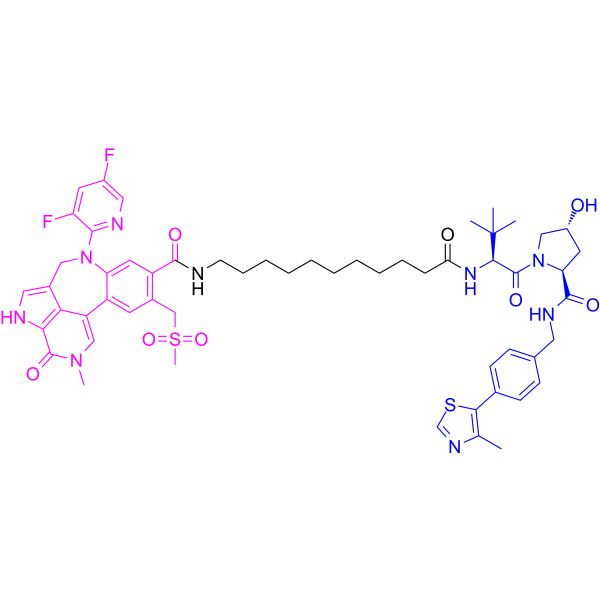 0 ImagesGNE-987 is a VHL-dependent BRD4 PROTAC degrader. GNE-987 exhibits picomolar cell BRD4 degradation activity (DC50=0.03 nM for EOL-1 AML cell line). GNE-987 binds equipotently to the BD1 and BD2 bromodomains of BRD4 with low nanomolar affinities (IC50=4.7 and 4.4 nM, respectively). GNE-987 can be used for the research of cancer.
-
ACBI2
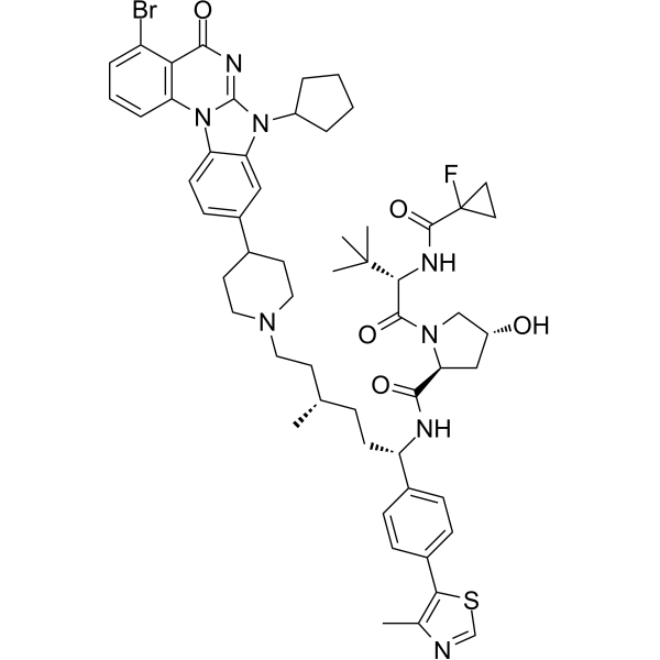 0 ImagesACBI2 is a highly potent and orally active VHL PROTAC SMARCA2 degrader (EC50: 7 nM), which selectively degrades SMARCA2 with a DC50 value of 1 nM in RKO cells. ACBI2 can be used in the research of lung cancer.
-
XL01126
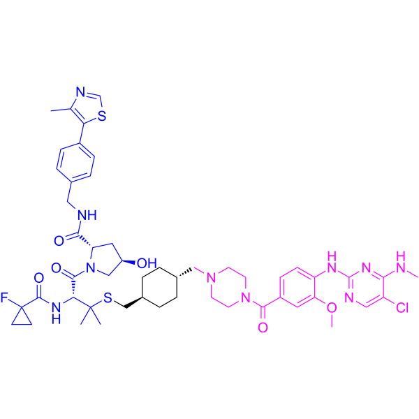 0 ImagesXL01126 is a blood-brain barrier-permeable, selective LRRK2 PROTAC degrader (DC50: 14 nM (G2019S LRRK2) and 32 nM (WT LRRK2)). XL01126 forms a positively cooperative ternary complex with VHL E3 ubiquitin ligase and LRRK2, mediating the polyubiquitination and proteasomal degradation of LRRK2. XL01126 induces Mitophagy. XL01126 is applicable to research related to Parkinson's disease.
-
ARCC-4
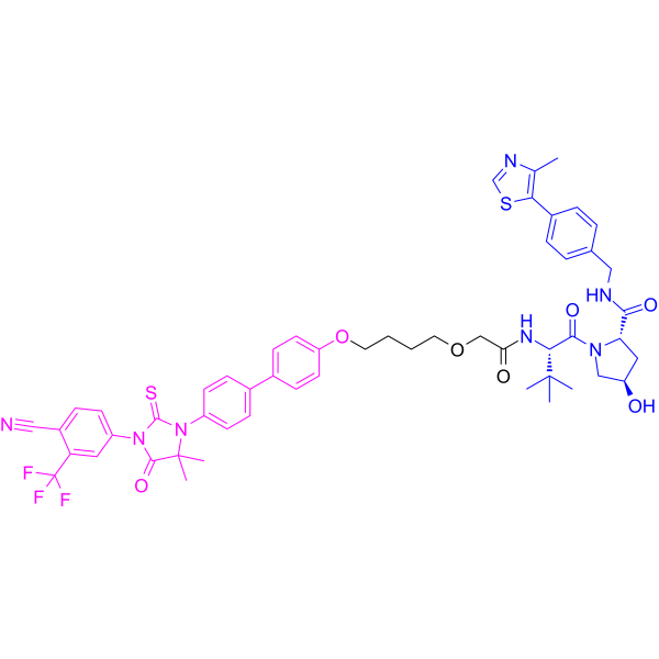 0 ImagesARCC-4 is a low-nanomolar Androgen Receptor (AR) degrader based on PROTAC, with a DC50 of 5?nM. ARCC-4 is an enzalutamide-based von Hippel-Lindau (VHL)-recruiting AR PROTAC and outperforms enzalutamide. ARCC-4 effectively degrades clinically relevant AR mutants associated with antiandrogen therapy.
-
MS8847
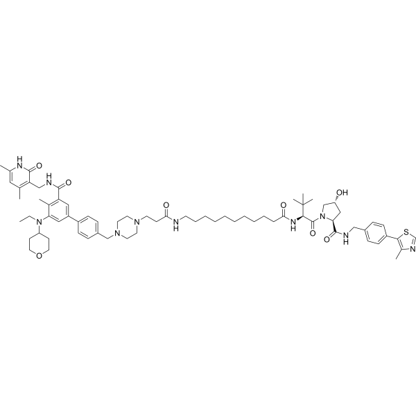 0 ImagesMS8847 is a PROTAC degrader and antiproliferative agent targeting EZH2 (DC50=34.4 nM in EOL-1 MLL-rAML cells). MS8847 recruits the E3 ligase von Hippel-Lindau (VHL) to mediate the degradation of EZH2 via the ubiquitin-proteasome system. MS8847 induces antiproliferative effects in MLL-rearranged acute myeloid leukemia cells and inhibits the growth of triple-negative breast cancer cell lines or 3D triple-negative breast cancer models. MS8847 is applicable to research related to MLL-rearranged acute myeloid leukemia and triple-negative breast cancer.
-
PROTAC KRAS G12D degrader 1
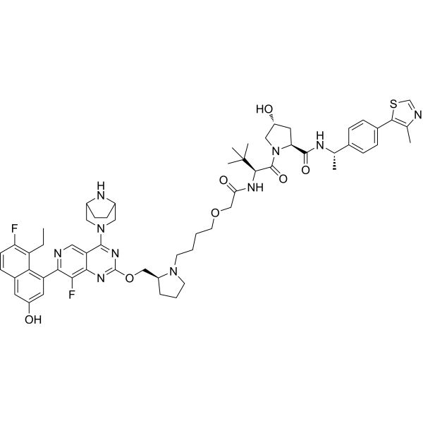 0 ImagesPROTAC KRAS G12D degrader 1 is a selective PROTAC KRASG12D degrader. PROTAC KRAS G12D degrader 1 inhibits proliferation of KRASG12D-mutant cells and suppresses ERK phosphorylation. PROTAC KRAS G12D degrader 1 inhibits tumor growth in mice bearing AsPC-1 xenografts. PROTAC KRAS G12D degrader 1 can be used for the study of KRASG12D-driven cancers.(Pink: KRAS ligand (HY-175892), Blue: VHL Ligand (HY-112078), Black: Linker, E3 ligase ligand-linker conjugate (HY-175893)).
-
dTAGV-1
 0 ImagesdTAGV-1 is a selective FKBP12F36V PORTAC degrader. dTAGV-1 induces rapid degradation of FKBP12F36V-tagged oncogenic fusion proteins, triggering collapse of downstream cellular signaling pathways, reduced proliferative capacity of cancer cells, and decreased levels of target proteins. dTAGV-1 is applicable to functional validation studies of cancer and undegradable oncoproteins.
-
SJF-0628
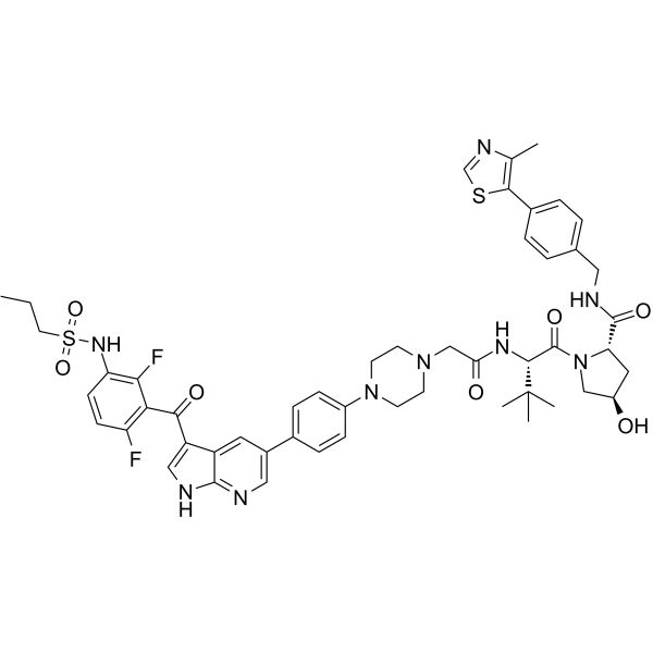 0 ImagesSJF-0628 is a PROTAC degrader of BRAF mutants, with a DC50 of 6.8 nM against BRAFV600E. SJF-0628 induces the ubiquitination and degradation of BRAF mutants in various cancer cell lines. SJF-0628 reduces pMEK and pErk levels. SJF-0628 exhibits antitumor activity. SJF-0628 can be used in research on colorectal cancer, melanoma, lung cancer, and triple-negative breast cancer.
-
PROTAC NCOA4 degrader-1
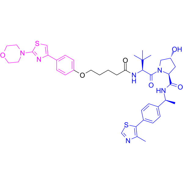
 0 ImagesPROTAC NCOA4 degrader-1 is a VHL-based PROTAC NCOA4 degrader. PROTAC NCOA4 degrader-1 is a ferroptosis inhibitor. PROTAC NCOA4 degrader-1 reduces Fe2+ elevation, ROS production, MDA content and PTGS2 mRNA expression. PROTAC NCOA4 degrader-1 ameliorates liver damage in a CCl4-induced acute liver injury model. PROTAC NCOA4 degrader-1 can be used for the researches of inflammation and immunology.
-
MS4322
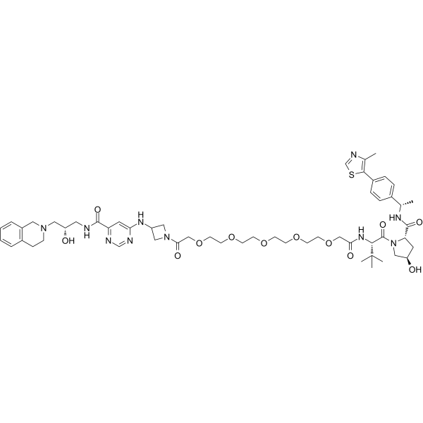 0 ImagesSynonyms: YS43-22MS4322 (YS43-22) is a specific PRMT5 PROTAC degrader. MS4322 reduces the PRMT5 protein level with a DC50 of 1.1 μM in MCF-7 cells. MS4322 inhibits the methyltransferase activity of PRMT5 with an IC50 of 18 nM. MS4322 promotes ubiquitination and degradation of PRMT5. MS4322 can be used for the research of breast cancer, lung cancer, and hepatocellular cancer. (Pink: PRMT5 ligand (HY-173092); Blue: E3 ligase ligand HY-112078); Black: linker (HY-124780); E3+linker (HY-173093 )).



