



LC-2
Based on 2 publication(s) in Google Scholar
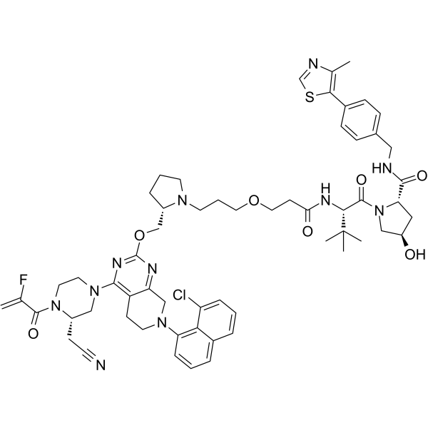
LC-2 is an orally active PROTAC-class degrader targeting KRASG12C and a MAPK inhibitor. LC-2 shows higher selectivity for KRASG12C than wild-type KRAS and other KRAS mutants. LC-2 covalently binds to KRASG12C via the MRTX849 (HY-130149) warhead, recruits the VHL E3 ligase to form a ternary complex, and induces ubiquitination and degradation of KRASG12C. Meanwhile, LC-2 inhibits the MAPK signaling pathway, reduces the phosphorylation levels of CRAF and AKT, thereby inducing apoptosis and suppressing cancer cell and tumor growth. LC-2 can be used for the research of KRASG12C-positive cancers, including non-small cell lung cancer and colorectal cancer.
(Pink: KRas G12C ligand (HY-130149); Blue: VHL ligand (HY-125845); Black: linker).
For research use only. We do not sell to patients.
- Purity : 98.48%
- CAS No.: 2502156-03-6
- Formula: C59H71ClFN11O7S
- Molecular Weight:1132.78
-
Storage:Powder -20°C, 3 years , 4°C, 2 years ; In solvent -80°C, 2 years , -20°C, 1 year

To place orders, for customer services and technical support, please contact: MedChemExpress USA
Tel: 609-228-6898 E-mail: [email protected] [email protected]


-
 Biological Activity
-
Chemical Information
-
Solvent & Solubility
- Protocols
- Purity & Documentation
- References
-
 Help & FAQs


-
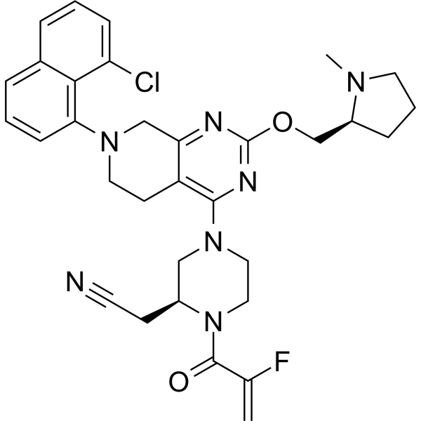
HY-130149
AdagrasibKRAS G12C Inhibitor -
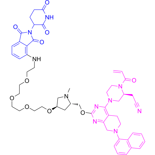
HY-129523
PROTAC K-Ras Degrader-1K-Ras PROTAC Degrader -
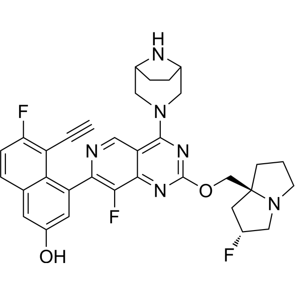
HY-134813
MRTX1133KRAS G12D Inhibitor -
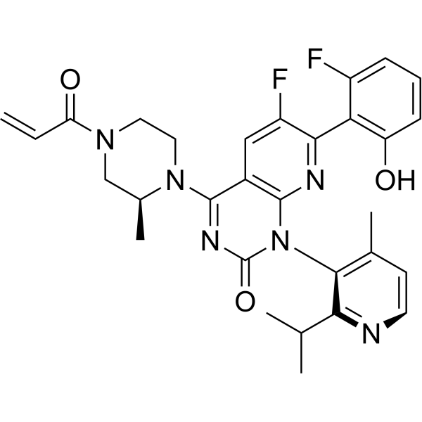
HY-114277
SotorasibKRAS G12C Inhibitor -
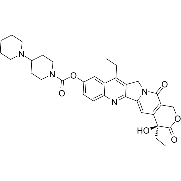
HY-16562
IrinotecanTopoisomerase I Inhibitor -
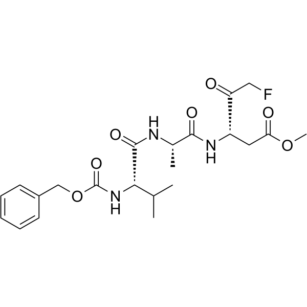
HY-16658
Z-VAD(OMe)-FMKPan-caspase Inhibitor -
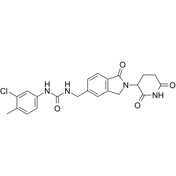
HY-101488
CC-885CRBN Modulator/GSPT1 Molecular Glue Degrader -
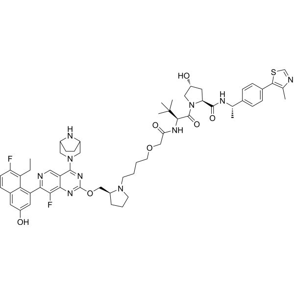
HY-161176
PROTAC KRAS G12D degrader 1PROTAC KRAS G12D Degrader -
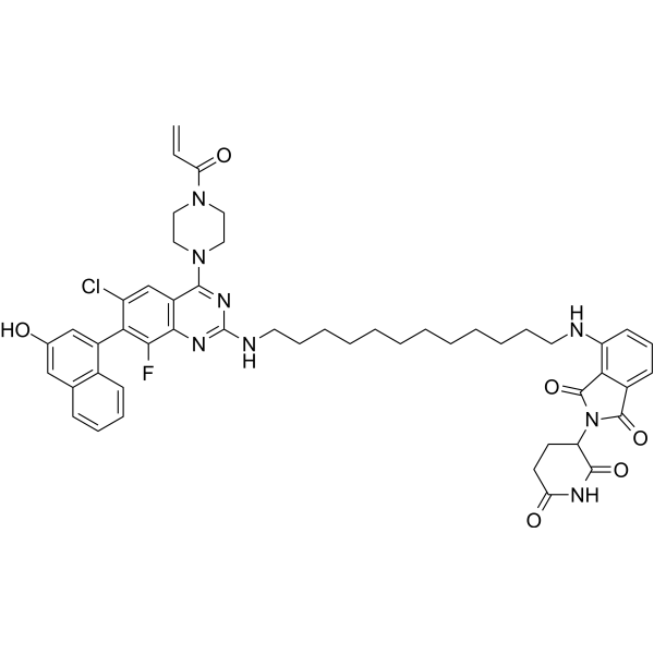
HY-139186
PROTAC KRAS G12C degrader-1PROTAC KRAS G12C Degrader
Publications Citing Use of MedChemExpress (MCE) LC-2
MoreAll PROTACs Isoforms
More

Biological Activity
Description
IC50 & Target
[1]|
KRAS(G12C) 0.25-0.76 μM (DC50) |
VHL |
MCE Validation Data
Cellular Effect
|
Cell Line

|
Type | Value | Description | References |
|---|---|---|---|---|
| NCI-H2030 | DC50 |
0.59 μM
|
Endogenous KRASG12C degradation in human NCI-H2030 cancer cells measured by immunoblot assay after 24 h incubation.
Endogenous KRASG12C degradation in human NCI-H2030 cancer cells measured by immunoblot assay after 24 h incubation.
|
32875077 |
| MIA PaCa-2 | DC50 |
0.32 μM
|
Endogenous KRASG12C degradation in human MIA PaCa-2 cancer cells measured by immunoblot assay after 24 h incubation.
Endogenous KRASG12C degradation in human MIA PaCa-2 cancer cells measured by immunoblot assay after 24 h incubation.
|
32875077 |
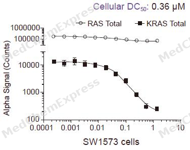
| SW1573 | DC50 |
0.76 μM
|
Endogenous KRASG12C degradation in human SW1573 cancer cells measured by immunoblot assay after 24 h incubation.
Endogenous KRASG12C degradation in human SW1573 cancer cells measured by immunoblot assay after 24 h incubation.
|
32875077 |
| NCI-H23 | DC50 |
0.25 μM
|
Endogenous KRASG12C degradation in human NCI-H23 cancer cells measured by immunoblot assay after 24 h incubation.
Endogenous KRASG12C degradation in human NCI-H23 cancer cells measured by immunoblot assay after 24 h incubation.
|
32875077 |
| NCI-H358 | DC50 |
0.52 μM
|
Endogenous KRASG12C degradation in human NCI-H358 cancer cells measured by immunoblot assay after 24 h incubation.
Endogenous KRASG12C degradation in human NCI-H358 cancer cells measured by immunoblot assay after 24 h incubation.
|
32875077 |
In Vitro
LC-2 (0.10-10 μM; 24 h) potently degrades endogenous KRASG12C in NCI-H2030 cells, with a DC50 of 0.59 μM and a maximum degradation rate of approximately 80% after 24 h[1].
LC-2 (24 h) degrades endogenous KRASG12C in MIA PaCa-2, SW1573, NCI-H23 and NCI-H358 cells, with DC50 values ranging from 0.25 to 0.76 μM and maximum degradation rates of ~40% to ~90% after 24 h[1].
The degradation of KRASG12C induced by LC-2 in NCI-H2030 and NCI-H23 cells occurs through a bona fide PROTAC mechanism dependent on VHL recruitment, proteasome function, and neddylation, but independent of lysosomal acidification[1].
LC-2 (2.5 μM) induces rapid and sustained degradation of endogenous KRASG12C in NCI-H2030, SW1573, MIA PaCa-2 and NCI-H23 cells. In most cell lines, the degradation reaches a peak at 8 to 24 h and can persist for up to 72 h[1].
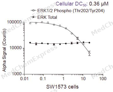
LC-2 (0.10-10 μM; 24 h) induces KRASG12C degradation, thereby regulating the downstream Erk signaling pathway in NCI-H2030, NCI-H23, MIA PaCa-2 and SW1573 cells. It exerts a dose-dependent inhibitory effect on pErk, and the signal kinetics are cell line-dependent[1].
LC-2 (1 μM; 14 days) shows intrinsic resistance in KRASG12C colorectal cancer cells SW1463 and SW837 with MDR1 overexpression[2].
The combination of LC-2 (1 μM; 14 days) and 2 μM Lapatinib exerts a more significant inhibitory effect on colony formation in KRASG12C colorectal cancer cells SW1463 and SW837 with MDR1 overexpression than LC-2 alone or LC-2 combined with Tariquidar[2].
LC-2 (1 μM; 48 h) alone does not reduce the protein level of KRASG12C in MDR1-overexpressing colorectal cancer cells SW1463, but combined treatment with Lapatinib (HY-50898) or Tariquidar (HY-10550) restores the degradation of KRASG12C, with Lapatinib additionally inhibiting downstream signaling and inducing apoptosis[2].
LC-2 (1 μM; 48 h) alone does not alter the protein level of KRASG12C in SW837 colorectal cancer cells overexpressing MDR1, but combined treatment with Lapatinib inhibits the downstream KRAS effector signaling pathway and induces apoptosis[2].
MedChemExpress (MCE) has not independently confirmed the accuracy of these methods. They are for reference only. Further protocols information, click here.
-
Cell Line:NCI-H2030 (homozygous KRASG12C)
-
Concentration:0.10, 0.25, 1.0, 2.5, 10 μM
-
Incubation Time:24 h
-
Result:Induced dose-dependent degradation of endogenous KRASG12C, with maximal degradation of ~80% achieved at 2.5 μM.
Measured a DC50 of 0.59 μM.
Observed an undegraded higher molecular weight band corresponding to PROTAC-bound KRASG12C at 10 μM, indicating the onset of a PROTAC "hook-effect".
-
Cell Line:NCI-H2030 (homozygous KRASG12C), NCI-H23 (heterozygous KRASG12C)
-
Concentration:2.5 μM (LC-2); 2.5 μM (LC-2 Epimer); 1 μM (epoxomicin), 1 μM (MLN4924), 0.1 μM (MLN4924), 0.1 μM (bafilomycin A1, pretreatment); molar excess (VHL ligand, pretreatment)
-
Incubation Time:4 h (LC-2/LC-2 Epimer incubation); 1 h (VHL ligand pretreatment)
-
Result:Failed to induce KRASG12C degradation at 2.5 μM when using LC-2 Epimer, while induced ~65% degradation in NCI-H2030 cells when using LC-2.
Rescued KRASG12C levels in NCI-H2030 cells treated with LC-2 after pretreatment with VHL ligand, epoxomicin, or MLN4924.
Failed to rescue KRASG12C levels in NCI-H23 cells treated with LC-2 after pretreatment with bafilomycin A1.
-
Cell Line:NCI-H2030 (homozygous KRASG12C), SW1573 (homozygous KRASG12C), MIA PaCa-2 (homozygous KRASG12C), NCI-H23 (heterozygous KRASG12C)
-
Concentration:2.5 μM
-
Incubation Time:1, 2, 4, 8, 12, 24 h (NCI-H2030, SW1573); 6, 24, 48, 72 h (MIA PaCa-2, NCI-H23)
-
Result:Detected KRASG12C binding in NCI-H2030 cells by 1 h, reached maximal degradation by 8 h, and degradation persisted up to 24 h.
Achieved near-maximal engagement in SW1573 cells by 1 h, reached maximal degradation by 12 h, and degradation persisted up to 24 h.
Reached maximal degradation in MIA PaCa-2 cells within 24 h, which was sustained up to 72 h.
Induced degradation in NCI-H23 cells within 6 h, reached maximum at 24 h, and degradation began to rebound by 72 h.
-
Cell Line:NCI-H2030 (homozygous KRASG12C), NCI-H23 (heterozygous KRASG12C), MIA PaCa-2 (homozygous KRASG12C), SW1573 (homozygous KRASG12C)
-
Concentration:0.10, 0.25, 1.0, 2.5, 10 μM (dose-response assays); 2.5 μM (time-course assays)
-
Incubation Time:24 h (dose-response assays); 6, 24 h (MIA PaCa-2, NCI-H23); 1, 4, 8, 24 h (SW1573); 1, 4, 8, 24 h (NCI-H2030)
-
Result:Induced a dose-dependent decrease in pErk signaling after 24 h in NCI-H2030 and NCI-H23 cells, corresponding to dose-dependent KRASG12C degradation.
Suppressed pErk signaling by 2.5 μM LC-2 at both 6 h and 24 h in MIA PaCa-2 and NCI-H23 cells.
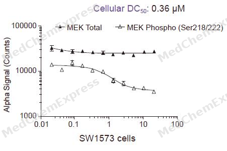
Inhibited pErk by 2.5 μM LC-2 between 1 and 4 h in SW1573 cells, with pErk rebounding between 8 and 24 h but remaining significantly lower than DMSO-treated cells at 24 h;
Increased total Erk levels in LC-2-treated SW1573 cells across all time points.
-
Cell Line:SW1463 homozygous KRASG12C colorectal cancer cells
-
Concentration:1 μM (LC-2 alone); 1 μM LC-2 + 0.1 μM tariquidar; 1 μM LC-2 + 5 μM lapatinib
-
Incubation Time:48 h
-
Result:Had no effect on KRASG12C protein levels when used alone.
Reduced KRASG12C protein levels when combined with 0.1 μM tariquidar or 5 μM lapatinib.
Reduced phosphorylation of CRAF, AKT, MEK, and ERK, and induced apoptosis when combined with 5 μM lapatinib.
-
Cell Line:SW837 heterozygous KRASG12C colorectal cancer cells
-
Concentration:1 μM (LC-2 alone); 1 μM LC-2 + 5 μM lapatinib
-
Incubation Time:48 h
-
Result:Did not reduce KRASG12C protein levels when used alone.
Reduced phosphorylation of CRAF, AKT, MEK, and ERK, and induced apoptosis when combined with 5 μM lapatinib.
-
Cell Line:SW1463 homozygous KRASG12C colorectal cancer cells, SW837 heterozygous KRASG12C colorectal cancer cells (both MDR1-overexpressing)
-
Concentration:Increasing concentrations
-
Incubation Time:5 days
-
Result:Exhibited synergy with lapatinib, with Bliss synergy scores of 27.0 for SW1463 cells and 25.0 for SW837 cells.
In Vivo
LC-2 (50 mg/kg; p.o.; every 3 days) inhibits growth of KRASG12C mutant non-small cell lung cancer xenografts and reduces KRAS expression in these tumors[3].
MedChemExpress (MCE) has not independently confirmed the accuracy of these methods. They are for reference only.
-
Animal Model:BALB/c nude (4-6 weeks old)[3]
-
Dosage:50 mg/kg
-
Administration:p.o.; every 3 days
-
Result:Failed to suppress growth of HCT116-derived tumors.
Did not reduce KRAS expression in these tumors.
-
Animal Model:BALB/c nude (4-6 weeks old)[3]
-
Dosage:50 mg/kg
-
Administration:p.o.; every 3 days
-
Result:Suppressed growth of H358-derived tumors.
Reduced KRAS expression in these tumors.


Chemical Information
-
CAS No. 2502156-03-6
-
Appearance Solid
-
Molecular Weight 1132.78
-
Formula C59H71ClFN11O7S
-
Color Off-white to brown
-
SMILES
O=C(NCC1=CC=C(C=C1)C2=C(N=CS2)C)[C@H]3N(C[C@@H](C3)O)C([C@H](C(C)(C)C)NC(CCOCCCN4[C@@H](CCC4)COC5=NC(N6C[C@@H](N(CC6)C(C(F)=C)=O)CC#N)=C7C(CN(CC7)C8=C9C(Cl)=CC=CC9=CC=C8)=N5)=O)=O
-
Shipping
Room temperature in continental US; may vary elsewhere.
-
Storage
Powder -20°C 3 years 4°C 2 years In solvent -80°C 2 years -20°C 1 year


Publications (2)
-
Journal Impact Factor
-
Most Recent
-
Biochem Biophys Res Commun
Accelerating PROTAC drug discovery: Establishing a relationship between ubiquitination and target protein degradation. [Abstract]2022 Nov 5:628:68-75. PMID: 36084553 -


Solvent & Solubility
In Vitro:
DMSO : 100 mg/mL (88.28 mM; Need ultrasonic; Hygroscopic DMSO has a significant impact on the solubility of product, please use newly opened DMSO)
Please refer to the solubility information to select the appropriate solvent. Once prepared, please aliquot and store the solution to prevent product inactivation from repeated freeze-thaw cycles.
Storage method and period of stock solution: -80°C, 2 years; -20°C, 1 year. When stored at -80°C, please use it within 2 years. When stored at -20°C, please use it within 1 year.
Please refer to the solubility information to select the appropriate solvent. Once prepared, please aliquot and store the solution to prevent product inactivation from repeated freeze-thaw cycles.
Storage method and period of stock solution: -80°C, 2 years; -20°C, 1 year. When stored at -80°C, please use it within 2 years. When stored at -20°C, please use it within 1 year.
Concentration (start) × Volume (start) = Concentration (final) × Volume (final)
In Vivo:
Select the appropriate dissolution method based on your experimental animal and administration route.
- For the following dissolution methods, please ensure to first prepare a clear stock solution using an In Vitro approach and then sequentially add co-solvents:
- To ensure reliable experimental results, the clarified stock solution can be appropriately stored based on storage conditions. As for the working solution for In Vivo experiments, it is recommended to prepare freshly and use it on the same day.
- The percentages shown for the solvents indicate their volumetric ratio in the final prepared solution. If precipitation or phase separation occurs during preparation, heat and/or sonication can be used to aid dissolution.
Add each solvent one by one: 10% DMSO 40% PEG300 5% Tween-80 45% Saline
Solubility: ≥ 2.5 mg/mL (2.21 mM); Clear solution
This protocol yields a clear solution of ≥ 2.5 mg/mL (saturation unknown).
Taking 1 mL working solution as an example, add 100 μL DMSO stock solution (25.0 mg/mL) to 400 μL PEG300, and mix evenly; then add 50 μL Tween-80 and mix evenly; then add 450 μL Saline to adjust the volume to 1 mL.
Preparation of Saline: Dissolve 0.9 g sodium chloride in ddH₂O and dilute to 100 mL to obtain a clear Saline solution.
Add each solvent one by one: 10% DMSO 90% (20% SBE-β-CD in Saline)
Solubility: 2.08 mg/mL (1.84 mM); Suspended solution; Need ultrasonic
This protocol yields a suspended solution of 2.08 mg/mL. Suspended solution can be used for oral and intraperitoneal injection.
Taking 1 mL working solution as an example, add 100 μL DMSO stock solution (20.8 mg/mL) to 900 μL 20% SBE-β-CD in Saline, and mix evenly.
Preparation of 20% SBE-β-CD in Saline (4°C, storage for one week): 2 g SBE-β-CD powder is dissolved in 10 mL Saline, completely dissolve until clear.
In Vivo Dissolution Calculator
Please enter the basic information of animal experiments:
-
-
-
-
Recommended: Prepare an additional quantity of animals to account for potential losses during experiments.
Please enter your animal formula composition:
-
%DMSO +
Recommended: Keep the proportion of DMSO in working solution below 2% if your animal is weak.
-
%+
-
+%Tween-80 + +
-
%Saline +
The co-solvents required include: DMSO, . All of co-solvents are available by MedChemExpress (MCE). , Tween 80. All of co-solvents are available by MedChemExpress (MCE).
Working solution concentration: 0.22 mg/mL
Method for preparing stock solution: mg drug dissolved in μL DMSO. Stock solution concentration: mg/mL.
1. Take μL DMSO stock solution;
2. Add μL .
μL , mix evenly;
3. Then add μL Tween 80, mix evenly;
4. Then add μL
Please ensure that the stock solution in the first step is dissolved to a clear state, and add co-solvents in sequence. You can use ultrasonic heating (ultrasonic cleaner, recommended frequency 20-40 kHz), vortexing, etc. to assist dissolution.

Protocols
-
Kinase activity and phosphorylation assays
Kinase activity assays measure the ability of kinases to transfer phosphate groups from ATP to specific substrates, while phosphorylation assays detect the presence and levels of phosphorylated proteins. Common methods include radiolabeled ATP incorporation (e. g. ,), ADP release detection via bioluminescence (e. g. ,[3]), enzyme-linked immunosorbent assays (ELISA) for phospho-specific epitopes (e. g. ,[6]), and microtiter-based formats for high-throughput screening (e. g. ,[8]). The ADP-Glo assay quantifies kinase activity by measuring ADP produced during phosphorylation using a luciferase-based system. Radiometric assays involve autoradiography or scintillation counting after incorporation of 32P-labeled ATP into substrate proteins. ELISA-based approaches rely on phospho-specific antibodies to detect activated kinases in cell lysates or purified samples.
-
Western Blot
Western blotting (WB) is a commonly used experimental method in molecular biology, biochemistry, and immunogenetics for identifying and quantifying target proteins. It combines gel electrophoresis with immunoassay, enabling researchers to analyze protein expression, post-translational modifications, and molecular weight.
-
Apoptosis
Apoptosis, also called programmed cell death, is generally characterized by distinct morphological characteristics.
-
TUNEL staining for apoptotic DNA fragmentation
TUNEL staining detects DNA strand breaks by using terminal deoxynucleotidyl transferase to add labeled nucleotides to exposed 3′-OH DNA termini, generating either microscopic staining in fixed cells or tissue sections, or fluorescence/cytometric signal in cell suspensions. TUNEL positivity reflects DNA fragmentation but should not be interpreted alone as definitive apoptosis, because TUNEL can also label necrotic, autolytic, mechanically damaged, or DNA-repair-associated DNA breaks.
-
Annexin V plus membrane-impermeant dye apoptosis staining
Annexin V-based apoptosis assays rely on the detection of phosphatidylserine (PS) externalization from the inner leaflet of the plasma membrane to the outer leaflet, an early biochemical hallmark of apoptosis. Fluorescently labeled Annexin V binds PS in a calcium-dependent manner, enabling identification of early apoptotic cells by flow cytometry or fluorescence microscopy. When combined with a membrane-impermeant DNA-binding dye (e. g. , propidium iodide), this approach allows discrimination between viable (Annexin V−/dye−), early apoptotic (Annexin V+/dye−), and late apoptotic or necrotic (Annexin V+/dye+) cell populations by assessing membrane integrity and PS exposure.
-
Patient-Derived Xenograft (PDX)
Patient-derived xenograft (PDX) models are generated by engrafting primary human tumor tissue directly into immunodeficient mice, allowing in vivo propagation of patient tumor biology without initial in vitro adaptation. These models are used to preserve key histopathological and molecular characteristics of the original tumor and enable assessment of tumor growth dynamics and therapeutic response in a living organism. The biological readout is tumor engraftment and subsequent growth in the murine host, which reflects the ability of human tumor cells to survive, vascularize, and expand in an immunocompromised microenvironment.
-
Apoptosis Solutions
Apoptosis is a regulated, generally non-lytic cell-death pathway that removes unwanted, damaged, infected, or abnormal cells through coordinated morphological changes, caspase activation, DNA fragmentation, and membrane remodeling. The intrinsic apoptosis pathway is controlled mainly by mitochondrial outer membrane permeabilization, BCL-2 family proteins, cytochrome c release, apoptosome formation, caspase-9 activation, and downstream executioner caspase-3/7 activation. The extrinsic apoptosis pathway is initiated by death receptors such as Fas, TNFR, and TRAIL receptors, which recruit adaptor proteins and activate caspase-8 before engaging executioner caspases or mitochondrial amplification through BID cleavage. Apoptosis is linked to many phenotypes, including cancer cell killing, tissue homeostasis, immune regulation, neurodegeneration, infection response, and treatment-induced cytotoxicity; unresolved questions include how apoptosis interacts with necroptosis, pyroptosis, ferroptos
-
Patient-Derived Orthotopic Xenograft (PDOX)
Patient-derived orthotopic xenograft (PDOX) modeling implants fresh patient tumor tissue or patient-derived tumor cells into the anatomically corresponding organ or tissue site of immunodeficient mice, usually by surgical orthotopic implantation, to preserve patient tumor histology, local microenvironmental context, invasion, metastatic behavior, and treatment-response features better than subcutaneous implantation. PDOX readouts include tumor engraftment, orthotopic tumor growth, local invasion, metastasis, recurrence after resection, histologic similarity to the donor tumor, biomarker retention, molecular concordance, survival, and response or resistance to therapy. PDOX models are used for preclinical drug testing and individualized therapy evaluation, but engraftment success varies by tumor type and specimen quality.
-
Protocol for Kinase activity and phosphorylation assays
Kinase activity assays measure transfer of phosphate from ATP to a protein or peptide substrate, generating phosphorylated substrate, ADP, or incorporated radiolabeled phosphate as the readout; phosphorylation assays measure site-specific phosphorylation in cells or tissues as a proxy for kinase-pathway activation, inhibition, or substrate regulation. Phosphorylation can be detected by phospho-specific Western blot, immunoprecipitation kinase assay, phospho-immunofluorescence, phospho-flow cytometry, luminescent ADP detection, radiolabeled ATP incorporation, or reporter-based pathway assays, and these readouts can be applied to cancer cells, primary neurons, mouse tumors, organoids, inflammatory macrophages, ferroptosis studies, and mitophagy studies when the kinase target is biologically relevant.
-
Subcutaneous Cell-Line-Derived Xenograft
Subcutaneous cell-line-derived xenograft (CDX) models are established by implanting cultured human cancer cell lines into immunodeficient mice, where the injected cells form localized tumors that can be monitored in vivo as a measure of tumorigenic potential, growth kinetics, and treatment response. These models are widely used in oncology research because they allow reproducible tumor formation and enable comparative assessment of tumor growth between different cell lines or genetic manipulations in a controlled in vivo microenvironment. Subcutaneous implantation of cancer cells in immunodeficient mice is a standard approach for evaluating tumor growth behavior and therapeutic response across multiple cancer types, including prostate, esophageal, pancreatic, and colon cancer models.
-
Orthotopic Cell-Line Xenograft
Orthotopic cell-line xenograft models involve implantation of human cancer cell lines into the anatomically corresponding organ of immunodeficient mice to reproduce tumor growth within a native microenvironment, enabling more clinically relevant tumor behavior compared with subcutaneous models. These models are widely used because orthotopic placement better recapitulates tumor progression, including invasion and metastatic spread, which are often underrepresented in heterotopic implantation systems. Compared with conventional xenografts, orthotopic implantation is described as more technically complex but provides improved simulation of tumor-microenvironment interactions and metastatic behavior, making it particularly valuable for translational oncology research. Surgical orthotopic implantation approaches have been emphasized as enabling faithful reproduction of clinical cancer features, including metastasis and disease progression patterns that align with the tumor’s organ of origi


Purity & Documentation
-
Data Sheet (302 KB)
-
SDS (396 KB)
- English - EN (396 KB)
- Français - FR (396 KB)
- Deutsch - DE (396 KB)
- Norwegian - NO (396 KB)
- Español - ES (396 KB)
- Swedish - SV (396 KB)
- Italian - IT (396 KB)
- Korean - KR (396 KB)
- Portuguese - PT (396 KB)
-
Handling Instructions (2659 KB)
References
[1]. Bond MJ, et al. Targeted Degradation of Oncogenic KRAS by VHL-Recruiting PROTACs. ACS central science. 2020 Aug 26;6(8):1367-1375. [Content Brief]
[2]. Kurimchak AM, et al. The drug efflux pump MDR1 promotes intrinsic and acquired resistance to PROTACs in cancer cells. Science signaling. 2022 Aug 30;15(749):eabn2707. [Content Brief]
[3]. Yang J, et al. A pan-KRAS degrader for the treatment of KRAS-mutant cancers. Cell discovery. 2024 Jun 28;10(1):70. [Content Brief]


Complete Stock Solution Preparation Table
Please refer to the solubility information to select the appropriate solvent. Once prepared, please aliquot and store the solution to prevent product inactivation from repeated freeze-thaw cycles.
Storage method and period of stock solution: -80°C, 2 years; -20°C, 1 year. When stored at -80°C, please use it within 2 years. When stored at -20°C, please use it within 1 year.
| Optional Solvent | Concentration Solvent Mass | 1 mg | 5 mg | 10 mg | 25 mg |
|---|---|---|---|---|---|
| DMSO | 1 mM | 0.8828 mL | 4.4139 mL | 8.8278 mL | 22.0696 mL |
| 5 mM | 0.1766 mL | 0.8828 mL | 1.7656 mL | 4.4139 mL | |
| 10 mM | 0.0883 mL | 0.4414 mL | 0.8828 mL | 2.2070 mL | |
| 15 mM | 0.0589 mL | 0.2943 mL | 0.5885 mL | 1.4713 mL | |
| 20 mM | 0.0441 mL | 0.2207 mL | 0.4414 mL | 1.1035 mL | |
| 25 mM | 0.0353 mL | 0.1766 mL | 0.3531 mL | 0.8828 mL | |
| 30 mM | 0.0294 mL | 0.1471 mL | 0.2943 mL | 0.7357 mL | |
| 40 mM | 0.0221 mL | 0.1103 mL | 0.2207 mL | 0.5517 mL | |
| 50 mM | 0.0177 mL | 0.0883 mL | 0.1766 mL | 0.4414 mL | |
| 60 mM | 0.0147 mL | 0.0736 mL | 0.1471 mL | 0.3678 mL | |
| 80 mM | 0.0110 mL | 0.0552 mL | 0.1103 mL | 0.2759 mL |


LC-2 Related Classifications
HY-137516 Related Classifications
 Powered by Bioz