


Teniposide
Based on 13 publication(s) in Google Scholar
Teniposide is a podophyllotoxin derivative, acts as a topoisomerase II inhibitor, and used as a chemotherapeutic agent.
For research use only. We do not sell to patients.
- Purity: 98.40%
- CAS No.: 29767-20-2
- Formula: C32H32O13S
- Molecular Weight:656.65
-
Storage:
4°C, protect from light
* In solvent : -80°C, 6 months; -20°C, 1 month (protect from light)

To place orders, for customer services and technical support, please contact: MedChemExpress USA
Tel: 609-228-6898 E-mail: [email protected] [email protected]


-
 Biological Activity
-
Chemical Information
-
Solvent & Solubility
- Protocol
- Purity & Documentation
- References
-
 Help & FAQs
-


Cell Cycle/DNA Damage Compound Library
HY-L004
-
FDA-Approved Drug Library
HY-L022
-
Anti-Cancer Compound Library
HY-L025
-
Anti-Aging Compound Library
HY-L034
-
Drug Repurposing Compound Library
HY-L035
-
NMPA-Approved Drug Library
HY-L053
-
FDA Approved & Pharmacopeial Drug Library
HY-L066
-
Anti-Lung Cancer Compound Library
HY-L075
-
Drug-Induced Liver Injury (DILI) Compound Library
HY-L076
-
Anti-Blood Cancer Compound Library
HY-L079
-
Targeted Diversity Library
HY-L099
-
Tumorigenesis-Related Compound Library
HY-L100
-
Rare Diseases Drug Library
HY-L102
-
Chemotherapy Drug Library
HY-L112
-
FDA-Approved Anticancer Drug Library
HY-L122
-
Human Metabolite Library
HY-L123
-
Off-patent Drug Library
HY-L141
-
Highly Selective Inhibitors Library
HY-L158
-
Anti-Hematopathy Compound Library
HY-L171
-
Bioactive Compound Library Max
HY-L181
-
MCE Bioactive Compound Library
HY-L001V
-
Drug Repurposing Compound Library Plus
HY-L035P
-
FDA-Approved Drug Library Plus
HY-L022P
-
FDA-Approved Drug Library Mini
HY-L022M
-
Bioactive Compound Library
HY-L001
-
High-Throughput Bioactive Compound Library
HY-L205
-
Anti-Cancer Approved Drug Library
HY-L213
-
Mass Spectrometry Human Metabolite Library
HY-L215
-
Biotoxin Library
HY-L220


Publications Citing Use of MedChemExpress (MCE) Teniposide
More- Cell Mol Immunol. 2024 Aug;21(8):856-872. [Abstract]
- Cell Death Dis. 2020 Nov 12;11(11):976. [Abstract]
- J Immunother Cancer. 2022 Aug;10(8):e004006. [Abstract]
- Acta Pharmacol Sin. 2021 Jan;42(1):108-114. [Abstract]
- Cell Rep Methods. 2023 Oct 23;3(10):100599. [Abstract]
- Int Immunopharmacol. 2021 Dec;101(Pt A):108264. [Abstract]
- J Mol Med (Berl). 2019 Aug;97(8):1183-1193. [Abstract]
- Mol Pharm. 2022 Nov 7;19(11):4320-4332. [Abstract]
- J Cell Mol Med. 2026 Apr;30(7):e71101. [Abstract]
- BMC Cancer. 2024 Apr 22;24(1):504. [Abstract]
- Mol Biol Cell. 2023 May 1;34(5):ar47. [Abstract]
- Biomed Pharmacother. 2025 Jun 20:189:118246. [Abstract]
- Biomed Pharmacother. 2024 Jul 19:178:117167. [Abstract]
 Customer Validation & Images

-
ELISA
-
RT-PCR
-
WB
-
Histological Imaging/Staining
-
RT-PCR
All Topoisomerase Isoforms
More

Biological Activity
|
Topoisomerase II |
|
Cell Line
|
Type | Value | Description | References |
|---|---|---|---|---|
| A549 | IC50 |
15.8 nM
Compound: Ref 34, Cpd 3
|
Antiproliferative activity against human A549 cells incubated for 72 hrs by MTT assay
Antiproliferative activity against human A549 cells incubated for 72 hrs by MTT assay
|
[PMID: 32992133] |
| A549 | IC50 |
8.2 μM
Compound: Teniposide
|
Anticancer activity against human A549 cells assessed as inhibition of cell proliferation measured after 48 hrs by MTT assay
Anticancer activity against human A549 cells assessed as inhibition of cell proliferation measured after 48 hrs by MTT assay
|
[PMID: 32084316] |
| CWR22R | IC50 |
0.082 μM
Compound: 3
|
Antiproliferative activity against human 22Rv1 cells after 96 hrs by propidium iodide-based monolayer assay
Antiproliferative activity against human 22Rv1 cells after 96 hrs by propidium iodide-based monolayer assay
|
[PMID: 26854430] |
| GLC4 cell line | IC50 |
0.48 μM
Compound: Teniposide
|
Cytotoxic effect against GLC4 (human small cell lung carcinoma cell line) using the microculture tetrazolium (MTT) assay based on continuous incubation
Cytotoxic effect against GLC4 (human small cell lung carcinoma cell line) using the microculture tetrazolium (MTT) assay based on continuous incubation
|
[PMID: 7783142] |
| GLC4 cell line | IC50 |
2.8 μM
Compound: Teniposide
|
Cytotoxic effect against GLC4 (human small cell lung carcinoma cell line) using the microculture tetrazolium (MTT) assay based on 2 hr incubation
Cytotoxic effect against GLC4 (human small cell lung carcinoma cell line) using the microculture tetrazolium (MTT) assay based on 2 hr incubation
|
[PMID: 7783142] |
| HeLa | ED50 |
0.3 μg/mL
Compound: 5
|
Cytotoxicity against human HeLa cells after 4 days by trypan blue assay
Cytotoxicity against human HeLa cells after 4 days by trypan blue assay
|
[PMID: 1602298] |
| HeLa | IC50 |
14.9 μM
Compound: Teniposide
|
Anticancer activity against human HeLa cells assessed as inhibition of cell proliferation measured after 48 hrs by MTT assay
Anticancer activity against human HeLa cells assessed as inhibition of cell proliferation measured after 48 hrs by MTT assay
|
[PMID: 32084316] |
| HepG2 | ED50 |
40.46 mg/kg
Compound: Teniposide
|
Antitumor activity against human HepG2 xenografted in ip dosed BALB/c nude mouse administered for 20 days and measured after 20 days by electronic caliper method
Antitumor activity against human HepG2 xenografted in ip dosed BALB/c nude mouse administered for 20 days and measured after 20 days by electronic caliper method
|
[PMID: 32084316] |
| HepG2 | IC50 |
4.3 μM
Compound: Teniposide
|
Anticancer activity against human HepG2 cells assessed as inhibition of cell proliferation measured after 48 hrs by MTT assay
Anticancer activity against human HepG2 cells assessed as inhibition of cell proliferation measured after 48 hrs by MTT assay
|
[PMID: 32084316] |
| HepG2 | IC50 |
62.7 μM
Compound: Teniposide
|
Poison activity at topoisomerase-2 in human HepG2 cell lysate assessed as topoisomerase-2 band depletion after 3 hrs by Western blot analysis
Poison activity at topoisomerase-2 in human HepG2 cell lysate assessed as topoisomerase-2 band depletion after 3 hrs by Western blot analysis
|
[PMID: 32084316] |
| HMEC | IC50 |
12.4 μM
Compound: Teniposide
|
Cytotoxicity against human HMEC cells assessed as inhibition of cell proliferation measured after 48 hrs by MTT assay
Cytotoxicity against human HMEC cells assessed as inhibition of cell proliferation measured after 48 hrs by MTT assay
|
[PMID: 32084316] |
| L02 | IC50 |
11.5 μM
Compound: Teniposide
|
Cytotoxicity against human HL7702 cells assessed as inhibition of cell proliferation measured after 48 hrs by MTT assay
Cytotoxicity against human HL7702 cells assessed as inhibition of cell proliferation measured after 48 hrs by MTT assay
|
[PMID: 32084316] |
| MCF7 | IC50 |
0.125 μM
Compound: 3
|
Antiproliferative activity against human MCF7 cells after 96 hrs by propidium iodide-based monolayer assay
Antiproliferative activity against human MCF7 cells after 96 hrs by propidium iodide-based monolayer assay
|
[PMID: 26854430] |
| MCF7 | IC50 |
11.1 μM
Compound: Teniposide
|
Anticancer activity against human MCF7 cells assessed as inhibition of cell proliferation measured after 48 hrs by MTT assay
Anticancer activity against human MCF7 cells assessed as inhibition of cell proliferation measured after 48 hrs by MTT assay
|
[PMID: 32084316] |
| MCF7 | IC50 |
15.8 nM
Compound: Ref 34, Cpd 3
|
Antiproliferative activity against human MCF7 cells incubated for 72 hrs by MTT assay
Antiproliferative activity against human MCF7 cells incubated for 72 hrs by MTT assay
|
[PMID: 32992133] |
| MRC5 | IC50 |
12.2 μM
Compound: Teniposide
|
Cytotoxicity against human MRC5 cells assessed as inhibition of cell proliferation measured after 48 hrs by MTT assay
Cytotoxicity against human MRC5 cells assessed as inhibition of cell proliferation measured after 48 hrs by MTT assay
|
[PMID: 32084316] |
| PC-3 | IC50 |
15.8 nM
Compound: Ref 34, Cpd 3
|
Antiproliferative activity against human PC3 cells incubated for 72 hrs by MTT assay
Antiproliferative activity against human PC3 cells incubated for 72 hrs by MTT assay
|
[PMID: 32992133] |
| RPMI 8402 | IC50 |
0.22 μM
Compound: VM-26
|
Cytotoxic activity against human lymphoblast tumor cell line RPMI8402 after 4 days of treatment
Cytotoxic activity against human lymphoblast tumor cell line RPMI8402 after 4 days of treatment
|
[PMID: 12747798] |
| RPMI 8402 | IC50 |
0.22 μM
Compound: VM-26
|
Cytotoxicity against human lymphoblast tumor cell line RPM18402
Cytotoxicity against human lymphoblast tumor cell line RPM18402
|
[PMID: 12392745] |
| RPMI 8402 | IC50 |
0.28 μM
Compound: VM-26
|
Cytotoxicity using camptothecin-resistant variant of RPM18402 (CPT-K5) possessing functional, but mutant TOP-1
Cytotoxicity using camptothecin-resistant variant of RPM18402 (CPT-K5) possessing functional, but mutant TOP-1
|
[PMID: 12392745] |
Teniposide is a topoisomerase II inhibitor. Teniposide (VM-26, 0.15-45 mg/L) inhibits the proliferation of Tca8113 cells in a dose-dependent manner, with an IC50 of 0.35 mg/L. Teniposide (5 mg/L) induces apoptosis of Tca8113 cells. Teniposide (5.0 mg/L) causes cell arrested at G2/M phase in Tca8113 cells[2]. Teniposide is active on primary cultured glioma cells from patients, when the level of miR-181b is high in the cells, with an IC50 of 1.3 ± 0.34 μg/mL. Cells treated with teniposide with low MDM2 have decreased viability compared with control cells, and the IC50 decreases from 5.86 ± 0.36 μg/mL to 2.90 ± 0.35 μg/mL upon MDM2 suppression. Teniposide also inhibits the viability of glioma cell with high level of miR-181b, through mediation of MDM2[3].
MedChemExpress (MCE) has not independently confirmed the accuracy of these methods. They are for reference only.
MedChemExpress (MCE) has not independently confirmed the accuracy of these methods. They are for reference only.
| NCT Number | Sponsor | Condition | Start Date |
Phase
|
|---|---|---|---|---|
| NCT01329991 | Plexxikon| | 2011-05 | PHASE1 |


Chemical Information
-
CAS No. 29767-20-2
-
Appearance Solid
-
Molecular Weight 656.65
-
Formula C32H32O13S
-
Color White to off-white
-
SMILES
O=C1OC[C@]2([H])[C@H](O[C@H]3[C@@H]([C@H]([C@@H]([C@@H](CO4)O3)O[C@@H]4C5=CC=CS5)O)O)C6=C(C=C7OCOC7=C6)[C@@H](C8=CC(OC)=C(O)C(OC)=C8)[C@]21[H]
-
Synonyms
VM26
-
Shipping
Room temperature in continental US; may vary elsewhere.
-
Storage
4°C, protect from light
* In solvent : -80°C, 6 months; -20°C, 1 month (protect from light)


Publications (13)
-
Journal Impact Factor
-
Most Recent
-
Cell Mol Immunol
Chaperone- and PTM-mediated activation of IRF1 tames radiation-induced cell death and the inflammatory response. [Abstract]2024 Aug;21(8):856-872. PMID: 38849539 -
Cell Death Dis
2020 Nov 12;11(11):976. PMID: 33184290 -
J Immunother Cancer
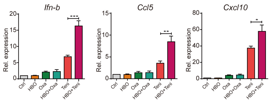
Hyperbaric oxygen facilitates teniposide-induced cGAS-STING activation to enhance the antitumor efficacy of PD-1 antibody in HCC. [Abstract]2022 Aug;10(8):e004006. PMID: 36002188
Teniposide purchased from MedChemExpress. Usage Cited in: J Immunother Cancer. 2022 Aug;10(8):e004006. [Abstract]
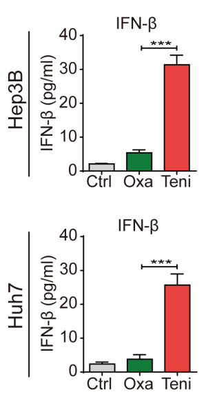
Teniposide (10.7 μM for Hep3B cells; 26.15 μM for Huh7 cells; 24 h) significantly increased the IFN-β secretion in Hep3B and Huh7 cells.
Teniposide purchased from MedChemExpress. Usage Cited in: J Immunother Cancer. 2022 Aug;10(8):e004006. [Abstract]
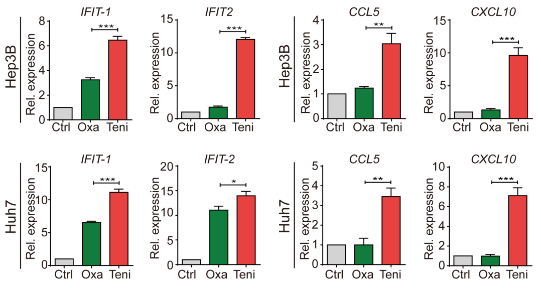
Teniposide (10.7 μM for Hep3B cells; 26.15 μM for Huh7 cells; 24 h) significantly increased the mRNA expression of IFIT1, IFIT2, CCL5 and CXCL10 in Hep3B and Huh7 cells.
Teniposide purchased from MedChemExpress. Usage Cited in: J Immunother Cancer. 2022 Aug;10(8):e004006. [Abstract]
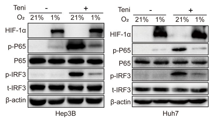
Teniposide (10.7 μM for Hep3B cells; 26.15 μM for Huh7 cells; 24 h) increased the cellular protein expression of p-IRF3 and p-P65 in Hep3B and Huh7 cells under normoxic conditions.
Teniposide purchased from MedChemExpress. Usage Cited in: J Immunother Cancer. 2022 Aug;10(8):e004006. [Abstract]
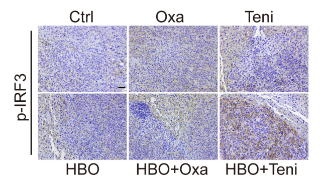
Teniposide (Teni) (10 mg/kg; i.p.; every other day for twice) increased tumor p-IRF3 expression in mouse HCC tumor model.
Teniposide purchased from MedChemExpress. Usage Cited in: J Immunother Cancer. 2022 Aug;10(8):e004006. [Abstract]
Teniposide (Teni) (10 mg/kg; i.p.; every other day for twice)significantly increased Ifn-b, Ccl5 and Cxcl10 mRNA expression in the tumor tissues ofmouse HCC tumor model.
-
Acta Pharmacol Sin
Osimertinib successfully combats EGFR-negative glioblastoma cells by inhibiting the MAPK pathway. [Abstract]2021 Jan;42(1):108-114. PMID: 32398685 -
Cell Rep Methods
RECOVER identifies synergistic drug combinations in vitro through sequential model optimization. [Abstract]2023 Oct 23;3(10):100599. PMID: 37797618 -
Int Immunopharmacol
Topoisomerase 2 inhibitor etoposide promotes interleukin-10 production in LPS-induced macrophages via upregulating transcription factor Maf and activating PI3K/Akt pathway. [Abstract]2021 Dec;101(Pt A):108264. PMID: 34715493 -
J Mol Med (Berl)
2019 Aug;97(8):1183-1193. PMID: 31201471 -
Mol Pharm
2022 Nov 7;19(11):4320-4332. PMID: 36269563 -
J Cell Mol Med
2026 Apr;30(7):e71101. PMID: 41896195 -
BMC Cancer
PROTAC EZH2 degrader-1 overcomes the resistance of podophyllotoxin derivatives in refractory small cell lung cancer with leptomeningeal metastasis. [Abstract]2024 Apr 22;24(1):504. PMID: 38644473 -
Mol Biol Cell
Mitotic DNA damage promotes chromokinesin-mediated missegregation of polar chromosomes in cancer cells. [Abstract]2023 May 1;34(5):ar47. PMID: 36989031 -
Biomed Pharmacother
Identification of nsp16 inhibitors of SARS -CoV-2, SARS -CoV-1 and MERS-CoV from FDA-approved drugs using in silico and in vitro methods. [Abstract]2025 Jun 20:189:118246. PMID: 40543162 -
Biomed Pharmacother
Characterization of a deazaflavin analog as a potent inhibitor of multidrug resistance-associated protein 1. [Abstract]2024 Jul 19:178:117167. PMID: 39032285


Solvent & Solubility
DMSO : ≥ 30 mg/mL (45.69 mM; Hygroscopic DMSO has a significant impact on the solubility of product, please use newly opened DMSO)
* "≥" means soluble, but saturation unknown.
Please refer to the solubility information to select the appropriate solvent. Once prepared, please aliquot and store the solution to prevent product inactivation from repeated freeze-thaw cycles.
Storage method and period of stock solution: -80°C, 6 months; -20°C, 1 month (protect from light). When stored at -80°C, please use it within 6 months. When stored at -20°C, please use it within 1 month.
Concentration (start) × Volume (start) = Concentration (final) × Volume (final)
Select the appropriate dissolution method based on your experimental animal and administration route.
- For the following dissolution methods, please ensure to first prepare a clear stock solution using an In Vitro approach and then sequentially add co-solvents:
- To ensure reliable experimental results, the clarified stock solution can be appropriately stored based on storage conditions. As for the working solution for In Vivo experiments, it is recommended to prepare freshly and use it on the same day.
- The percentages shown for the solvents indicate their volumetric ratio in the final prepared solution. If precipitation or phase separation occurs during preparation, heat and/or sonication can be used to aid dissolution.
Add each solvent one by one: 10% DMSO 40% PEG300 5% Tween-80 45% Saline
Solubility: ≥ 2.5 mg/mL (3.81 mM); Clear solution
This protocol yields a clear solution of ≥ 2.5 mg/mL (saturation unknown).
Taking 1 mL working solution as an example, add 100 μL DMSO stock solution (25.0 mg/mL) to 400 μL PEG300, and mix evenly; then add 50 μL Tween-80 and mix evenly; then add 450 μL Saline to adjust the volume to 1 mL.
Preparation of Saline: Dissolve 0.9 g sodium chloride in ddH₂O and dilute to 100 mL to obtain a clear Saline solution.
Please enter the basic information of animal experiments:
-
-
-
-
Recommended: Prepare an additional quantity of animals to account for potential losses during experiments.
Please enter your animal formula composition:
-
%DMSO +
Recommended: Keep the proportion of DMSO in working solution below 2% if your animal is weak.
-
%+
-
+%Tween-80 + +
-
%Saline +
The co-solvents required include: DMSO, . All of co-solvents are available by MedChemExpress (MCE). , Tween 80. All of co-solvents are available by MedChemExpress (MCE).
Working solution concentration: 0.22 mg/mL
Method for preparing stock solution: mg drug dissolved in μL DMSO. Stock solution concentration: mg/mL. * In solvent : -80°C, 6 months; -20°C, 1 month (protect from light)
1. Take μL DMSO stock solution;
2. Add μL .
μL , mix evenly;
3. Then add μL Tween 80, mix evenly;
4. Then add μL
Please ensure that the stock solution in the first step is dissolved to a clear state, and add co-solvents in sequence. You can use ultrasonic heating (ultrasonic cleaner, recommended frequency 20-40 kHz), vortexing, etc. to assist dissolution.

Protocol
Logarithmically growing Tca8113 cells are trypsinized and made into single cell suspension then plated in 96-well culture plate at a concentration of 5 × 10⁴ cells/well, eight columns for Teniposide and seven columns for CDDP in each plate, 3 wells in each column. After 24 hours of incubation, the medium of the 3 wells in each column are replaced with medium containing Teniposide of 0.15 mg/L, 0.5 mg/L, 1.5 mg/L, 5.0 mg/L, 15 mg/L and 45 mg/L or CDDP of 0.1 mg/L, 0.3 mg/L, 1.0 mg/L, 3.0 mg/L and 9.0 mg/L, respectively. Blank control wells are added medium without drugs. Cells are then cultured for another 24 hours, 48 hours, 72 hours, 96 hours and 120 hours. The supernatants are removed and 20 μL MTT solution is added in each well, followed with another 4 hours of culture. The supernatants are discarded carefully and 200 μL dimethyl sulphoxide (DMSO) is added and shaken vigorously to dissolve the purple precipitation formation. Optical density (OD) of each well is tested using Spectrophotometer with a wavelength of 450 nm. The experiment is repeated in triplicate[2].
MedChemExpress (MCE) has not independently confirmed the accuracy of these methods. They are for reference only.
Animals (mice) are treated with 0.5 mg/kg teniposide and bone marrow is sampled 24 h after treatment. Colchicine and mitomycin C are used as a positive control aneugen and clastogen, respectively, at the dose of 2 mg/kg each. Bone marrow smears are prepared and stained with May-Gruenwald/Giemsa solutions. At least four slides are made for each animal and allowed to dry overnight. One slide per animal is stained with May-Gruenwald/Giemsa solutions for conventional assessment of the micronuclei (MN) frequencies in polychromatic erythrocytes (PCEs) and normochromatic erythrocytes (NCEs). The remaining unstained slides are stored at −20°C for the distinction between the clastogenic and aneugenic effects by identifying the origin of MN with the mouse DNA probes. Per animal, 1000 PCE of coded slides are scored for the presence of MN. In addition, the number of PCEs among 1000 NCE per animal is recorded to evaluate bone marrow suppression and mitotic activity is calculated as %PCE = [PCE/(PCE + NCE)] × 100[1].
MedChemExpress (MCE) has not independently confirmed the accuracy of these methods. They are for reference only.


Purity & Documentation
-
Data Sheet (290 KB)
-
SDS (418 KB)
- English - EN (418 KB)
- Français - FR (418 KB)
- Deutsch - DE (418 KB)
- Norwegian - NO (418 KB)
- Español - ES (418 KB)
- Swedish - SV (418 KB)
- Italian - IT (418 KB)
- Korean - KR (418 KB)
- Portuguese - PT (418 KB)
-
Handling Instructions (2659 KB)
References
[1]. Attia SM, et al. Molecular cytogenetic evaluation of the aneugenic effects of teniposide in somatic and germinal cells of male mice. Mutagenesis. 2012 Jan;27(1):31-9. [Content Brief]
[2]. Li J, et al. Topoisomerase II trapping agent teniposide induces apoptosis and G2/M or S phase arrest of oral squamous cell carcinoma. World J Surg Oncol. 2006 Jul 6;4:41. [Content Brief]
[3]. Sun YC, et al. MiR-181b sensitizes glioma cells to teniposide by targeting MDM2. BMC Cancer. 2014 Aug 25;14:611. [Content Brief]


Complete Stock Solution Preparation Table
Please refer to the solubility information to select the appropriate solvent. Once prepared, please aliquot and store the solution to prevent product inactivation from repeated freeze-thaw cycles.
Storage method and period of stock solution: -80°C, 6 months; -20°C, 1 month (protect from light). When stored at -80°C, please use it within 6 months. When stored at -20°C, please use it within 1 month.
| Optional Solvent | Concentration Solvent Mass | 1 mg | 5 mg | 10 mg | 25 mg |
|---|---|---|---|---|---|
| DMSO | 1 mM | 1.5229 mL | 7.6144 mL | 15.2288 mL | 38.0720 mL |
| 5 mM | 0.3046 mL | 1.5229 mL | 3.0458 mL | 7.6144 mL | |
| 10 mM | 0.1523 mL | 0.7614 mL | 1.5229 mL | 3.8072 mL | |
| 15 mM | 0.1015 mL | 0.5076 mL | 1.0153 mL | 2.5381 mL | |
| 20 mM | 0.0761 mL | 0.3807 mL | 0.7614 mL | 1.9036 mL | |
| 25 mM | 0.0609 mL | 0.3046 mL | 0.6092 mL | 1.5229 mL | |
| 30 mM | 0.0508 mL | 0.2538 mL | 0.5076 mL | 1.2691 mL | |
| 40 mM | 0.0381 mL | 0.1904 mL | 0.3807 mL | 0.9518 mL |


 Powered by Bioz