


(+)-JQ-1
Based on 301 publication(s) in Google Scholar
(+)-JQ-1 (JQ1), a chemical probe, is a potent, specific, CNS-penetrant and reversible BET bromodomain inhibitor, with IC50s of 77 and 33 nM for the first and second bromodomain (BRD4(1/2)). (+)-JQ-1 also activates autophagy.
For research use only. We do not sell to patients.
- Purity: 99.91%
- CAS No.: 1268524-70-4
- Formula: C23H25ClN4O2S
- Molecular Weight:456.99
-
Storage:Powder -20°C, 3 years , 4°C, 2 years ; In solvent -80°C, 1 year , -20°C, 6 months
-JQ-1 Chemical Structure")


-
 Biological Activity
-
Chemical Information
-
Solvent & Solubility
- Protocol
- Purity & Documentation
- References
-
 Help & FAQs
-


Epigenetics Compound Library
HY-L005
-
Histone Modification Research Compound Library
HY-L024
-
Anti-Cancer Compound Library
HY-L025
-
Clinical Compound Library
HY-L026
-
CNS-Penetrant Compound Library
HY-L028
-
Autophagy Compound Library
HY-L029
-
Reprogramming Compound Library
HY-L039
-
Chemical Probe Library
HY-L063
-
Anti-Blood Cancer Compound Library
HY-L079
-
Target Protein Ligand Library
HY-L129
-
Heterocyclic Compound Library
HY-L138
-
Highly Selective Inhibitors Library
HY-L158
-
Highly Selective Activators Library
HY-L159
-
Cell Death Library
HY-L162
-
Anti-Hematopathy Compound Library
HY-L171
-
Multi-Target Compound Library
HY-L176
-
Bioactive Compound Library Max
HY-L181
-
Epigenetics Compound Library Mini
HY-L005M
-
MCE Bioactive Compound Library
HY-L001V
-
Clinical Compound Library Plus
HY-L026P
-
Bioactive Compound Library
HY-L001
-
High-Throughput Bioactive Compound Library
HY-L205
-
RNA Binding Bioactive Compound Library
HY-L248


Publications Citing Use of MedChemExpress (MCE) (+)-JQ-1
More- Cancer Cell. 2018 Feb 12;33(2):274-291.e8. [Abstract]
- Cell. 2025 Oct 2;188(20):5632-5652.e25. [Abstract]
- Cell. 2018 Sep 20;175(1):186-199.e19. [Abstract]
- Nat Biotechnol. 2025 Nov 6. [Abstract]
- Mol Cancer. 2023 Mar 30;22(1):64. [Abstract]
- Mol Cancer. 2020 Sep 9;19(1):139. [Abstract]
- Nat Genet. 2024 Jun;56(6):1213-1224. [Abstract]
- Nat Genet. 2024 Apr;56(4):663-674. [Abstract]
- Nat Genet. 2022 Apr;54(4):481-491. [Abstract]
- Nat Cancer. 2021 Nov;2(11):1185-1203. [Abstract]
- Cell Res. 2022 Sep;32(9):801-813. [Abstract]
- Blood. 2022 Jan 6;139(1):73-86. [Abstract]
- Nat Aging. 2026 Feb;6(2):316-328. [Abstract]
- Nat Cell Biol. 2020 Apr;22(4):465-475. [Abstract]
- Mol Cell. 2024 Dec 5:S1097-2765(24)00949-3. [Abstract]
- Mol Cell. 2024 Jan 18;84(2):202-220.e15. [Abstract]
- Mol Cell. 2022 May 5;82(9):1708-1723.e10. [Abstract]
- Cancer Res. 2021 Jun 1;81(11):3105-3120. [Abstract]
- Cancer Res. 2020 Jun 1;80(11):2380-2393. [Abstract]
- Cancer Res. 2019 May 15;79(10):2761-2774. [Abstract]
- Cancer Res. 2019 Jan 1;79(1):251-262. [Abstract]
- Nat Commun. 2026 Feb 12;17(1):1214. [Abstract]
- Nat Commun. 2025 Mar 25;16(1):2890. [Abstract]
- Nat Commun. 2024 May 13;15(1):4018. [Abstract]
- Nat Commun. 2024 Mar 22;15(1):2567. [Abstract]
- Nat Commun. 2023 Jan 20;14(1):336. [Abstract]
- Nat Commun. 2022 Aug 4;13(1):4534. [Abstract]
- Nat Commun. 2022 Apr 22;13(1):2195. [Abstract]
- Nat Commun. 2021 Jul 21;12(1):4441. [Abstract]
- Nat Commun. 2020 Jun 26;11(1):3243. [Abstract]
- Nat Commun. 2020 Apr 22;11(1):1935. [Abstract]
- Nat Commun. 2020 Apr 14;11(1):1833. [Abstract]
- Nat Commun. 2018 Nov 19;9(1):4866. [Abstract]
- Cell Death Differ. 2023 May;30(5):1260-1278. [Abstract]
- Acta Pharm Sin B. 2025 Dec;15(12):6461-6477. [Abstract]
- Adv Sci (Weinh). 2026 Apr;13(21):e18975. [Abstract]
- Adv Sci (Weinh). 2022 Mar;9(8):e2104344. [Abstract]
- Nat Chem Biol. 2024 Dec;20(12):1640-1649. [Abstract]
- Nat Chem Biol. 2016 Jul;12(7):504-10. [Abstract]
- J Clin Invest. 2024 Jan 23;134(6):e171166. [Abstract]
- J Clin Invest. 2020 Dec 1;130(12):6354-6365. [Abstract]
- Leukemia. 2017 Oct;31(10):2037-2047. [Abstract]
- Theranostics. 2024 Jan 1;14(2):593-607. [Abstract]
- Chem Eng J. 2024 Nov 1.
- Nucleic Acids Res. 2026 Jun 22;54(12):gkag631. [Abstract]
- J Exp Clin Cancer Res. 2022 Nov 11;41(1):321. [Abstract]
- J Exp Clin Cancer Res. 2022 Oct 22;41(1):311. [Abstract]
- Sci Adv. 2025 Dec 12;11(50):eaea9074. [Abstract]
- Sci Adv. 2025 Jul 11;11(28):eadv0079. [Abstract]
- Sci Adv. 2022 Aug 5;8(31):eabo5633. [Abstract]
- Sci Adv. 2022 Dec 14;8(50):eabq2216. [Abstract]
- Sci Adv. 2021 Mar 5;7(10):eabe7853. [Abstract]
- J Biomed Sci. 2025 Mar 17;32(1):36. [Abstract]
- Metabolism. 2016 Oct;65(10):1478-88. [Abstract]
- J Control Release. 2026 May 10:393:114828. [Abstract]
- J Control Release. 2020 Jul 10;323:463-474. [Abstract]
- J Hazard Mater. 2022 Feb 5;423(Pt A):127110. [Abstract]
- Genome Med. 2020 Jul 15;12(1):63. [Abstract]
- EBioMedicine. 2019 Jun:44:419-430. [Abstract]
- Cell Rep Med. 2025 Dec 16;6(12):102467. [Abstract]
- Cell Rep Med. 2025 Jun 17;6(6):102146. [Abstract]
- Clin Cancer Res. 2019 Jun 1;25(11):3404-3416. [Abstract]
- Cancer Lett. 2024 Jul 22:217129. [Abstract]
- Cancer Lett. 2024 Jun 28:592:216919. [Abstract]
- Cancer Lett. 2024 May 1:589:216811. [Abstract]
- Cancer Lett. 2020 Apr 10;475:53-64. [Abstract]
- Cancer Lett. 2020 Jan 1;468:48-58. [Abstract]
- Cancer Lett. 2018 Oct 28:435:44-54. [Abstract]
- Cancer Lett. 2018 Apr 28:420:195-207. [Abstract]
- Cancer Lett. 2018 Apr 10:419:64-74. [Abstract]
- Cancer Lett. 2017 Aug 28:402:100-109. [Abstract]
- Int J Biol Sci. 2019 Jun 10;15(8):1733-1742. [Abstract]
- Acta Biomater. 2025 Sep 10:S1742-7061(25)00677-4. [Abstract]
- Biomater Res. 2025 Aug 8:29:0237. [Abstract]
- Acta Biomater. 2021 Oct 15:134:633-648. [Abstract]
- Cell Death Dis. 2021 Jan 25;12(1):121. [Abstract]
- Cell Death Dis. 2019 Apr 15;10(5):331. [Abstract]
- Cell Death Dis. 2018 Jan 26;9(2):129. [Abstract]
- Proc Natl Acad Sci U S A. 2019 Jun 25;116(26):12986-12995. [Abstract]
- Proc Natl Acad Sci U S A. 2019 Feb 19;116(8):2961-2966. [Abstract]
- Cell Genom. 2024 Sep 4:100651. [Abstract]
- Acta Pharmacol Sin. 2025 Aug 19. [Abstract]
- Acta Pharmacol Sin. 2022 Feb;43(2):457-469. [Abstract]
- Acta Pharmacol Sin. 2019 Jan;40(1):98-110. [Abstract]
- Phytomedicine. 2025 Aug 13:147:157156. [Abstract]
- Phytomedicine. 2025 Jun:141:156667. [Abstract]
- EMBO Mol Med. 2023 Dec 7;15(12):e18459. [Abstract]
- Phytomedicine. 2021 Jan;80:153383. [Abstract]
- EMBO Mol Med. 2019 Oct;11(10):e9930. [Abstract]
- EMBO Mol Med. 2018 Apr;10(4). pii: e8163. [Abstract]
- NPJ Precis Oncol. 2023 Feb 18;7(1):20. [Abstract]
- Neoplasia. 2024 Nov:57:101046. [Abstract]
- Neoplasia. 2024 Jul 10:55:101020. [Abstract]
- Sens Actuators B Chem. 2023 Apr 8,133784.
- Br J Pharmacol. 2020 Jul;177(13):2959-2973. [Abstract]
- Genes Dev. 2017 Jun 15;31(12):1212-1227. [Abstract]
- J Transl Med. 2024 Sep 27;22(1):871. [Abstract]
- J Transl Med. 2022 Jul 28;20(1):336. [Abstract]
- Biomed Pharmacother. 2020 May;125:109818. [Abstract]
- Oncogene. 2023 Jan;42(1):11-25. [Abstract]
- Stem Cell Res Ther. 2022 Jul 26;13(1):340. [Abstract]
- Oncogene. 2021 Apr;40(15):2711-2724. [Abstract]
- Oncogene. 2018 Aug;37(33):4611-4625. [Abstract]
- Cell Chem Biol. 2025 Jul 17;32(7):969-981.e7. [Abstract]
- Cell Death Discov. 2024 May 1;10(1):206. [Abstract]
- Cell Death Discov. 2022 Mar 15;8(1):117. [Abstract]
- Cell Rep. 2025 Aug 12;44(8):116147. [Abstract]
- Cell Rep. 2024 Jul 17:114496. [Abstract]
- Cell Rep. 2024 Jul 3;43(7):114431. [Abstract]
- Cell Rep. 2024 May 8;43(5):114211. [Abstract]
- Cell Rep. 2020 Oct 20;33(3):108290. [Abstract]
- J Med Chem. 2024 Sep 12;67(17):15098-15117. [Abstract]
- J Med Chem. 2024 Apr 25;67(8):6658-6672. [Abstract]
- Clin Transl Med. 2023 Sep;13(9):e1361. [Abstract]
- J Med Chem. 2020 Jul 9;63(13):7186-7210. [Abstract]
- J Med Chem. 2018 Jan 25;61(2):504-513. [Abstract]
- Anal Chem. 2023 Sep 12;95(36):13575-13585. [Abstract]
- Int J Nanomedicine. 2024 Jul 3:19:6717-6730. [Abstract]
- Elife. 2019 Sep 3:8:e47327. [Abstract]
- Oncoimmunology. 2019 Mar 1;8(5):e1581529. [Abstract]
- J Agric Food Chem. 2025 Dec 3;73(48):30904-30921. [Abstract]
- EMBO Rep. 2024 Dec;25(12):5352-5382. [Abstract]
- EMBO Rep. 2023 Feb 6;24(2):e54977. [Abstract]
- J Agric Food Chem. 2017 Jun 7;65(22):4384-4394. [Abstract]
- JCI Insight. 2026 Feb 3:e194683. [Abstract]
- Mol Ther Nucleic Acids. 2025 Dec 15.
- JCI Insight. 2022 May 9;7(9):e150871. [Abstract]
- Ecotoxicol Environ Saf. 2021 Aug 13:224:112620. [Abstract]
- JCI Insight. 2020 Dec 8;6(1):e141640. [Abstract]
- Cancer Cell Int. 2021 Apr 15;21(1):216. [Abstract]
- Eur J Med Chem. 2023 Dec 5:261:115858. [Abstract]
- Front Immunol. 2022 Jan 26;13:837977. [Abstract]
- Front Immunol. 2021 Feb 18:12:609319. [Abstract]
- Biochem Pharmacol. 2025 May:235:116843. [Abstract]
- Biochem Pharmacol. 2024 Jun 27:116387. [Abstract]
- Biochem Pharmacol. 2022 Mar:197:114901. [Abstract]
- Biochem Pharmacol. 2021 Mar:185:114435. [Abstract]
- Biochem Pharmacol. 2020 Oct;180:114126. [Abstract]
- Biochem Pharmacol. 2020 Jan;171:113691. [Abstract]
- Biochem Pharmacol. 2019 Jun:164:237-251. [Abstract]
- Biochem Pharmacol. 2018 Oct:156:511-523. [Abstract]
- ACS Biomater Sci Eng. 2025 Jul 14;11(7):4381-4393. [Abstract]
- Mol Cancer Ther. 2020 Dec;19(12):2542-2553. [Abstract]
- Mol Cancer Ther. 2020 Jun;19(6):1351-1362. [Abstract]
- Genome Res. 2017 Nov;27(11):1830-1842. [Abstract]
- Mol Cancer Ther. 2016 Jun;15(6):1217-26. [Abstract]
- J Mol Liq.
- Cancer Drug Resist. 2024 Oct 18:7:40. [Abstract]
- Drug Des Devel Ther. 2026 Feb 25:20:578815. [Abstract]
- Commun Biol. 2022 Sep 2;5(1):904. [Abstract]
- J Clin Endocrinol Metab. 2022 Jul 14;107(8):2296-2306. [Abstract]
- Commun Biol. 2021 Jul 15;4(1):878. [Abstract]
- Respir Res. 2023 Jan 31;24(1):37. [Abstract]
- Int J Mol Sci. 2025 Nov 6;26(21):10805. [Abstract]
- Int J Mol Sci. 2025 Aug 21;26(16):8074. [Abstract]
- J Nutr Biochem. 2024 Apr:126:109584. [Abstract]
- Int J Mol Sci. 2023 Apr 12;24(8):7130. [Abstract]
- Int J Mol Sci. 2022 Dec 2;23(23):15163. [Abstract]
- PLoS Pathog. 2020 Mar 24;16(3):e1008429. [Abstract]
- Int J Oncol. 2019 Oct;55(4):879-895. [Abstract]
- PLoS Pathog. 2016 Oct 20;12(10):e1005950. [Abstract]
- Pharmaceuticals (Basel). 2022 Mar 10;15(3):338. [Abstract]
- Pharmaceuticals (Basel). 2021 Dec 15;14(12):1308. [Abstract]
- Front Cell Infect Microbiol. 2021 Jul 19:11:686035. [Abstract]
- Int Immunopharmacol. 2025 Mar 24:153:114538. [Abstract]
- Eur J Pharmacol. 2024 Aug 15.
- Eur J Pharmacol. 2020 Oct 15;885:173500. [Abstract]
- ACS Appl Bio Mater. 2020 Jul 20;3(7):4543-4555. [Abstract]
- Mol Cancer Res. 2019 Nov;17(11):2208-2220. [Abstract]
- Eur J Pharmacol. 2019 Jun 5:852:189-197. [Abstract]
- J Mol Cell Cardiol. 2019 Feb:127:83-96. [Abstract]
- Cancer Biol Ther. 2022 Dec 31;23(1):1-15. [Abstract]
- Cancer Biol Ther. 2018 May 4;19(5):407-415. [Abstract]
- J Infect Dis. 2025 Jul 16:jiaf373. [Abstract]
- Antimicrob Agents Chemother. 2021 Nov 17;65(12):e0047021. [Abstract]
- Cancers (Basel). 2022 Sep 15;14(18):4474. [Abstract]
- Transcription. 2021 Oct;12(5):277-293. [Abstract]
- Cancers (Basel). 2021 Mar 22;13(6):1438. [Abstract]
- ACS Omega. 2026 Feb 11;11(7):11739-11752. [Abstract]
- Hum Genomics. 2023 Mar 31;17(1):33. [Abstract]
- Eur J Pharm Biopharm. 2022 Feb:171:39-49. [Abstract]
- Eur J Pharm Biopharm. 2019 Jan:134:96-106. [Abstract]
- Cancer Sci. 2018 Nov;109(11):3602-3610. [Abstract]
- Drug Dev Res. 2025 Feb;86(1):e70049. [Abstract]
- Stem Cell Rev Rep. 2020 Dec;16(6):1280-1291. [Abstract]
- iScience. 2024 May 16;27(6):110011. [Abstract]
- iScience. 2023 Jan 31;26(2):106096. [Abstract]
- Tissue Eng Regen Med. 2021 Oct;18(5):841-850. [Abstract]
- Sci Rep. 2025 Oct 23;15(1):37099. [Abstract]
- Sci Rep. 2025 Oct 27;15(1):37493. [Abstract]
- Sci Rep. 2025 Jul 13;15(1):25338. [Abstract]
- Sci Rep. 2025 Jan 6;15(1):954. [Abstract]
- Oral Oncol. 2024 Sep 7:159:107016. [Abstract]
- Sci Rep. 2022 May 11;12(1):7779. [Abstract]
- J Biol Chem. 2022 Apr;298(4):101794. [Abstract]
- Oncol Rep. 2021 May;45(5):70. [Abstract]
- J Biol Chem. 2019 Jan 4;294(1):116-129. [Abstract]
- Sci Rep. 2018 Aug 1;8(1):11554. [Abstract]
- J Biol Chem. 2016 Nov 4;291(45):23756-23768. [Abstract]
- Clin Transl Immunology. 2026 Mar 11;15(3):e70085. [Abstract]
- ACS Infect Dis. 2023 Nov 10;9(11):2105-2118. [Abstract]
- ACS Chem Biol. 2019 Oct 18;14(10):2215-2223. [Abstract]
- ACS Chem Biol. 2015 Aug 21;10(8):1770-7. [Abstract]
- Biochim Biophys Acta. 2016 Dec;1859(12):1527-1537. [Abstract]
- J Proteome Res. 2025 Oct 23. [Abstract]
- Heliyon. 2024 Feb 9;10(4):e25725. [Abstract]
- Heliyon. 2024 Jan 9;10(2):e24389. [Abstract]
- Parasit Vectors. 2025 Jul 4;18(1):259. [Abstract]
- Med Oncol. 2025 Jul 22;42(8):362. [Abstract]
- Exp Cell Res. 2023 Sep 15;430(2):113718. [Abstract]
- Toxicol Appl Pharmacol. 2026 Feb:507:117704. [Abstract]
- Cell Cycle. 2019 Aug;18(15):1784-1797. [Abstract]
- Cancer Res Commun. 2023 Jul 3;3(7):1152-1165. [Abstract]
- Front Oncol. 2021 Apr 16:11:656628. [Abstract]
- Front Neurosci. 2022 Jun 21;16:896322. [Abstract]
- Funct Integr Genomics. 2024 Jul 12;24(4):125. [Abstract]
- Cell Biol Int. 2023 Mar;47(3):622-633. [Abstract]
- Noncoding RNA. 2025 Apr 29;11(3):33. [Abstract]
- Bioorg Med Chem. 2024 Oct 26:115:117970. [Abstract]
- Discov Oncol. 2025 Oct 29;16(1):1985. [Abstract]
- Discov Oncol. 2023 Aug 21;14(1):152. [Abstract]
- Anal Sens. 22 June 2022.
- Am J Cancer Res. 2022 Feb 15;12(2):779-792. [Abstract]
- Mol Biol Rep. 2022 Sep;49(9):8197-8207. [Abstract]
- J Cell Biochem. 2017 Aug;118(8):2182-2192. [Abstract]
- Invest New Drugs. 2025 Jun;43(3):621-633. [Abstract]
- Mol Biol Cell. 2024 Jun 1;35(6):ar88. [Abstract]
- SLAS Discov. 2024 Feb 12;29(2):100147. [Abstract]
- Exp Eye Res. 2021 Jan;202:108304. [Abstract]
- Invest New Drugs. 2021 Apr;39(2):362-376. [Abstract]
- Brain Res. 2022 Dec 15:1797:148126. [Abstract]
- Livers. 2025 Dec 5;5(4):63.
- Int J Stem Cells. 2023 May 30;16(2):156-167. [Abstract]
- Eur J Neurosci. 2024 Sep;60(6):5266-5283. [Abstract]
- Oncol Lett. 2024 Dec 19;29(3):106. [Abstract]
- Biochem Biophys Res Commun. 2023 Sep 3:671:75-79. [Abstract]
- Biochem Biophys Res Commun. 2022 Jan 15:588:147-153. [Abstract]
- Biochem Biophys Res Commun. 2021 Jun 18:558:216-223. [Abstract]
- Biochem Bioph Res Co. 2020 Nov 5;532(2):271-279. [Abstract]
- Biochem Biophys Res Commun. 2019 Dec 3;520(2):250-256. [Abstract]
- Exp Hematol. 2023 Mar-Apr:119-120:28-41. [Abstract]
- Neurosci Lett. 2023 Aug 24:812:137385. [Abstract]
- Hematology. 2023 Dec;28(1):2247253. [Abstract]
- Biosci Biotechnol Biochem. 2024 Nov 22;88(12):1432-1441. [Abstract]
- Bio Protoc. 2017 Jul 5;7(13):e2368. [Abstract]
- Harvard University. 2026.
- bioRxiv. 2026 May 25.
- bioRxiv. 2026 May 18.
- Res Sq. 2026 May 20.
- bioRxiv. 2026 May 26:2026.05.22.726988. [Abstract]
- bioRxiv. 2026 Feb 18:2026.02.16.706212. [Abstract]
- Royal College of Surgeons in Ireland
- bioRxiv. 2025 Dec 12:2025.12.12.693952. [Abstract]
- University of Innsbruck. 2025.
- bioRxiv. 2025 Sep 18:2025.09.17.676776. [Abstract]
- bioRxiv. 2025 Sep 7.
- bioRxiv. 2025 Sep 3:2025.08.30.673282. [Abstract]
- SSRN. 2025 Jul 8.
- bioRxiv. 2025 Jun 26:2025.06.25.661543. [Abstract]
- bioRxiv. 2025 May 4:2025.04.29.651320. [Abstract]
- bioRxiv. 2025 May 13:2025.05.10.653074. [Abstract]
- Res Sq. 2025 Apr 20.
- bioRxiv. 2025 Feb 17.
- Research Square Preprint. 2024 Nov 27.
- Blood Neoplasia. 2025 Jan 20;2(2):100069. [Abstract]
- bioRxiv. 2024 Sep 27:2024.09.25.615094. [Abstract]
- Patent. US20240239787A1.
- Preprints. 2024 Apr 15.
- SSRN. 2024 Apr 6.
- bioRxiv. 2024 Feb 7.
- bioRxiv. 2023 Oct 10:2023.10.07.561308. [Abstract]
- University of California. 2023 Sep.
- Research Square Preprint. 2023 Aug 11.
- bioRxiv. 2023 Jul 7:2023.07.07.548101. [Abstract]
- Research Square Preprint. 2023 Jul 11.
- University of Texas. 2023 Jun 13.
- SSRN. 2023 Jun 22.
- Harvard University. 2023 Mar. 30487357.
- bioRxiv. 2023 Feb 15:2023.02.14.528208. [Abstract]
- Research Square Print. 2023 Feb 13.
- bioRxiv. 2023.
- bioRxiv. 2023 Jan 2:2023.01.01.522436. [Abstract]
- Patent. US20220347186A1.
- Patent. US20220340976A1.
- Oxid Med Cell Longev. 2022 Sep 6;2022:4813571. [Abstract]
- Research Square Preprint. 2022 Feb.
- Research Square Preprint. 2021 Oct.
- Chemrxiv. 2021 Jul.
- Università degli Studi di CAGLIARI. 2021 May.
- University of Kentucky. 2021 May.
- Patent. US20200376102A1.
- Patent. US20200376146A1.
- SSRN. 23 Nov 2020.
- bioRxiv. 2020 Mar.
- University of Groningen. 2020 Jan.
- bioRxiv. 2019 Dec.
- Patent. US20190255066A1.
- bioRxiv. 2019 May.
- Oncotarget. 2018 May 1;9(33):23003-23017. [Abstract]
- Patent. US20180050021A1.
- Oncotarget. 2017 Dec 2;8(68):112313-112329. [Abstract]
 Customer Validation & Images

-
WB
-
WB
-
WB
-
WB
-
WB


Biological Activity
|
BRD4(BD1BD2) |
BRD4 |
BET |
BRD3 BD2 |
BRD3 |
|
Cell Line
|
Type | Value | Description | References |
|---|---|---|---|---|
| 697 | EC50 |
0.09 μM
Compound: 1; (+)-JQ1
|
Cytotoxicity against human 697 cells assessed as reduction in cell viability after 5 days by CellTiter-Glo assay
Cytotoxicity against human 697 cells assessed as reduction in cell viability after 5 days by CellTiter-Glo assay
|
[PMID: 29170024] |
| A2780 | IC50 |
0.1 μM
Compound: Chemical probe: JQ1
|
Cytotoxicity against human A2780 cells assessed as reduction in cell viability after 3 days by XTT assay
Cytotoxicity against human A2780 cells assessed as reduction in cell viability after 3 days by XTT assay
|
[PMID: 28881656] |
| A-375 | IC50 |
7.5 μM
Compound: (+)-JQ1
|
Antiproliferative activity against human A375 cells assessed as reduction in cell growth measured after 48 hrs by MTT assay
Antiproliferative activity against human A375 cells assessed as reduction in cell growth measured after 48 hrs by MTT assay
|
[PMID: 30926312] |
| A549 | IC50 |
1.67 μM
Compound: 1; (+)-JQ-1
|
Antiproliferative activity against human A549 cells treated at 12 hrs post cell seeding measured at 72 to 144 hrs post cell seeding by Cell-Titer Glo assay
Antiproliferative activity against human A549 cells treated at 12 hrs post cell seeding measured at 72 to 144 hrs post cell seeding by Cell-Titer Glo assay
|
[PMID: 29758518] |
| A549 | IC50 |
13.1 μM
Compound: (+)-JQ1
|
Antiproliferative activity against human A549 cells assessed as reduction in cell growth measured after 48 hrs by MTT assay
Antiproliferative activity against human A549 cells assessed as reduction in cell growth measured after 48 hrs by MTT assay
|
[PMID: 30926312] |
| A549 | EC50 |
17 μM
Compound: (+)-JQ1
|
Cytotoxicity against human A549 cells assessed as viable cells incubated for 23 hrs by alamar blue assay
Cytotoxicity against human A549 cells assessed as viable cells incubated for 23 hrs by alamar blue assay
|
[PMID: 35007061] |
| A549 | IC50 |
18.28 μM
Compound: (+)-JQ1; 1
|
Antiproliferative activity against human A549 cells incubated for 72 hrs by MTT assay
Antiproliferative activity against human A549 cells incubated for 72 hrs by MTT assay
|
[PMID: 37379621] |
| ASPC1 | IC50 |
3.39 μM
Compound: JQ1
|
Antiproliferative activity against human ASPC1 cells assessed as cell growth inhibition incubated for 6 days by MTS assay
Antiproliferative activity against human ASPC1 cells assessed as cell growth inhibition incubated for 6 days by MTS assay
|
[PMID: 35500243] |
| BCPAP | IC50 |
254 nM
Compound: JQ1
|
Anti-proliferative activity against human BCPAP cells assessed as reduction in cell proliferation incubated for 72 hrs
Anti-proliferative activity against human BCPAP cells assessed as reduction in cell proliferation incubated for 72 hrs
|
[PMID: 28981843] |
| BT-474 | IC50 |
1.7 μM
Compound: Cpd X1; (+)-JQ1
|
Antiproliferative activity against human BT474 cells
Antiproliferative activity against human BT474 cells
|
[PMID: 31421967] |
| BT-549 | IC50 |
3.89 μM
Compound: (+)-JQ1; 1
|
Antiproliferative activity against human BT-549 cells incubated for 72 hrs by MTT assay
Antiproliferative activity against human BT-549 cells incubated for 72 hrs by MTT assay
|
[PMID: 37379621] |
| CAPAN-1 | IC50 |
0.8 μM
Compound: Cpd X1; (+)-JQ1
|
Antiproliferative activity against human Capan1 cells
Antiproliferative activity against human Capan1 cells
|
[PMID: 31421967] |
| CAPAN-1 | IC50 |
8.6 μM
Compound: (+)-JQ1
|
Anticancer activity against human CAPAN-1 cells
Anticancer activity against human CAPAN-1 cells
|
[PMID: 34107385] |
| CAPAN-1 | IC50 |
2.44 μM
Compound: JQ-1
|
Antiproliferative activity against human CAPAN-1 cells assessed as reduction in cell viability after 4 days by MTT assay
Antiproliferative activity against human CAPAN-1 cells assessed as reduction in cell viability after 4 days by MTT assay
|
[PMID: 34813314] |
| CAPAN-1 | IC50 |
0.45 μM
Compound: (+)-JQ1
|
Antiproliferative activity against human CAPAN-1 cells assessed as inhibition of cell growth measured after 7 days by MTT assay
Antiproliferative activity against human CAPAN-1 cells assessed as inhibition of cell growth measured after 7 days by MTT assay
|
[PMID: 35091172] |
| CAPAN-1 | IC50 |
0.58 μM
Compound: (+)-JQ1
|
Antiproliferative activity against human CAPAN-1 cells assessed as inhibition of cell growth measured after 5 days by MTT assay
Antiproliferative activity against human CAPAN-1 cells assessed as inhibition of cell growth measured after 5 days by MTT assay
|
[PMID: 35091172] |
| CAPAN-1 | IC50 |
2.44 μM
Compound: (+)-JQ1
|
Antiproliferative activity against human CAPAN-1 cells assessed as inhibition of cell growth measured after 4 days by MTT assay
Antiproliferative activity against human CAPAN-1 cells assessed as inhibition of cell growth measured after 4 days by MTT assay
|
[PMID: 35091172] |
| CAPAN-1 | IC50 |
24.1 μM
Compound: (+)-JQ1
|
Antiproliferative activity against human CAPAN-1 cells assessed as inhibition of cell growth measured after 3 days by MTT assay
Antiproliferative activity against human CAPAN-1 cells assessed as inhibition of cell growth measured after 3 days by MTT assay
|
[PMID: 35091172] |
| CFPAC-1 | IC50 |
1.56 μM
Compound: JQ-1
|
Antiproliferative activity against human CFPAC-1 cells assessed as reduction in cell viability after 4 days by MTT assay
Antiproliferative activity against human CFPAC-1 cells assessed as reduction in cell viability after 4 days by MTT assay
|
[PMID: 34813314] |
| CFPAC-1 | IC50 |
0.35 μM
Compound: (+)-JQ1
|
Antiproliferative activity against human CFPAC-1 cells assessed as inhibition of cell growth measured after 7 days by MTT assay
Antiproliferative activity against human CFPAC-1 cells assessed as inhibition of cell growth measured after 7 days by MTT assay
|
[PMID: 35091172] |
| CFPAC-1 | IC50 |
1.56 μM
Compound: (+)-JQ1
|
Antiproliferative activity against human CFPAC-1 cells assessed as inhibition of cell growth measured after 5 days by MTT assay
Antiproliferative activity against human CFPAC-1 cells assessed as inhibition of cell growth measured after 5 days by MTT assay
|
[PMID: 35091172] |
| CFPAC-1 | IC50 |
1.86 μM
Compound: (+)-JQ1
|
Antiproliferative activity against human CFPAC-1 cells assessed as inhibition of cell growth measured after 4 days by MTT assay
Antiproliferative activity against human CFPAC-1 cells assessed as inhibition of cell growth measured after 4 days by MTT assay
|
[PMID: 35091172] |
| CFPAC-1 | IC50 |
13.9 μM
Compound: (+)-JQ1
|
Antiproliferative activity against human CFPAC-1 cells assessed as inhibition of cell growth measured after 3 days by MTT assay
Antiproliferative activity against human CFPAC-1 cells assessed as inhibition of cell growth measured after 3 days by MTT assay
|
[PMID: 35091172] |
| CT26 | IC50 |
28.67 μM
Compound: 1; JQ1
|
Antiproliferative activity against human CT26 cells assessed as inhibition of cell proliferation incubated for 48 hrs by MTT assay
Antiproliferative activity against human CT26 cells assessed as inhibition of cell proliferation incubated for 48 hrs by MTT assay
|
[PMID: 33862375] |
| CWR22R | IC50 |
0.071 μM
Compound: 1; (+)-JQ1
|
Antiproliferative activity against human 22Rv1 cells after 96 hrs by Cell-Titer glo reagent based luminescence assay
Antiproliferative activity against human 22Rv1 cells after 96 hrs by Cell-Titer glo reagent based luminescence assay
|
[PMID: 29541371] |
| CWR22R | IC50 |
0.071 μM
Compound: 1; (+)-JQ-1
|
Antiproliferative activity against human 22Rv1 cells treated at 12 hrs post cell seeding measured at 72 to 144 hrs post cell seeding by Cell-Titer Glo assay
Antiproliferative activity against human 22Rv1 cells treated at 12 hrs post cell seeding measured at 72 to 144 hrs post cell seeding by Cell-Titer Glo assay
|
[PMID: 29758518] |
| CWR22R | IC50 |
0.033 μM
Compound: 1, JQ1
|
Antiproliferative activity against human 22Rv1 cells assessed as inhibition of cell growth incubated for 96 to 120 hrs by celltiter glo assay
Antiproliferative activity against human 22Rv1 cells assessed as inhibition of cell growth incubated for 96 to 120 hrs by celltiter glo assay
|
[PMID: 34999525] |
| CWR22R | IC50 |
0.63 μM
Compound: (+)-JQ1
|
Cytotoxicity against human androgen-independent/androgen-responsive 22Rv1 cells assessed as cell viability incubated for 96 hrs by MTT assay
Cytotoxicity against human androgen-independent/androgen-responsive 22Rv1 cells assessed as cell viability incubated for 96 hrs by MTT assay
|
[PMID: 38153295] |
| D283 Med | IC50 |
>1 μM
Compound: Chemical probe: JQ1
|
Anti-proliferative activity against human D283 cells assessed as reduction in cell viability incubated for 48 hrs by MTT assay
Anti-proliferative activity against human D283 cells assessed as reduction in cell viability incubated for 48 hrs by MTT assay
|
[PMID: 24231268] |
| D283 Med | IC50 |
256 nM
Compound: Chemical probe: JQ1
|
Anti-proliferative activity against human D283 cells assessed as reduction in cell viability incubated for 96 hrs by MTT assay
Anti-proliferative activity against human D283 cells assessed as reduction in cell viability incubated for 96 hrs by MTT assay
|
[PMID: 24231268] |
| D283 Med | IC50 |
316 nM
Compound: Chemical probe: JQ1
|
Anti-proliferative activity against human D283 cells assessed as reduction in cell viability incubated for 120 hrs by MTT assay
Anti-proliferative activity against human D283 cells assessed as reduction in cell viability incubated for 120 hrs by MTT assay
|
[PMID: 24231268] |
| D283 Med | IC50 |
477 nM
Compound: Chemical probe: JQ1
|
Anti-proliferative activity against human D283 cells assessed as reduction in cell viability incubated for 72 hrs by MTT assay
Anti-proliferative activity against human D283 cells assessed as reduction in cell viability incubated for 72 hrs by MTT assay
|
[PMID: 24231268] |
| D283 Med | IC50 |
0.06 μM
Compound: 1a; JQ1
|
Antiproliferative activity against human D283 Med cells assessed as inhibition of cell growth measured after 72 hrs by CellTiter-Glo assay
Antiproliferative activity against human D283 Med cells assessed as inhibition of cell growth measured after 72 hrs by CellTiter-Glo assay
|
[PMID: 36898329] |
| Daoy | IC50 |
>1 μM
Compound: Chemical probe: JQ1
|
Anti-proliferative activity against human Daoy cells assessed as reduction in cell viability incubated for 48 hrs by MTT assay
Anti-proliferative activity against human Daoy cells assessed as reduction in cell viability incubated for 48 hrs by MTT assay
|
[PMID: 24231268] |
| Daoy | IC50 |
>1 μM
Compound: Chemical probe: JQ1
|
Anti-proliferative activity against human Daoy cells assessed as reduction in cell viability incubated for 72 hrs by MTT assay
Anti-proliferative activity against human Daoy cells assessed as reduction in cell viability incubated for 72 hrs by MTT assay
|
[PMID: 24231268] |
| Daoy | IC50 |
>1 μM
Compound: Chemical probe: JQ1
|
Anti-proliferative activity against human Daoy cells assessed as reduction in cell viability incubated for 96 hrs by MTT assay
Anti-proliferative activity against human Daoy cells assessed as reduction in cell viability incubated for 96 hrs by MTT assay
|
[PMID: 24231268] |
| Daoy | IC50 |
544 nM
Compound: Chemical probe: JQ1
|
Anti-proliferative activity against human Daoy cells assessed as reduction in cell viability incubated for 120 hrs by MTT assay
Anti-proliferative activity against human Daoy cells assessed as reduction in cell viability incubated for 120 hrs by MTT assay
|
[PMID: 24231268] |
| DU-145 | IC50 |
2.52 μM
Compound: 1; (+)-JQ1
|
Antiproliferative activity against human DU145 cells after 96 hrs by Cell-Titer glo reagent based luminescence assay
Antiproliferative activity against human DU145 cells after 96 hrs by Cell-Titer glo reagent based luminescence assay
|
[PMID: 29541371] |
| DU-145 | IC50 |
2.52 μM
Compound: 1; (+)-JQ-1
|
Antiproliferative activity against human DU145 cells treated at 12 hrs post cell seeding measured at 72 to 144 hrs post cell seeding by Cell-Titer Glo assay
Antiproliferative activity against human DU145 cells treated at 12 hrs post cell seeding measured at 72 to 144 hrs post cell seeding by Cell-Titer Glo assay
|
[PMID: 29758518] |
| DU-145 | IC50 |
0.2 μM
Compound: Cpd X1; (+)-JQ1
|
Antiproliferative activity against human DU145 cells
Antiproliferative activity against human DU145 cells
|
[PMID: 31421967] |
| DU-145 | EC50 |
>2000 nM
Compound: (+)-JQ1
|
Antiproliferative activity against AR-positive human DU-145 cells assessed as reduction in cell viability measured after 72 hrs by MTS assay
Antiproliferative activity against AR-positive human DU-145 cells assessed as reduction in cell viability measured after 72 hrs by MTS assay
|
[PMID: 36577036] |
| DU-145 | IC50 |
2.1 μM
Compound: (+)-JQ1
|
Cytotoxicity against human DU-145 cells assessed as cell viability incubated for 96 hrs by MTT assay
Cytotoxicity against human DU-145 cells assessed as cell viability incubated for 96 hrs by MTT assay
|
[PMID: 38153295] |
| GES1 | IC50 |
17.98 μM
Compound: (+)-JQ1
|
Antiproliferative activity against human GES1 cells incubated for 72 hrs MTT assay
Antiproliferative activity against human GES1 cells incubated for 72 hrs MTT assay
|
[PMID: 38749341] |
| HCCLM3 | IC50 |
0.4 μM
Compound: JQ1
|
Antiproliferative activity against human HCCLM3 cells incubated for 120 hrs in presence of FK866 by MTT assay
Antiproliferative activity against human HCCLM3 cells incubated for 120 hrs in presence of FK866 by MTT assay
|
[PMID: 38691889] |
| HCCLM3 | IC50 |
86.17 μM
Compound: JQ1
|
Antiproliferative activity against human HCCLM3 cells incubated for 120 hrs by MTT assay
Antiproliferative activity against human HCCLM3 cells incubated for 120 hrs by MTT assay
|
[PMID: 38691889] |
| HCT-116 | IC50 |
0.4 μM
Compound: Cpd X1; (+)-JQ1
|
Antiproliferative activity against human HCT116 cells
Antiproliferative activity against human HCT116 cells
|
[PMID: 31421967] |
| HCT-116 | IC50 |
4.212 μM
Compound: 1; JQ1
|
Antiproliferation activity against human HCT116 cells assessed as reduction in cell viability incubated for 48 hrs by MTT assay
Antiproliferation activity against human HCT116 cells assessed as reduction in cell viability incubated for 48 hrs by MTT assay
|
[PMID: 33214826] |
| HCT-116 | IC50 |
2.03 μM
Compound: (+)-JQ1
|
Antiproliferative activity against BRCA2-mutated human HCT-116 cells assessed as inhibition of cell growth measured after 7 days by MTT assay
Antiproliferative activity against BRCA2-mutated human HCT-116 cells assessed as inhibition of cell growth measured after 7 days by MTT assay
|
[PMID: 35091172] |
| HCT-116 | IC50 |
3.07 μM
Compound: (+)-JQ1; 1
|
Antiproliferative activity against human HCT-116 cells incubated for 72 hrs by MTT assay
Antiproliferative activity against human HCT-116 cells incubated for 72 hrs by MTT assay
|
[PMID: 37379621] |
| HEK-293T | IC50 |
107 nM
Compound: JQ1
|
Inhibition of cell growth in HEK293T cells incubated for 72 hrs by CellTiter-blue reagent based assay
Inhibition of cell growth in HEK293T cells incubated for 72 hrs by CellTiter-blue reagent based assay
|
[PMID: 34710325] |
| HEL | IC50 |
0.069 μM
Compound: 6; +-JQ-1
|
Antiproliferative activity against human HEL cells incubated for 48 hrs by CCK8 assay
Antiproliferative activity against human HEL cells incubated for 48 hrs by CCK8 assay
|
[PMID: 37037787] |
| HeLa | IC50 |
>100 μM
Compound: 1, (+)-JQ1
|
Cytotoxicity against human HeLa cells after 24 hrs by WST-1 assay
Cytotoxicity against human HeLa cells after 24 hrs by WST-1 assay
|
[PMID: 23517011] |
| HeLa | IC50 |
3.76 μM
Compound: 1; (+)-JQ-1
|
Antiproliferative activity against human HeLa cells treated at 12 hrs post cell seeding measured at 72 to 144 hrs post cell seeding by Cell-Titer Glo assay
Antiproliferative activity against human HeLa cells treated at 12 hrs post cell seeding measured at 72 to 144 hrs post cell seeding by Cell-Titer Glo assay
|
[PMID: 29758518] |
| Hep 3B2 | IC50 |
1.55 μM
Compound: JQ1
|
Synergistic antiproliferative activity against human Hep3B cells measured after 72 hrs in presence of FK866
Synergistic antiproliferative activity against human Hep3B cells measured after 72 hrs in presence of FK866
|
[PMID: 38691889] |
| Hep 3B2 | IC50 |
5 μM
Compound: JQ1
|
Antiproliferative activity against human Hep3B cells incubated for 120 hrs by MTT assay
Antiproliferative activity against human Hep3B cells incubated for 120 hrs by MTT assay
|
[PMID: 38691889] |
| Hep 3B2 | IC50 |
5 μM
Compound: JQ1
|
Antiproliferative activity against human Hep3B cells measured after 72 hrs
Antiproliferative activity against human Hep3B cells measured after 72 hrs
|
[PMID: 38691889] |
| HepG2 | IC50 |
11.3 μM
Compound: (+)-JQ1
|
Antiproliferative activity against human HepG2 cells assessed as reduction in cell growth measured after 48 hrs by MTT assay
Antiproliferative activity against human HepG2 cells assessed as reduction in cell growth measured after 48 hrs by MTT assay
|
[PMID: 30926312] |
| HepG2 | IC50 |
18.39 μM
Compound: 1; JQ1
|
Antiproliferative activity against human HepG2 cells assessed as inhibition of cell proliferation incubated for 48 hrs by MTT assay
Antiproliferative activity against human HepG2 cells assessed as inhibition of cell proliferation incubated for 48 hrs by MTT assay
|
[PMID: 33862375] |
| HFF | EC50 |
>2000 nM
Compound: (+)-JQ1
|
Cytotoxicity against HFF cells assessed as reduction in cell viability measured after 72 hrs by MTS assay
Cytotoxicity against HFF cells assessed as reduction in cell viability measured after 72 hrs by MTS assay
|
[PMID: 36577036] |
| HFL1 | IC50 |
0.29 μM
Compound: 1; (+)-JQ-1
|
Antiproliferative activity against HFL1 cells treated at 12 hrs post cell seeding measured at 72 to 144 hrs post cell seeding by Cell-Titer Glo assay
Antiproliferative activity against HFL1 cells treated at 12 hrs post cell seeding measured at 72 to 144 hrs post cell seeding by Cell-Titer Glo assay
|
[PMID: 29758518] |
| HL-60 | IC50 |
0.06 μM
Compound: 1; (+)-JQ1
|
Antiproliferative activity against human HL60 cells after 72 hrs by CCK8 assay
Antiproliferative activity against human HL60 cells after 72 hrs by CCK8 assay
|
[PMID: 26731490] |
| HL-60 | IC50 |
5.3 μM
Compound: 3; (+)-JQ1
|
Cytotoxicity against human HL60 cells assessed as decrease in cell viability after 24 hrs by MTT assay
Cytotoxicity against human HL60 cells assessed as decrease in cell viability after 24 hrs by MTT assay
|
[PMID: 27266999] |
| HL-60 | IC50 |
0.11 μM
Compound: (+)-JQ1
|
Antiproliferative activity against BRD4-sensitive human HL60 cells after 72 hrs by CCK8 assay
Antiproliferative activity against BRD4-sensitive human HL60 cells after 72 hrs by CCK8 assay
|
[PMID: 28314513] |
| HL-60 | EC50 |
0.2 μM
Compound: (+)-JQ1
|
Induction of ATRA-induced human HL60 cell differentiation pretreated for 1 hr followed by ATRA addition measured after 3 days by Wright-Giemsa staining based microscopic analysis
Induction of ATRA-induced human HL60 cell differentiation pretreated for 1 hr followed by ATRA addition measured after 3 days by Wright-Giemsa staining based microscopic analysis
|
[PMID: 28549889] |
| HL-60 | IC50 |
2.91 μM
Compound: (+)-JQ-1
|
Antiproliferative activity against human HL60 cells incubated for 72 hrs by CCK-8 assay
Antiproliferative activity against human HL60 cells incubated for 72 hrs by CCK-8 assay
|
[PMID: 31079968] |
| HL-60 | IC50 |
0.87 μM
Compound: 2; JQ1
|
Antiproliferative activity against human HL60 cells assessed as reduction in cell viability by MTT assay
Antiproliferative activity against human HL60 cells assessed as reduction in cell viability by MTT assay
|
[PMID: 31857846] |
| HL-60 | IC50 |
3.46 μM
Compound: (+)-JQ-1
|
Antiproliferative activity against human HL60 cells assessed as reduction in cell viability incubated for 72 hrs by CCK-8 assay
Antiproliferative activity against human HL60 cells assessed as reduction in cell viability incubated for 72 hrs by CCK-8 assay
|
[PMID: 32631570] |
| HL-60 | IC50 |
0.16 μM
Compound: 1; (+)-jQ1
|
Antiproliferative activity against human HL60 cells incubated for 72 hrs by CCK8 assay
Antiproliferative activity against human HL60 cells incubated for 72 hrs by CCK8 assay
|
[PMID: 32883643] |
| HL-60 | GI50 |
0.021 μM
Compound: 15; (+)-JQ1
|
Growth inhibition of human HL60 cells after 72 hrs by CellTiter 96S AQeous non-radioactive cell proliferation assay
Growth inhibition of human HL60 cells after 72 hrs by CellTiter 96S AQeous non-radioactive cell proliferation assay
|
[PMID: 33275431] |
| HL-60 | IC50 |
0.15 μM
Compound: 6; +-JQ-1
|
Antiproliferative activity against human HL-60 cells incubated for 48 hrs by CCK8 assay
Antiproliferative activity against human HL-60 cells incubated for 48 hrs by CCK8 assay
|
[PMID: 37037787] |
| HL-60 | IC50 |
0.07 μM
Compound: (+)-JQ1
|
Antiproliferative activity against human HL-60 cells incubated for 72 hrs CCK-8 assay
Antiproliferative activity against human HL-60 cells incubated for 72 hrs CCK-8 assay
|
[PMID: 38749341] |
| Hs-578T | IC50 |
0.16 μM
Compound: 1; (+)-JQ-1
|
Antiproliferative activity against human Hs578T cells treated at 12 hrs post cell seeding measured at 72 to 144 hrs post cell seeding by Cell-Titer Glo assay
Antiproliferative activity against human Hs578T cells treated at 12 hrs post cell seeding measured at 72 to 144 hrs post cell seeding by Cell-Titer Glo assay
|
[PMID: 29758518] |
| HT-29 | IC50 |
0.104 μM
Compound: 1, (+)-JQ1
|
Growth inhibition of human HT-29 cells after 72 hrs by SRB assay
Growth inhibition of human HT-29 cells after 72 hrs by SRB assay
|
[PMID: 25559428] |
| HT-29 | IC50 |
0.28 μM
Compound: 1; (+)-JQ1
|
Antiproliferative activity against human HT-29 cells after 72 hrs by CCK8 assay
Antiproliferative activity against human HT-29 cells after 72 hrs by CCK8 assay
|
[PMID: 26731490] |
| HT-29 | IC50 |
0.02 μM
Compound: 1; (+)-JQ-1
|
Antiproliferative activity against human HT-29 cells treated at 12 hrs post cell seeding measured at 72 to 144 hrs post cell seeding by Cell-Titer Glo assay
Antiproliferative activity against human HT-29 cells treated at 12 hrs post cell seeding measured at 72 to 144 hrs post cell seeding by Cell-Titer Glo assay
|
[PMID: 29758518] |
| HT-29 | IC50 |
19.6 μM
Compound: (+)-JQ1
|
Antiproliferative activity against human HT-29 cells assessed as reduction in cell growth measured after 48 hrs by MTT assay
Antiproliferative activity against human HT-29 cells assessed as reduction in cell growth measured after 48 hrs by MTT assay
|
[PMID: 30926312] |
| HT-29 | IC50 |
0.48 μM
Compound: 1; (+)-jQ1
|
Antiproliferative activity against human HT29 cells incubated for 72 hrs by MTT assay
Antiproliferative activity against human HT29 cells incubated for 72 hrs by MTT assay
|
[PMID: 32883643] |
| HT-29 | IC50 |
4.58 μM
Compound: (+)-JQ1
|
Antiproliferative activity against human HT-29 cells incubated for 72 hrs MTT assay
Antiproliferative activity against human HT-29 cells incubated for 72 hrs MTT assay
|
[PMID: 38749341] |
| Huh-7 | IC50 |
0.67 μM
Compound: JQ1
|
Antiproliferative activity against human Huh-7 cells incubated for 120 hrs in presence of FK866 by MTT assay
Antiproliferative activity against human Huh-7 cells incubated for 120 hrs in presence of FK866 by MTT assay
|
[PMID: 38691889] |
| Huh-7 | IC50 |
62.85 μM
Compound: JQ1
|
Antiproliferative activity against human Huh-7 cells incubated for 120 hrs by MTT assay
Antiproliferative activity against human Huh-7 cells incubated for 120 hrs by MTT assay
|
[PMID: 38691889] |
| HUVEC | IC50 |
0.58 μM
Compound: 6; +-JQ-1
|
Cytotoxicity against HUVECs assessed as inhibition of cell growth incubated for 48 hrs by CCK8 assay
Cytotoxicity against HUVECs assessed as inhibition of cell growth incubated for 48 hrs by CCK8 assay
|
[PMID: 37037787] |
| Jurkat | IC50 |
16.29 μM
Compound: (+)-JQ-1
|
Antiproliferative activity against human Jurkat cells assessed as inhibition of cell growth incubated for 48 hrs by MTT assay
Antiproliferative activity against human Jurkat cells assessed as inhibition of cell growth incubated for 48 hrs by MTT assay
|
[PMID: 37992518] |
| K562 | IC50 |
>2 μM
Compound: Chemical Probe: JQ1
|
Antiproliferative activity against human K562 cells assessed as decrease in cell viability after 96 hrs by WST-1 assay
Antiproliferative activity against human K562 cells assessed as decrease in cell viability after 96 hrs by WST-1 assay
|
[PMID: 24403256] |
| K562 | IC50 |
>2000 nM
Compound: 1, JQ-1
|
Cytotoxicity against human K562 cells harboring BCR-ABL fusion gene assessed as growth inhibition after 4 days by CellTiter-Glo luminescent assay
Cytotoxicity against human K562 cells harboring BCR-ABL fusion gene assessed as growth inhibition after 4 days by CellTiter-Glo luminescent assay
|
[PMID: 26080064] |
| K562 | IC50 |
0.64 μM
Compound: 2; (+-)JQ1
|
Antiproliferative activity against human K562 cells assessed as reduction in cell viability incubated for 72 hrs by Alamar blue assay
Antiproliferative activity against human K562 cells assessed as reduction in cell viability incubated for 72 hrs by Alamar blue assay
|
[PMID: 27142751] |
| K562 | IC50 |
9.12 μM
Compound: (+)-JQ1
|
Antiproliferative activity against BRD4-independent human K562 cells after 72 hrs by CCK8 assay
Antiproliferative activity against BRD4-independent human K562 cells after 72 hrs by CCK8 assay
|
[PMID: 28314513] |
| K562 | IC50 |
1.4 μM
Compound: 6; +-JQ-1
|
Antiproliferative activity against human K562 cells incubated for 48 hrs by CCK8 assay
Antiproliferative activity against human K562 cells incubated for 48 hrs by CCK8 assay
|
[PMID: 37037787] |
| L02 | IC50 |
54.2 μM
Compound: (+)-JQ1
|
Cytotoxicity against human LO2 cells assessed as reduction in cell growth measured after 24 hrs by MTT assay
Cytotoxicity against human LO2 cells assessed as reduction in cell growth measured after 24 hrs by MTT assay
|
[PMID: 30926312] |
| LNCaP | IC50 |
0.16 μM
Compound: 1; (+)-JQ1
|
Antiproliferative activity against human LNCAP cells after 96 hrs by Cell-Titer glo reagent based luminescence assay
Antiproliferative activity against human LNCAP cells after 96 hrs by Cell-Titer glo reagent based luminescence assay
|
[PMID: 29541371] |
| LNCaP | IC50 |
0.16 μM
Compound: 1; (+)-JQ-1
|
Antiproliferative activity against human LNCAP cells treated at 12 hrs post cell seeding measured at 72 to 144 hrs post cell seeding by Cell-Titer Glo assay
Antiproliferative activity against human LNCAP cells treated at 12 hrs post cell seeding measured at 72 to 144 hrs post cell seeding by Cell-Titer Glo assay
|
[PMID: 29758518] |
| LNCaP | IC50 |
109.6 nM
Compound: 1; (+)-JQ-1
|
Antiproliferative activity against human LNCAP cells
Antiproliferative activity against human LNCAP cells
|
[PMID: 29758518] |
| LNCaP | EC50 |
162 nM
Compound: (+)-JQ1
|
Antiproliferative activity against androgen-sensitive human LNCaP cells assessed as reduction in cell viability measured after 72 hrs by MTS assay
Antiproliferative activity against androgen-sensitive human LNCaP cells assessed as reduction in cell viability measured after 72 hrs by MTS assay
|
[PMID: 36577036] |
| LNCaP | IC50 |
0.05 μM
Compound: (+)-JQ1
|
Cytotoxicity against human AR-dependent LNCaP cells assessed as cell viability incubated for 96 hrs by MTT assay
Cytotoxicity against human AR-dependent LNCaP cells assessed as cell viability incubated for 96 hrs by MTT assay
|
[PMID: 38153295] |
| LNCaP C4-2B | IC50 |
0.19 μM
Compound: 1; (+)-JQ1
|
Antiproliferative activity against human C4-2B cells after 96 hrs by Cell-Titer glo reagent based luminescence assay
Antiproliferative activity against human C4-2B cells after 96 hrs by Cell-Titer glo reagent based luminescence assay
|
[PMID: 29541371] |
| LNCaP C4-2B | IC50 |
0.19 μM
Compound: 1; (+)-JQ-1
|
Antiproliferative activity against human C4-2B cells treated at 12 hrs post cell seeding measured at 72 to 144 hrs post cell seeding by Cell-Titer Glo assay
Antiproliferative activity against human C4-2B cells treated at 12 hrs post cell seeding measured at 72 to 144 hrs post cell seeding by Cell-Titer Glo assay
|
[PMID: 29758518] |
| LOUCY | EC50 |
0.09 μM
Compound: 1; (+)-JQ1
|
Cytotoxicity against human Loucy cells assessed as reduction in cell viability after 5 days by CellTiter-Glo assay
Cytotoxicity against human Loucy cells assessed as reduction in cell viability after 5 days by CellTiter-Glo assay
|
[PMID: 29170024] |
| LX-2 | IC50 |
<0.032 μM
Compound: JQ1
|
Antiproliferative activity against human LX2 cells incubated for 120 hrs by MTT assay
Antiproliferative activity against human LX2 cells incubated for 120 hrs by MTT assay
|
[PMID: 38691889] |
| LX-2 | IC50 |
<0.032 μM
Compound: JQ1
|
Antiproliferative activity against human LX2 cells incubated for 120 hrs in presence of FK866 by MTT assay
Antiproliferative activity against human LX2 cells incubated for 120 hrs in presence of FK866 by MTT assay
|
[PMID: 38691889] |
| MCF7 | IC50 |
12 μM
Compound: JQ1
|
Anti-proliferative activity against human MCF7 cells assessed as reduction in cell proliferation incubated for 72 hrs
Anti-proliferative activity against human MCF7 cells assessed as reduction in cell proliferation incubated for 72 hrs
|
[PMID: 28981843] |
| MCF7 | IC50 |
0.2 μM
Compound: 1; (+)-JQ-1
|
Antiproliferative activity against human MCF7 cells treated at 12 hrs post cell seeding measured at 72 to 144 hrs post cell seeding by Cell-Titer Glo assay
Antiproliferative activity against human MCF7 cells treated at 12 hrs post cell seeding measured at 72 to 144 hrs post cell seeding by Cell-Titer Glo assay
|
[PMID: 29758518] |
| MCF7 | IC50 |
38 μM
Compound: (+)-JQ1
|
Antiproliferative activity against human MCF7 cells assessed as reduction in cell growth measured after 48 hrs by MTT assay
Antiproliferative activity against human MCF7 cells assessed as reduction in cell growth measured after 48 hrs by MTT assay
|
[PMID: 30926312] |
| MCF7 | GI50 |
>50 μM
Compound: 15; (+)-JQ1
|
Growth inhibition of human MCF7 cells after 72 hrs by CellTiter 96S AQeous non-radioactive cell proliferation assay
Growth inhibition of human MCF7 cells after 72 hrs by CellTiter 96S AQeous non-radioactive cell proliferation assay
|
[PMID: 33275431] |
| MCF7:CFR | IC50 |
90 nM
Compound: 1; JQ1
|
Growth inhibition of fulvestrant-resistant human MCF7:CFR cells measured after 5 days by Hoechst 33342 dye based imaging analysis
Growth inhibition of fulvestrant-resistant human MCF7:CFR cells measured after 5 days by Hoechst 33342 dye based imaging analysis
|
[PMID: 32453591] |
| MDA-MB-231 | IC50 |
507 nM
Compound: JQ1
|
Anti-proliferative activity against human MDA-MB-231 cells assessed as reduction in cell proliferation incubated for 72 hrs
Anti-proliferative activity against human MDA-MB-231 cells assessed as reduction in cell proliferation incubated for 72 hrs
|
[PMID: 28981843] |
| MDA-MB-231 | IC50 |
0.64 μM
Compound: JQ-1
|
Antiproliferative activity against human MDA-MB-231 cells assessed as reduction in cell viability after 7 days
Antiproliferative activity against human MDA-MB-231 cells assessed as reduction in cell viability after 7 days
|
[PMID: 34813314] |
| MDA-MB-231 | IC50 |
29.51 μM
Compound: 1; JQ1
|
Antiproliferative activity against human MDA-MB-231 cells incubated for 24 hrs by MTT assay
Antiproliferative activity against human MDA-MB-231 cells incubated for 24 hrs by MTT assay
|
[PMID: 34908415] |
| MDA-MB-231 | IC50 |
0.64 μM
Compound: (+)-JQ1
|
Antiproliferative activity against BRCA-proficient human MDA-MB-231 cells assessed as inhibition of cell growth measured after 7 days by MTT assay
Antiproliferative activity against BRCA-proficient human MDA-MB-231 cells assessed as inhibition of cell growth measured after 7 days by MTT assay
|
[PMID: 35091172] |
| MDA-MB-231 | IC50 |
6.17 μM
Compound: (+)-JQ1; 1
|
Antiproliferative activity against human MDA-MB-231 cells incubated for 72 hrs by MTT assay
Antiproliferative activity against human MDA-MB-231 cells incubated for 72 hrs by MTT assay
|
[PMID: 37379621] |
| MDA-MB-231 | IC50 |
3.148 μM
Compound: JQ1; HY-13030
|
Antiproliferative activity against human MDA-MB-231 cells assessed as inhibition of cell growth incubated for 48 hrs by MTT assay
Antiproliferative activity against human MDA-MB-231 cells assessed as inhibition of cell growth incubated for 48 hrs by MTT assay
|
[PMID: 37837671] |
| MDA-MB-231 | IC50 |
0.896 μM
Compound: 6; JQ1
|
Antiproliferative activity against human MDA-MB-231 cells assessed as inhibition of cell viability incubated for 72 hrs by CCK8 assay
Antiproliferative activity against human MDA-MB-231 cells assessed as inhibition of cell viability incubated for 72 hrs by CCK8 assay
|
[PMID: 38070430] |
| MDA-MB-468 | IC50 |
30.19 μM
Compound: 1; JQ1
|
Antiproliferative activity against human MDA-MB-468 cells incubated for 24 hrs by MTT assay
Antiproliferative activity against human MDA-MB-468 cells incubated for 24 hrs by MTT assay
|
[PMID: 34908415] |
| MDA-MB-468 | IC50 |
0.54 μM
Compound: (+)-JQ1
|
Antiproliferative activity against BRCA-proficient human MDA-MB-468 cells assessed as inhibition of cell growth measured after 7 days by MTT assay
Antiproliferative activity against BRCA-proficient human MDA-MB-468 cells assessed as inhibition of cell growth measured after 7 days by MTT assay
|
[PMID: 35091172] |
| Medulloblastoma cell | IC50 |
~ 50 nM
Compound: Chemical Probe: JQ1
|
Cytotoxicity in Ptch-mutated in human medulloblastoma cells expressing wildtype SMO by CellTiter-Glo analysis
Cytotoxicity in Ptch-mutated in human medulloblastoma cells expressing wildtype SMO by CellTiter-Glo analysis
|
[PMID: 24973920] |
| MM1.S | IC50 |
0.109 μM
Compound: 1, (+)-JQ1
|
Growth inhibition of human MM1S cells after 72 hrs by SRB assay
Growth inhibition of human MM1S cells after 72 hrs by SRB assay
|
[PMID: 25559428] |
| MM1.S | IC50 |
0.02 μM
Compound: 2; (+)-JQ1
|
Cytotoxicity against human MM1S cells assessed as reduction in cell viability after 72 hrs by MTT assay
Cytotoxicity against human MM1S cells assessed as reduction in cell viability after 72 hrs by MTT assay
|
[PMID: 28535045] |
| MM1.S | IC50 |
0.019 μM
Compound: 1; (+)-JQ1
|
Antiproliferative activity against human MM1S cells after 72 hrs by CCK8 assay
Antiproliferative activity against human MM1S cells after 72 hrs by CCK8 assay
|
[PMID: 28586718] |
| MM1.S | IC50 |
69.1 nM
Compound: 1; (+)-JQ-1
|
Antiproliferative activity against human MM1S cells after 72 hrs by CCK8 assay
Antiproliferative activity against human MM1S cells after 72 hrs by CCK8 assay
|
[PMID: 29525435] |
| MM1.S | IC50 |
29.8 nM
Compound: (+)-JQ-1; 1
|
Antiproliferative activity against human MM1S cells after 72 hrs by CCK8 or SRB assay
Antiproliferative activity against human MM1S cells after 72 hrs by CCK8 or SRB assay
|
[PMID: 31490070] |
| MM1.S | IC50 |
33 nM
Compound: JQ1
|
Inhibition of cell growth in human MM1.S cells incubated for 72 hrs by CellTiter-blue reagent based assay
Inhibition of cell growth in human MM1.S cells incubated for 72 hrs by CellTiter-blue reagent based assay
|
[PMID: 34710325] |
| MM1.S | IC50 |
0.062 μM
Compound: JQ1
|
Antiproliferative activity against human MM1.S cells assessed as inhibition of cell growth incubated for 72 hrs MTS assay
Antiproliferative activity against human MM1.S cells assessed as inhibition of cell growth incubated for 72 hrs MTS assay
|
[PMID: 36563515] |
| MM1.S | IC50 |
2.84 μM
Compound: (+)-JQ-1
|
Antiproliferative activity against human MM1.S cells assessed as inhibition of cell growth incubated for 48 hrs by MTT assay
Antiproliferative activity against human MM1.S cells assessed as inhibition of cell growth incubated for 48 hrs by MTT assay
|
[PMID: 37992518] |
| MOLM-13 | IC50 |
56 nM
Compound: 1, JQ-1
|
Cytotoxicity against human MOLM13 cells harboring MLL1 fusion gene assessed as growth inhibition after 4 days by CellTiter-Glo luminescent assay
Cytotoxicity against human MOLM13 cells harboring MLL1 fusion gene assessed as growth inhibition after 4 days by CellTiter-Glo luminescent assay
|
[PMID: 26080064] |
| MOLM-13 | IC50 |
56 nM
Compound: 1; JQ-1
|
Growth inhibition of human MOLM13 cells after 4 days by WST-8 assay
Growth inhibition of human MOLM13 cells after 4 days by WST-8 assay
|
[PMID: 28463487] |
| MOLM-13 | IC50 |
0.024 μM
Compound: 1a; JQ1
|
Antiproliferative activity against human MOLM-13 cells assessed as inhibition of cell growth measured after 72 hrs by CellTiter-Glo assay
Antiproliferative activity against human MOLM-13 cells assessed as inhibition of cell growth measured after 72 hrs by CellTiter-Glo assay
|
[PMID: 36898329] |
| MOLM-13 | IC50 |
1.73 μM
Compound: (+)-JQ-1
|
Antiproliferative activity against human MOLM-13 cells assessed as inhibition of cell growth incubated for 48 hrs by MTT assay
Antiproliferative activity against human MOLM-13 cells assessed as inhibition of cell growth incubated for 48 hrs by MTT assay
|
[PMID: 37992518] |
| Monocyte | IC50 |
0.002 μM
Compound: JQ-1
|
Inhibition of LPS-induced MCP-1 production in mouse Monocyte
Inhibition of LPS-induced MCP-1 production in mouse Monocyte
|
[PMID: 38876177] |
| MV4-11 | IC50 |
0.242 μM
Compound: 1, (+)-JQ1
|
Cytotoxicity against human MV4-11 cells after 72 hrs by MTS assay
Cytotoxicity against human MV4-11 cells after 72 hrs by MTS assay
|
[PMID: 23517011] |
| MV4-11 | IC50 |
0.023 μM
Compound: 1, (+)-JQ1
|
Growth inhibition of human MV4-11 cells after 72 hrs by SRB assay
Growth inhibition of human MV4-11 cells after 72 hrs by SRB assay
|
[PMID: 25559428] |
| MV4-11 | IC50 |
24 nM
Compound: 1, JQ-1
|
Cytotoxicity against human MV4-11 cells harboring MLL1 fusion gene assessed as growth inhibition after 4 days by CellTiter-Glo luminescent assay
Cytotoxicity against human MV4-11 cells harboring MLL1 fusion gene assessed as growth inhibition after 4 days by CellTiter-Glo luminescent assay
|
[PMID: 26080064] |
| MV4-11 | GI50 |
0.08 μM
Compound: (+)-JQ-1
|
Cytotoxicity against human MV4-11 cells after 24 hrs by CellTiter-Blue assay
Cytotoxicity against human MV4-11 cells after 24 hrs by CellTiter-Blue assay
|
[PMID: 26191363] |
| MV4-11 | IC50 |
0.012 μM
Compound: 1; (+)-JQ1
|
Antiproliferative activity against human MV4-11 cells after 72 hrs by CCK8 assay
Antiproliferative activity against human MV4-11 cells after 72 hrs by CCK8 assay
|
[PMID: 26731490] |
| MV4-11 | IC50 |
0.24 μM
Compound: 2; (+-)JQ1
|
Antiproliferative activity against human MV4-11 cells assessed as reduction in cell viability incubated for 72 hrs by Alamar blue assay
Antiproliferative activity against human MV4-11 cells assessed as reduction in cell viability incubated for 72 hrs by Alamar blue assay
|
[PMID: 27142751] |
| MV4-11 | IC50 |
6.4 μM
Compound: 3; (+)-JQ1
|
Cytotoxicity against human MV4-11 cells assessed as decrease in cell viability after 24 hrs by MTT assay
Cytotoxicity against human MV4-11 cells assessed as decrease in cell viability after 24 hrs by MTT assay
|
[PMID: 27266999] |
| MV4-11 | IC50 |
72 nM
Compound: 7; (+)-JQ1
|
Antiproliferative activity against human MV4-11 cells
Antiproliferative activity against human MV4-11 cells
|
[PMID: 28195723] |
| MV4-11 | IC50 |
0.08 μM
Compound: (+)-JQ1
|
Antiproliferative activity against BRD4-sensitive human MV411 cells after 72 hrs by CCK8 assay
Antiproliferative activity against BRD4-sensitive human MV411 cells after 72 hrs by CCK8 assay
|
[PMID: 28314513] |
| MV4-11 | IC50 |
24 nM
Compound: 1; JQ-1
|
Growth inhibition of human MV4-11 cells after 4 days by WST-8 assay
Growth inhibition of human MV4-11 cells after 4 days by WST-8 assay
|
[PMID: 28463487] |
| MV4-11 | IC50 |
0.16 μM
Compound: 1; (+)-JQ-1
|
Antiproliferative activity against human MV4-11 cells treated at 12 hrs post cell seeding measured at 72 to 144 hrs post cell seeding by Cell-Titer Glo assay
Antiproliferative activity against human MV4-11 cells treated at 12 hrs post cell seeding measured at 72 to 144 hrs post cell seeding by Cell-Titer Glo assay
|
[PMID: 29758518] |
| MV4-11 | IC50 |
0.57 μM
Compound: (+)-JQ-1
|
Antiproliferative activity against human MV4-11 cells incubated for 72 hrs by CCK-8 assay
Antiproliferative activity against human MV4-11 cells incubated for 72 hrs by CCK-8 assay
|
[PMID: 31079968] |
| MV4-11 | IC50 |
0.06 μM
Compound: Cpd X1; (+)-JQ1
|
Antiproliferative activity against human MV4-11 cells assessed as reduction in cell viability incubated for 72 hrs by CellTiter-Glo luminescent cell viability assay
Antiproliferative activity against human MV4-11 cells assessed as reduction in cell viability incubated for 72 hrs by CellTiter-Glo luminescent cell viability assay
|
[PMID: 31421967] |
| MV4-11 | IC50 |
0.08082 μM
Compound: 1; JQ1
|
Antiproliferative activity against human MV4-11 cells assessed as reduction in cell viability measured after 48 hrs by CellTiter-Glo assay
Antiproliferative activity against human MV4-11 cells assessed as reduction in cell viability measured after 48 hrs by CellTiter-Glo assay
|
[PMID: 31767403] |
| MV4-11 | IC50 |
72 nM
Compound: (+)-JQ1
|
Anticancer activity against human MV4-11 cells assessed as cell growth inhibition
Anticancer activity against human MV4-11 cells assessed as cell growth inhibition
|
[PMID: 32502863] |
| MV4-11 | IC50 |
0.16 μM
Compound: (+)-JQ-1
|
Antiproliferative activity against human MV4-11 cells assessed as reduction in cell viability incubated for 72 hrs by CCK-8 assay
Antiproliferative activity against human MV4-11 cells assessed as reduction in cell viability incubated for 72 hrs by CCK-8 assay
|
[PMID: 32631570] |
| MV4-11 | IC50 |
0.157 μM
Compound: (+)-JQ-1
|
Antiproliferative activity against human MV4-11 cells assessed as cell growth inhibition incubated for 72 hrs by CellTiter-Glo assay
Antiproliferative activity against human MV4-11 cells assessed as cell growth inhibition incubated for 72 hrs by CellTiter-Glo assay
|
[PMID: 34731760] |
| MV4-11 | IC50 |
0.017 μM
Compound: 1, JQ1
|
Antiproliferative activity against human MV4-11 cells assessed as inhibition of cell growth incubated for 96 to 120 hrs by celltiter glo assay
Antiproliferative activity against human MV4-11 cells assessed as inhibition of cell growth incubated for 96 to 120 hrs by celltiter glo assay
|
[PMID: 34999525] |
| MV4-11 | IC50 |
0.042 μM
Compound: JQ1
|
Antiproliferative activity against human MM4-11 cells assessed as inhibition of cell growth incubated for 72 hrs MTS assay
Antiproliferative activity against human MM4-11 cells assessed as inhibition of cell growth incubated for 72 hrs MTS assay
|
[PMID: 36563515] |
| MV4-11 | IC50 |
0.099 μM
Compound: 1a; JQ1
|
Antiproliferative activity against human MV4-11 cells assessed as inhibition of cell growth measured after 72 hrs by CellTiter-Glo assay
Antiproliferative activity against human MV4-11 cells assessed as inhibition of cell growth measured after 72 hrs by CellTiter-Glo assay
|
[PMID: 36898329] |
| MV4-11 | IC50 |
1.3 μM
Compound: (+)-JQ-1
|
Antiproliferative activity against human MV4-11 cells assessed as inhibition of cell growth incubated for 48 hrs by MTT assay
Antiproliferative activity against human MV4-11 cells assessed as inhibition of cell growth incubated for 48 hrs by MTT assay
|
[PMID: 37992518] |
| MV4-11 | IC50 |
0.14 μM
Compound: (+)-JQ1
|
Antiproliferative activity against human MV4-11 cells incubated for 72 hrs CCK-8 assay
Antiproliferative activity against human MV4-11 cells incubated for 72 hrs CCK-8 assay
|
[PMID: 38749341] |
| MX1 | EC50 |
254 nM
Compound: 1b; JQ1
|
Antiproliferative activity against human MX1 cells after 72 hrs by Cell Titer Glo assay
Antiproliferative activity against human MX1 cells after 72 hrs by Cell Titer Glo assay
|
[PMID: 28949521] |
| MX1 | EC50 |
0.144 μM
Compound: 1; (+)-JQ1
|
Inhibition of BRD4 in human MX1 cells assessed as decrease in cell proliferation after 3 days by Celltiter-Glo assay
Inhibition of BRD4 in human MX1 cells assessed as decrease in cell proliferation after 3 days by Celltiter-Glo assay
|
[PMID: 31303996] |
| NALM-6 | EC50 |
0.06 μM
Compound: 1; (+)-JQ1
|
Cytotoxicity against human NALM6 cells assessed as reduction in cell viability after 5 days by CellTiter-Glo assay
Cytotoxicity against human NALM6 cells assessed as reduction in cell viability after 5 days by CellTiter-Glo assay
|
[PMID: 29170024] |
| NCI-H1975 | IC50 |
1.23 μM
Compound: 1; (+)-JQ-1
|
Antiproliferative activity against human NCI-H1975 cells treated at 12 hrs post cell seeding measured at 72 to 144 hrs post cell seeding by Cell-Titer Glo assay
Antiproliferative activity against human NCI-H1975 cells treated at 12 hrs post cell seeding measured at 72 to 144 hrs post cell seeding by Cell-Titer Glo assay
|
[PMID: 29758518] |
| NRK-49F | IC50 |
21.1 μM
Compound: (+)-JQ1
|
Antifibrotic activity against rat NRK-49F cells assessed as reduction in cell growth measured after 48 hrs by MTT assay
Antifibrotic activity against rat NRK-49F cells assessed as reduction in cell growth measured after 48 hrs by MTT assay
|
[PMID: 30926312] |
| OCI-AML-3 | IC50 |
~ 500 nM
Compound: Chemical Probe: JQ1
|
Antiproliferative activity against human OCI-AML-3 cells harbouring DNMT3A-R882C and NPM1c mutants co-expressing wild-type p53 assessed as decrease in cell viability after 96 hrs by WST-1 assay
Antiproliferative activity against human OCI-AML-3 cells harbouring DNMT3A-R882C and NPM1c mutants co-expressing wild-type p53 assessed as decrease in cell viability after 96 hrs by WST-1 assay
|
[PMID: 24403256] |
| ONS-76 | IC50 |
123 nM
Compound: Chemical probe: JQ1
|
Anti-proliferative activity against human ONS-76 cells assessed as reduction in cell viability incubated for 120 hrs by MTT assay
Anti-proliferative activity against human ONS-76 cells assessed as reduction in cell viability incubated for 120 hrs by MTT assay
|
[PMID: 24231268] |
| ONS-76 | IC50 |
172 nM
Compound: Chemical probe: JQ1
|
Anti-proliferative activity against human ONS-76 cells assessed as reduction in cell viability incubated for 48 hrs by MTT assay
Anti-proliferative activity against human ONS-76 cells assessed as reduction in cell viability incubated for 48 hrs by MTT assay
|
[PMID: 24231268] |
| ONS-76 | IC50 |
175 nM
Compound: Chemical probe: JQ1
|
Anti-proliferative activity against human ONS-76 cells assessed as reduction in cell viability incubated for 96 hrs by MTT assay
Anti-proliferative activity against human ONS-76 cells assessed as reduction in cell viability incubated for 96 hrs by MTT assay
|
[PMID: 24231268] |
| ONS-76 | IC50 |
87 nM
Compound: Chemical probe: JQ1
|
Anti-proliferative activity against human ONS-76 cells assessed as reduction in cell viability incubated for 72 hrs by MTT assay
Anti-proliferative activity against human ONS-76 cells assessed as reduction in cell viability incubated for 72 hrs by MTT assay
|
[PMID: 24231268] |
| OVCAR-3 | IC50 |
0.1 μM
Compound: Chemical probe: JQ1
|
Cytotoxicity against human OVCAR-3 cells assessed as reduction in cell viability after 3 days by XTT assay
Cytotoxicity against human OVCAR-3 cells assessed as reduction in cell viability after 3 days by XTT assay
|
[PMID: 28881656] |
| OVCAR-5 | IC50 |
0.1 μM
Compound: Chemical probe: JQ1
|
Cytotoxicity against human OVCAR-5 cells assessed as reduction in cell viability after 3 days by XTT assay
Cytotoxicity against human OVCAR-5 cells assessed as reduction in cell viability after 3 days by XTT assay
|
[PMID: 28881656] |
| OVCAR-8 | IC50 |
0.1 μM
Compound: Chemical probe: JQ1
|
Cytotoxicity against human OVCAR-8 cells assessed as reduction in cell viability after 3 days by XTT assay
Cytotoxicity against human OVCAR-8 cells assessed as reduction in cell viability after 3 days by XTT assay
|
[PMID: 28881656] |
| PC-3 | IC50 |
3.01 μM
Compound: 1; (+)-JQ1
|
Antiproliferative activity against human PC3 cells after 96 hrs by Cell-Titer glo reagent based luminescence assay
Antiproliferative activity against human PC3 cells after 96 hrs by Cell-Titer glo reagent based luminescence assay
|
[PMID: 29541371] |
| PC-3 | IC50 |
3.01 μM
Compound: 1; (+)-JQ-1
|
Antiproliferative activity against human PC3 cells treated at 12 hrs post cell seeding measured at 72 to 144 hrs post cell seeding by Cell-Titer Glo assay
Antiproliferative activity against human PC3 cells treated at 12 hrs post cell seeding measured at 72 to 144 hrs post cell seeding by Cell-Titer Glo assay
|
[PMID: 29758518] |
| PC-3 | EC50 |
>2000 nM
Compound: (+)-JQ1
|
Antiproliferative activity against androgen-insensitive human PC-3 cells assessed as reduction in cell viability measured after 72 hrs by MTS assay
Antiproliferative activity against androgen-insensitive human PC-3 cells assessed as reduction in cell viability measured after 72 hrs by MTS assay
|
[PMID: 36577036] |
| PC-3 | IC50 |
1.28 μM
Compound: (+)-JQ1
|
Cytotoxicity against human PC-3 cells assessed as cell viability incubated for 96 hrs by MTT assay
Cytotoxicity against human PC-3 cells assessed as cell viability incubated for 96 hrs by MTT assay
|
[PMID: 38153295] |
| Raji | IC50 |
~ 1 μM
Compound: Chemical Probe: JQ1
|
Antiproliferative activity against human Raji cells assessed as decrease in cell viability after 96 hrs by WST-1 assay
Antiproliferative activity against human Raji cells assessed as decrease in cell viability after 96 hrs by WST-1 assay
|
[PMID: 24403256] |
| Raji | IC50 |
0.069 μM
Compound: 2, (+)-JQ1
|
Inhibition of BRD4 in human Raji cells assessed as reduction of MYC expression after 4 hrs
Inhibition of BRD4 in human Raji cells assessed as reduction of MYC expression after 4 hrs
|
[PMID: 24900758] |
| SK-MEL-5 | GI50 |
>40 μM
Compound: 15; (+)-JQ1
|
Growth inhibition of human SK-MEL-5 cells after 72 hrs by CellTiter 96S AQeous non-radioactive cell proliferation assay
Growth inhibition of human SK-MEL-5 cells after 72 hrs by CellTiter 96S AQeous non-radioactive cell proliferation assay
|
[PMID: 33275431] |
| SK-OV-3 | IC50 |
0.1 μM
Compound: Chemical probe: JQ1
|
Cytotoxicity against human SK-OV-3 cells assessed as reduction in cell viability after 3 days by XTT assay
Cytotoxicity against human SK-OV-3 cells assessed as reduction in cell viability after 3 days by XTT assay
|
[PMID: 28881656] |
| SW1990 | IC50 |
3.74 μM
Compound: JQ-1
|
Antiproliferative activity against human SW1990 cells assessed as reduction in cell viability after 4 days by MTT assay
Antiproliferative activity against human SW1990 cells assessed as reduction in cell viability after 4 days by MTT assay
|
[PMID: 34813314] |
| SW1990 | IC50 |
0.63 μM
Compound: (+)-JQ1
|
Antiproliferative activity against human SW1990 cells assessed as inhibition of cell growth measured after 7 days by MTT assay
Antiproliferative activity against human SW1990 cells assessed as inhibition of cell growth measured after 7 days by MTT assay
|
[PMID: 35091172] |
| SW1990 | IC50 |
1.11 μM
Compound: (+)-JQ1
|
Antiproliferative activity against human SW1990 cells assessed as inhibition of cell growth measured after 5 days by MTT assay
Antiproliferative activity against human SW1990 cells assessed as inhibition of cell growth measured after 5 days by MTT assay
|
[PMID: 35091172] |
| SW1990 | IC50 |
3.74 μM
Compound: (+)-JQ1
|
Antiproliferative activity against human SW1990 cells assessed as inhibition of cell growth measured after 4 days by MTT assay
Antiproliferative activity against human SW1990 cells assessed as inhibition of cell growth measured after 4 days by MTT assay
|
[PMID: 35091172] |
| SW1990 | IC50 |
31.6 μM
Compound: (+)-JQ1
|
Antiproliferative activity against human SW1990 cells assessed as inhibition of cell growth after 3 days by MTT assay
Antiproliferative activity against human SW1990 cells assessed as inhibition of cell growth after 3 days by MTT assay
|
[PMID: 35091172] |
| SW480 | IC50 |
14.1 μM
Compound: Cpd X1; (+)-JQ1
|
Antiproliferative activity against human SW480 cells
Antiproliferative activity against human SW480 cells
|
[PMID: 31421967] |
| T-cell | IC50 |
0.11 μM
Compound: 6m; JQ1
|
Immunosuppressive activity in BALB/c mouse splenic T cells assessed as inhibition of PMA/CD28 induced cell proliferation after 24 hrs by [methyl-3H]thymidine incorporation assay
Immunosuppressive activity in BALB/c mouse splenic T cells assessed as inhibition of PMA/CD28 induced cell proliferation after 24 hrs by [methyl-3H]thymidine incorporation assay
|
[PMID: 26869194] |
| THP-1 | GI50 |
15.5 μM
Compound: JQ1(+)
|
Growth inhibition of human THP-1 cells
Growth inhibition of human THP-1 cells
|
[PMID: 32320239] |
| THP-1 | IC50 |
0.33 μM
Compound: (+)-JQ1
|
Antiproliferative activity against human THP-1 cells expressing BRD4 assessed as inhibition of cell growth measured after 3 days by MTT assay
Antiproliferative activity against human THP-1 cells expressing BRD4 assessed as inhibition of cell growth measured after 3 days by MTT assay
|
[PMID: 35091172] |
| THP-1 | IC50 |
0.56 μM
Compound: JQ-1
|
Antiinflammatory activity against human THP-1 cells assessed as inhibition of LPS-induced IL-6 production incubated for 18 hrs by ELISA
Antiinflammatory activity against human THP-1 cells assessed as inhibition of LPS-induced IL-6 production incubated for 18 hrs by ELISA
|
[PMID: 35318165] |
| TPC1 | IC50 |
462 nM
Compound: JQ1
|
Anti-proliferative activity against human TPC1 cells assessed as reduction in cell proliferation incubated for 72 hrs
Anti-proliferative activity against human TPC1 cells assessed as reduction in cell proliferation incubated for 72 hrs
|
[PMID: 28981843] |
| U-266 | IC50 |
3.45 μM
Compound: 1; JQ1
|
Antiproliferative activity against human U-266 cells assessed as inhibition of cell proliferation incubated for 48 hrs by MTT assay
Antiproliferative activity against human U-266 cells assessed as inhibition of cell proliferation incubated for 48 hrs by MTT assay
|
[PMID: 33862375] |
| U2OS | IC50 |
>100 μM
Compound: 1, (+)-JQ1
|
Cytotoxicity against human U2OS cells after 24 hrs by WST-1 assay
Cytotoxicity against human U2OS cells after 24 hrs by WST-1 assay
|
[PMID: 23517011] |
| U2OS | IC50 |
1.62 μM
Compound: 1; (+)-JQ-1
|
Antiproliferative activity against human U2OS cells treated at 12 hrs post cell seeding measured at 72 to 144 hrs post cell seeding by Cell-Titer Glo assay
Antiproliferative activity against human U2OS cells treated at 12 hrs post cell seeding measured at 72 to 144 hrs post cell seeding by Cell-Titer Glo assay
|
[PMID: 29758518] |
| U-937 | IC50 |
>20 μM
Compound: 3; (+)-JQ1
|
Cytotoxicity against human U937 cells assessed as decrease in cell viability after 24 hrs by MTT assay
Cytotoxicity against human U937 cells assessed as decrease in cell viability after 24 hrs by MTT assay
|
[PMID: 27266999] |
| VCaP | IC50 |
0.012 μM
Compound: 1; (+)-JQ-1
|
Antiproliferative activity against human VCaP cells treated at 12 hrs post cell seeding measured at 72 to 144 hrs post cell seeding by Cell-Titer Glo assay
Antiproliferative activity against human VCaP cells treated at 12 hrs post cell seeding measured at 72 to 144 hrs post cell seeding by Cell-Titer Glo assay
|
[PMID: 29758518] |
| VCaP | EC50 |
160.3 nM
Compound: (+)-JQ1
|
Antiproliferative activity against AR-positive human VCaP cells assessed as reduction in cell viability measured after 72 hrs by MTS assay
Antiproliferative activity against AR-positive human VCaP cells assessed as reduction in cell viability measured after 72 hrs by MTS assay
|
[PMID: 36577036] |
| WI-38 | IC50 |
4.97 μM
Compound: 1; (+)-jQ1
|
Antiproliferative activity against human WI-38 cells incubated for 72 hrs by MTT assay
Antiproliferative activity against human WI-38 cells incubated for 72 hrs by MTT assay
|
[PMID: 32883643] |
(+)-JQ-1 represents a potent, highly specific and Kac competitive inhibitor for the BET family of bromodomains. (+)-JQ-1 (100 nM, 48 h) prompts squamous differentiation exhibited by cell spindling, flattening and increased expression of keratin. (+)-JQ-1 (250 nM) induces rapid expression of keratin in treated NMC 797 cells compared to (-)-JQ1 (250 nM) and vehicle controls, as determined by quantitative immunohistochemistry.(+)-JQ-1 (250 nM) elicits a time-dependent induction of strong (3+) keratin staining of treated NMC 797 cells, compared to (-)-JQ1 (250 nM)[1]. De-repression of autophagy genes is observed almost immediately after (+)-JQ-1 addition[2]. (+)-JQ-1 is a potent thienodiazepine inhibitor (Kd=90 nM) of the BET family coactivator protein BRD4, which is implicated in the pathogenesis of cancer via transcriptional control of the MYC oncogene. Dose-ranging studies of (+)-JQ-1 demonstrates potent inhibition of H4Kac4 binding with a IC50 value of 10 nM for murine BRDT(1) and 11 nM for human BRDT(1)[3].
MedChemExpress (MCE) has not independently confirmed the accuracy of these methods. They are for reference only.
MedChemExpress (MCE) has not independently confirmed the accuracy of these methods. They are for reference only.


Chemical Information
-
CAS No. 1268524-70-4
-
Appearance Solid
-
Molecular Weight 456.99
-
Formula C23H25ClN4O2S
-
Color White to yellow
-
SMILES
O=C(C[C@H]1C2=NN=C(N2C3=C(C(C4=CC=C(C=C4)Cl)=N1)C(C)=C(S3)C)C)OC(C)(C)C
-
Synonyms
JQ1
-
Shipping
Room temperature in continental US; may vary elsewhere.
-
Storage
Powder -20°C 3 years 4°C 2 years In solvent -80°C 1 year -20°C 6 months


Publications (301)
-
Journal Impact Factor
-
Most Recent
-
Cancer Cell
Ezh2 and Runx1 Mutations Collaborate to Initiate Lympho-Myeloid Leukemia in Early Thymic Progenitors. [Abstract]2018 Feb 12;33(2):274-291.e8. PMID: 29438697 -
Cell
2025 Oct 2;188(20):5632-5652.e25. PMID: 40680734 -
Cell
2018 Sep 20;175(1):186-199.e19. PMID: 30220457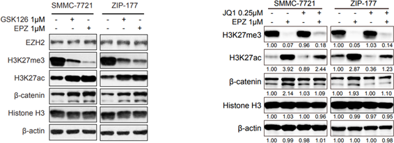
(+)-JQ-1 purchased from MedChemExpress. Usage Cited in: Cell. 2018 Sep 20;175(1):186-199.e19. [Abstract]
Cells are treated with EPZ-6438 (1 μM) or GSK126 (1 μM) for 6 days. Protein levels are analyzed by immunoblotting. Cells are treated with EPZ-6438 (1 μM), JQ1 (0.25 μM) alone or combination for 6 days. Protein levels are analyzed by immunoblotting.
-
Nat Biotechnol
CRISPR live-cell imaging reveals chromatin dynamics and enhancer interactions at multiple non-repetitive loci. [Abstract]2025 Nov 6. PMID: 41199023 -
Mol Cancer
CDK9 inhibition induces epigenetic reprogramming revealing strategies to circumvent resistance in lymphoma. [Abstract]2023 Mar 30;22(1):64. PMID: 36998071 -
Mol Cancer
Loss of FBXW7-mediated degradation of BRAF elicits resistance to BET inhibitors in adult T cell leukemia cells. [Abstract]2020 Sep 9;19(1):139. PMID: 32907612 -
Nat Genet
2024 Jun;56(6):1213-1224. PMID: 38802567 -
Nat Genet
MYC activity at enhancers drives prognostic transcriptional programs through an epigenetic switch. [Abstract]2024 Apr;56(4):663-674. PMID: 38454021 -
Nat Genet
2022 Apr;54(4):481-491. PMID: 35410381 -
Nat Cancer
TNF-α-producing macrophages determine subtype identity and prognosis via AP1 enhancer reprogramming in pancreatic cancer. [Abstract]2021 Nov;2(11):1185-1203. PMID: 35122059 -
Cell Res
Dynamic nucleosome organization after fertilization reveals regulatory factors for mouse zygotic genome activation. [Abstract]2022 Sep;32(9):801-813. PMID: 35428874 -
Blood
Oncogenic role of the SOX9-DHCR24-cholesterol biosynthesis axis in IGH-BCL2+ diffuse large B-cell lymphomas. [Abstract]2022 Jan 6;139(1):73-86. PMID: 34624089 -
Nat Aging
Comparative analysis of senolytic drugs reveals mitochondrial determinants of efficacy and resistance. [Abstract]2026 Feb;6(2):316-328. PMID: 41611832 -
Nat Cell Biol
Epigenetic remodelling shapes inflammatory renal cancer and neutrophil-dependent metastasis. [Abstract]2020 Apr;22(4):465-475. PMID: 32203421 -
Mol Cell
CRL3ARMC5 ubiquitin ligase and Integrator phosphatase form parallel mechanisms to control early stages of RNA Pol II transcription. [Abstract]2024 Dec 5:S1097-2765(24)00949-3. PMID: 39667934 -
Mol Cell
IDR-targeting compounds suppress HPV genome replication via disruption of phospho-BRD4 association with DNA damage response factors. [Abstract]2024 Jan 18;84(2):202-220.e15. PMID: 38103559 -
Mol Cell
Discovery of a large-scale, cell-state-responsive allosteric switch in the 7SK RNA using DANCE-MaP. [Abstract]2022 May 5;82(9):1708-1723.e10. PMID: 35320755 -
Cancer Res
Inhibition of the FACT Complex Targets Aberrant Hedgehog Signaling and Overcomes Resistance to Smoothened Antagonists. [Abstract]2021 Jun 1;81(11):3105-3120. PMID: 33853831 -
Cancer Res
BRD4 Levels Determine the Response of Human Lung Cancer Cells to BET Degraders That Potently Induce Apoptosis through Suppression of Mcl-1. [Abstract]2020 Jun 1;80(11):2380-2393. PMID: 32156781 -
Cancer Res
2019 May 15;79(10):2761-2774. PMID: 30877105 -
Cancer Res
MDM2-Recruiting PROTAC Offers Superior, Synergistic Antiproliferative Activity via Simultaneous Degradation of BRD4 and Stabilization of p53. [Abstract]2019 Jan 1;79(1):251-262. PMID: 30385614
(+)-JQ-1 purchased from MedChemExpress. Usage Cited in: Cancer Res. 2019 Jan 1;79(1):251-262. [Abstract]
Cleavage of PARP is assessed by western blotting with the treatment of JQ 1.
-
Nat Commun
Human iPSC-based Modeling of Pulmonary Fibrosis Reveals p300/CBP Inhibition Suppresses Alveolar Transitional Cell State. [Abstract]2026 Feb 12;17(1):1214. PMID: 41680175 -
Nat Commun
2025 Mar 25;16(1):2890. PMID: 40133308 -
Nat Commun
Gene editing for latent herpes simplex virus infection reduces viral load and shedding in vivo. [Abstract]2024 May 13;15(1):4018. PMID: 38740820 -
Nat Commun
BET inhibitors drive Natural Killer activation in non-small cell lung cancer via BRD4 and SMAD3. [Abstract]2024 Mar 22;15(1):2567. PMID: 38519469 -
Nat Commun
Polycomb deficiency drives a FOXP2-high aggressive state targetable by epigenetic inhibitors. [Abstract]2023 Jan 20;14(1):336. PMID: 36670102 -
Nat Commun
2022 Aug 4;13(1):4534. PMID: 35927228 -
Nat Commun
Chromatin profiling in human neurons reveals aberrant roles for histone acetylation and BET family proteins in schizophrenia. [Abstract]2022 Apr 22;13(1):2195. PMID: 35459277 -
Nat Commun
Stromal induction of BRD4 phosphorylation Results in Chromatin Remodeling and BET inhibitor Resistance in Colorectal Cancer. [Abstract]2021 Jul 21;12(1):4441. PMID: 34290255 -
Nat Commun
Spermine synthase and MYC cooperate to maintain colorectal cancer cell survival by repressing Bim expression. [Abstract]2020 Jun 26;11(1):3243. PMID: 32591507 -
Nat Commun
A BET family protein degrader provokes senolysis by targeting NHEJ and autophagy in senescent cells. [Abstract]2020 Apr 22;11(1):1935. PMID: 32321921 -
Nat Commun
2020 Apr 14;11(1):1833. PMID: 32286255 -
Nat Commun
TBX2 is a neuroblastoma core regulatory circuitry component enhancing MYCN/FOXM1 reactivation of DREAM targets. [Abstract]2018 Nov 19;9(1):4866. PMID: 30451831 -
Cell Death Differ
BAP18 facilitates CTCF-mediated chromatin accessible to regulate enhancer activity in breast cancer. [Abstract]2023 May;30(5):1260-1278. PMID: 36828916 -
Acta Pharm Sin B
THBru ameliorates atherosclerosis by inhibiting endothelial ferroptosis via regulation of the super-enhancer-associated ABCC1. [Abstract]2025 Dec;15(12):6461-6477. PMID: 41477343 -
Adv Sci (Weinh)
Targeting PRKCN, an Essential Driver Orchestrating mTOR-IRF4 Axis Independently of Kinase Activity, in Multiple Myeloma. [Abstract]2026 Apr;13(21):e18975. PMID: 41655233 -
Adv Sci (Weinh)
A Selective Small-Molecule c-Myc Degrader Potently Regresses Lethal c-Myc Overexpressing Tumors. [Abstract]2022 Mar;9(8):e2104344. PMID: 35048559 -
Nat Chem Biol
2024 Dec;20(12):1640-1649. PMID: 39075252 -
Nat Chem Biol
2016 Jul;12(7):504-10. PMID: 27159579 -
J Clin Invest
ZMYND8 protects breast cancer stem cells against oxidative stress and ferroptosis through activation of NRF2. [Abstract]2024 Jan 23;134(6):e171166. PMID: 38488001 -
J Clin Invest
Tendon-derived cathepsin K-expressing progenitor cells activate Hedgehog signaling to drive heterotopic ossification. [Abstract]2020 Dec 1;130(12):6354-6365. PMID: 32853181 -
Leukemia
Targeting BET proteins improves the therapeutic efficacy of BCL-2 inhibition in T-cell acute lymphoblastic leukemia. [Abstract]2017 Oct;31(10):2037-2047. PMID: 28074072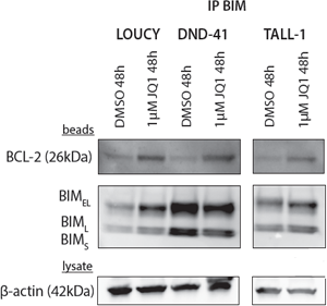
(+)-JQ-1 purchased from MedChemExpress. Usage Cited in: Leukemia. 2017 Oct;31(10):2037-2047. [Abstract]
Immunoprecipitation with anti-BIM antibody illustrates the increased binding of BIM to BCL-2 after JQ1 treatment.
-
Theranostics
2024 Jan 1;14(2):593-607. PMID: 38169595 -
-
Nucleic Acids Res
Quantitative limits of host-driven HIV transcription and host gene control by the viral transactivator Tat. [Abstract]2026 Jun 22;54(12):gkag631. PMID: 42328785 -
J Exp Clin Cancer Res
A novel dual epigenetic approach targeting BET proteins and HDACs in Group 3 (MYC-driven) Medulloblastoma. [Abstract]2022 Nov 11;41(1):321. PMID: 36357906
(+)-JQ-1 purchased from MedChemExpress. Usage Cited in: J Exp Clin Cancer Res. 2022 Nov 11;41(1):321. [Abstract]
The expression of BRD4 and MYC proteins are each downregulated by JQ1 (0.5 µM; 24 h) or panobinostat (PAN; 10 nM; 24 h) alone, and more profoundly by the combination of these two inhibitors in in MB HD-MB03 and D-283 cells.
-
J Exp Clin Cancer Res
Dissecting super-enhancer driven transcriptional dependencies reveals novel therapeutic strategies and targets for group 3 subtype medulloblastoma. [Abstract]2022 Oct 22;41(1):311. PMID: 36273157 -
Sci Adv
HERVH-derived eRNA activates a super-enhancer-driven ALDH1A3/SAT1 axis to promote ferroptosis escape and pancreatic cancer development. [Abstract]2025 Dec 12;11(50):eaea9074. PMID: 41370380 -
Sci Adv
NLRP3 inflammasome-driven hemophagocytic lymphohistiocytosis occurs independent of IL-1β and IL-18 and is targetable by BET inhibitors. [Abstract]2025 Jul 11;11(28):eadv0079. PMID: 40632844 -
Sci Adv
Identification of the NRF2 transcriptional network as a therapeutic target for trigeminal neuropathic pain. [Abstract]2022 Aug 5;8(31):eabo5633. PMID: 35921423 -
Sci Adv
Hypoxia deactivates epigenetic feedbacks via enzyme-derived clicking proteolysis-targeting chimeras. [Abstract]2022 Dec 14;8(50):eabq2216. PMID: 36516252 -
Sci Adv
2021 Mar 5;7(10):eabe7853. PMID: 33674313 -
J Biomed Sci
Metformin sensitizes triple-negative breast cancer to histone deacetylase inhibitors by targeting FGFR4. [Abstract]2025 Mar 17;32(1):36. PMID: 40091020 -
Metabolism
2016 Oct;65(10):1478-88. PMID: 27621183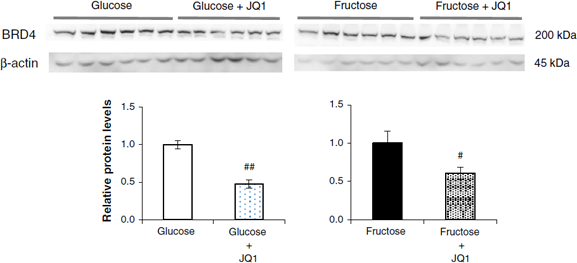
(+)-JQ-1 purchased from MedChemExpress. Usage Cited in: Metabolism. 2016 Oct;65(10):1478-88. [Abstract]
The BRD4 protein level in the liver is significantly suppressed by force-feeding fructose or glucose with (+)-JQ1. Protein levels of Brd4 in the liver of mice force-fed with glucose or fructose, with or without (+)-JQ1. Quantification of protein levels is performed by normalization against the level of β-actin (n = 6).
-
J Control Release
The HSP90-dependent bioorthogonal PROTAC prodrug system enables tumor-selective and enhanced protein degradation. [Abstract]2026 May 10:393:114828. PMID: 41839264 -
J Control Release
ApoE mimetic peptide targeted nanoparticles carrying a BRD4 inhibitor for treating Medulloblastoma in mice. [Abstract]2020 Jul 10;323:463-474. PMID: 32380205 -
J Hazard Mater
Epigenetic regulator BRD4 is involved in cadmium-induced acute kidney injury via contributing to lysosomal dysfunction, autophagy blockade and oxidative stress. [Abstract]2022 Feb 5;423(Pt A):127110. PMID: 34523489 -
Genome Med
ASXL3 bridges BRD4 to BAP1 complex and governs enhancer activity in small cell lung cancer. [Abstract]2020 Jul 15;12(1):63. PMID: 32669118 -
EBioMedicine
The BET inhibitor JQ1 attenuates double-strand break repair and sensitizes models of pancreatic ductal adenocarcinoma to PARP inhibitors. [Abstract]2019 Jun:44:419-430. PMID: 31126889
(+)-JQ-1 purchased from MedChemExpress. Usage Cited in: EBioMedicine. 2019 Jun:44:419-430. [Abstract]
Panc1 or MiaPaCa2 cells are exposed to JQ1 (0.5, 1, 5, or 10 μM) for 48 h, and immunoblotted for γH2AX.
(+)-JQ-1 purchased from MedChemExpress. Usage Cited in: EBioMedicine. 2019 Jun:44:419-430. [Abstract]
Immunoblots (IB) are done to detect γH2AX and DNA repair proteins, Ku80 and RAD51 using UAB-PA4 or UAB-PA16 tumors harvested from mice 24 h following final treatment. Quantitation by densitometry of results is shown below each IB image.
-
Cell Rep Med
2025 Dec 16;6(12):102467. PMID: 41308642 -
Cell Rep Med
DDX5 super-enhancer promotes vasculogenic mimicry formation and metastasis in nasopharyngeal carcinoma by enhancing ADAM10 transcription. [Abstract]2025 Jun 17;6(6):102146. PMID: 40412383 -
Clin Cancer Res
2019 Jun 1;25(11):3404-3416. PMID: 30796033 -
Cancer Lett
The Megacomplex protects ER-alpha from degradation by Fulvestrant in epithelial ovarian cancer. [Abstract]2024 Jul 22:217129. PMID: 39048045 -
Cancer Lett
BET inhibition decreases HMGCS2 and sensitizes resistant pancreatic tumors to gemcitabine. [Abstract]2024 Jun 28:592:216919. PMID: 38704133 -
Cancer Lett
Super-enhancer mediated upregulation of MYEOV suppresses ferroptosis in lung adenocarcinoma. [Abstract]2024 May 1:589:216811. PMID: 38490328 -
Cancer Lett
Targeting the EphB4 receptor tyrosine kinase sensitizes HER2-positive breast cancer cells to Lapatinib. [Abstract]2020 Apr 10;475:53-64. PMID: 32006616 -
Cancer Lett
The combination of BET and PARP inhibitors is synergistic in models of cholangiocarcinoma. [Abstract]2020 Jan 1;468:48-58. PMID: 31605774 -
Cancer Lett
Co-inhibition of BET and proteasome enhances ER stress and Bim-dependent apoptosis with augmented cancer therapeutic efficacy. [Abstract]2018 Oct 28:435:44-54. PMID: 30059709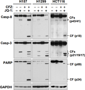
(+)-JQ-1 purchased from MedChemExpress. Usage Cited in: Cancer Lett. 2018 Oct 28:435:44-54. [Abstract]
H157, H1299, and HCT116 cells are harvested for preparation of whole-cell protein lysates and subsequent Western blotting to detect the indicated proteins.
-
Cancer Lett
Cynanbungeigenin C and D, a pair of novel epimers from Cynanchum bungei, suppress hedgehog pathway-dependent medulloblastoma by blocking signaling at the level of Gli. [Abstract]2018 Apr 28:420:195-207. PMID: 29425683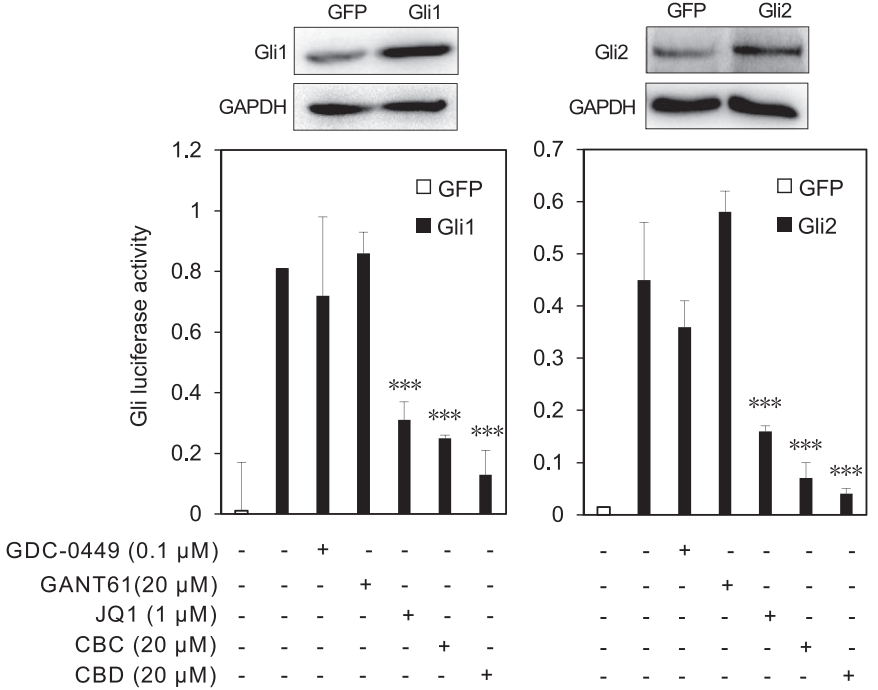
(+)-JQ-1 purchased from MedChemExpress. Usage Cited in: Cancer Lett. 2018 Apr 28:420:195-207. [Abstract]
Shh-Light 2 cells are transfected with Gli1 or Gli2 plasmids and the expression of proteins are analyzed by Western blot. Positive control JQ1, CBC and CBD inhibit Gli1 and Gli2 overexpression induced Gli luciferase activity, GDC-0499 and GANT61 have no effects.
-
Cancer Lett
Targeting c-Myc: JQ1 as a promising option for c-Myc-amplified esophageal squamous cell carcinoma. [Abstract]2018 Apr 10:419:64-74. PMID: 29366803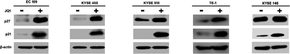
(+)-JQ-1 purchased from MedChemExpress. Usage Cited in: Cancer Lett. 2018 Apr 10:419:64-74. [Abstract]
ESCC cell lines are treated with 500 nM JQ1 for 48 h, whole cell lysates are analyzed by western blotting. The expressions of p21 and p27 are further upregulated by JQ1 concomitant with cell cycle arrest at G1 phase.
-
Cancer Lett
Targeting of super-enhancers and mutant BRAF can suppress growth of BRAF-mutant colon cancer cells via repression of MAPK signaling pathway. [Abstract]2017 Aug 28:402:100-109. PMID: 28576751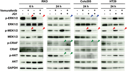
(+)-JQ-1 purchased from MedChemExpress. Usage Cited in: Cancer Lett. 2017 Aug 28:402:100-109. [Abstract]
Expression and activation statuses of signal proteins in three BRAFV600E-mutant colon cancer cell lines treated with either RG7204, JQ1, or their combination. Phosphorylation of CRAF and AKT is suppressed by JQ1 in RKO cells and possibly in Colo205 cells (green arrow heads).
-
Int J Biol Sci
Inhibition of super enhancer downregulates the expression of KLF5 in basal-like breast cancers. [Abstract]2019 Jun 10;15(8):1733-1742. PMID: 31360115 -
Acta Biomater
Engineering a multilayered 3D stromal barrier model for quantitative analysis of T cell infiltration and cytotoxicity. [Abstract]2025 Sep 10:S1742-7061(25)00677-4. PMID: 40939760 -
Biomater Res
Tryptamine-Functionalized Lipid Nanocarriers Co-delivering SMO/BRD4 Inhibitors for Synergistic Medulloblastoma Therapy. [Abstract]2025 Aug 8:29:0237. PMID: 40785845 -
Acta Biomater
2021 Oct 15:134:633-648. PMID: 34329780 -
Cell Death Dis
Sequential targeting of YAP1 and p21 enhances the elimination of senescent cells induced by the BET inhibitor JQ1. [Abstract]2021 Jan 25;12(1):121. PMID: 33495462 -
Cell Death Dis
Ferritinophagy is required for the induction of ferroptosis by the bromodomain protein BRD4 inhibitor (+)-JQ1 in cancer cells. [Abstract]2019 Apr 15;10(5):331. PMID: 30988278 -
Cell Death Dis
Targeting EHMT2 reverses EGFR-TKI resistance in NSCLC by epigenetically regulating the PTEN/AKT signaling pathway. [Abstract]2018 Jan 26;9(2):129. PMID: 29374157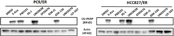
(+)-JQ-1 purchased from MedChemExpress. Usage Cited in: Cell Death Dis. 2018 Jan 26;9(2):129. [Abstract]
The effects of treatment with the indicated epigenetic inhibitors on cleaved PARP (Clv-PARP) expression in both PC9/ER and HCC827/ER cells. β-actin is used as a loading control.
-
Proc Natl Acad Sci U S A
CDK7 inhibition suppresses aberrant hedgehog pathway and overcomes resistance to smoothened antagonists. [Abstract]2019 Jun 25;116(26):12986-12995. PMID: 31182587 -
Proc Natl Acad Sci U S A
2019 Feb 19;116(8):2961-2966. PMID: 30718431 -
Cell Genom
Pooled endogenous protein tagging and recruitment for systematic profiling of protein function. [Abstract]2024 Sep 4:100651. PMID: 39255790 -
Acta Pharmacol Sin
DNMT inhibition epigenetically restores the cGAS-STING pathway and activates RIG-I/MDA5-MAVS to enhance antitumor immunity. [Abstract]2025 Aug 19. PMID: 40830678 -
Acta Pharmacol Sin
2022 Feb;43(2):457-469. PMID: 33850273 -
Acta Pharmacol Sin
The BET bromodomain inhibitor apabetalone induces apoptosis of latent HIV-1 reservoir cells following viral reactivation. [Abstract]2019 Jan;40(1):98-110. PMID: 29789664 -
Phytomedicine
The total xanthones from Gentianella acuta alleviate acute myocardial infarction by targeting BRD4-mediated cardiomyocyte pyroptosis and inflammation. [Abstract]2025 Aug 13:147:157156. PMID: 40845590 -
Phytomedicine
Wikstrol B reactivates latent human immunodeficiency virus (HIV-1) via the nuclear factor-κB (NF-κB) pathway. [Abstract]2025 Jun:141:156667. PMID: 40233507 -
EMBO Mol Med
Interactions between BRD4S, LOXL2, and MED1 drive cell cycle transcription in triple-negative breast cancer. [Abstract]2023 Dec 7;15(12):e18459. PMID: 37937685 -
Phytomedicine
Antileukemic effect of caffeic acid 3,4-dihydroxyphenetyl ester. Evidences for its mechanisms of action. [Abstract]2021 Jan;80:153383. PMID: 33091855 -
EMBO Mol Med
A genome-wide RNAi screen reveals essential therapeutic targets of breast cancer stem cells. [Abstract]2019 Oct;11(10):e9930. PMID: 31476112 -
EMBO Mol Med
An FGFR3/MYC positive feedback loop provides new opportunities for targeted therapies in bladder cancers. [Abstract]2018 Apr;10(4). pii: e8163. PMID: 29463565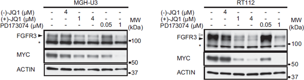
(+)-JQ-1 purchased from MedChemExpress. Usage Cited in: EMBO Mol Med. 2018 Apr;10(4). pii: e8163. [Abstract]
Western blot analysis of MYC and FGFR3 expression in lysates from MGH-U3 and RT112 cells treated with (+)-JQ1 (1 or 4 μM) for 48 h. Anti-actin antibody is used as a loading control. Pan-FGFR inhibitor, PD173074 (50 nM and 1 μM), and inactive enantiomer (-)-JQ1 (4 μM) are used as controls.
-
NPJ Precis Oncol
Targeting ACE2-BRD4 crosstalk in colorectal cancer and the deregulation of DNA repair and apoptosis. [Abstract]2023 Feb 18;7(1):20. PMID: 36801948 -
Neoplasia
Bromodomain inhibitor treatment leads to overexpression of multiple kinases in cancer cells. [Abstract]2024 Nov:57:101046. PMID: 39241280 -
Neoplasia
GRP78 inhibitor YUM70 upregulates 4E-BP1 and suppresses c-MYC expression and viability of oncogenic c-MYC tumors. [Abstract]2024 Jul 10:55:101020. PMID: 38991376 -
-
Br J Pharmacol
JQ1, a bromodomain inhibitor, suppresses Th17 effectors by blocking p300-mediated acetylation of RORγt. [Abstract]2020 Jul;177(13):2959-2973. PMID: 32060899 -
Genes Dev
Glioblastoma cellular cross-talk converges on NF-κB to attenuate EGFR inhibitor sensitivity. [Abstract]2017 Jun 15;31(12):1212-1227. PMID: 28724615 -
J Transl Med
Vascular restenosis following paclitaxel-coated balloon therapy is attributable to NLRP3 activation and LIN9 upregulation. [Abstract]2024 Sep 27;22(1):871. PMID: 39334121 -
J Transl Med
The BRD4 inhibitor JQ1 suppresses tumor growth by reducing c-Myc expression in endometrial cancer. [Abstract]2022 Jul 28;20(1):336. PMID: 35902869 -
Biomed Pharmacother
Inhibitory effects of JQ1 on listeria monocytogenes-induced acute liver injury by blocking BRD4/RIPK1 axis. [Abstract]2020 May;125:109818. PMID: 32106368 -
Oncogene
Therapeutic targeting the oncogenic driver EWSR1::FLI1 in Ewing sarcoma through inhibition of the FACT complex. [Abstract]2023 Jan;42(1):11-25. PMID: 36357572 -
Stem Cell Res Ther
Pretreatment of nucleus pulposus mesenchymal stem cells with appropriate concentration of H2O2 enhances their ability to treat intervertebral disc degeneration. [Abstract]2022 Jul 26;13(1):340. PMID: 35883157 -
Oncogene
2021 Apr;40(15):2711-2724. PMID: 33712705 -
Oncogene
2018 Aug;37(33):4611-4625. PMID: 29760405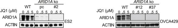
(+)-JQ-1 purchased from MedChemExpress. Usage Cited in: Oncogene. 2018 Aug;37(33):4611-4625. [Abstract]
ARID1A Western blot analysis of ARID1A protein levels in the ES2 polyclonal ARID1A knockout clone and ES2/OVCA429 monoclonal ARID1A knockout clones.
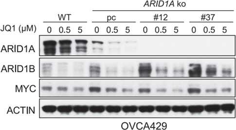
(+)-JQ-1 purchased from MedChemExpress. Usage Cited in: Oncogene. 2018 Aug;37(33):4611-4625. [Abstract]
The OVCA429 cells are subjected to Western blot analysis for the indicated proteins. ACTIN servea as a loading control.
-
Cell Chem Biol
2025 Jul 17;32(7):969-981.e7. PMID: 40614719 -
Cell Death Discov
The epigenetic regulators EP300/CREBBP represent promising therapeutic targets in MLL-rearranged acute myeloid leukemia. [Abstract]2024 May 1;10(1):206. PMID: 38693103 -
Cell Death Discov
MYC inhibition reprograms tumor immune microenvironment by recruiting T lymphocytes and activating the CD40/CD40L system in osteosarcoma. [Abstract]2022 Mar 15;8(1):117. PMID: 35292660 -
Cell Rep
SUCLG1 deficiency-induced histone succinylation impairs oncogene expression in acute myeloid leukemia. [Abstract]2025 Aug 12;44(8):116147. PMID: 40811057 -
Cell Rep
Citrate metabolism controls the senescent microenvironment via the remodeling of pro-inflammatory enhancers. [Abstract]2024 Jul 17:114496. PMID: 39043191 -
Cell Rep
Elevating PLK1 overcomes BETi resistance in prostate cancer via triggering BRD4 phosphorylation-dependent degradation in mitosis. [Abstract]2024 Jul 3;43(7):114431. PMID: 38968071 -
Cell Rep
Identification of WNK1 as a therapeutic target to suppress IgH/MYC expression in multiple myeloma. [Abstract]2024 May 8;43(5):114211. PMID: 38722741 -
Cell Rep
Temporal Analysis of Brd4 Displacement in the Control of B Cell Survival, Proliferation, and Differentiation. [Abstract]2020 Oct 20;33(3):108290. PMID: 33086063 -
J Med Chem
Discovery of a Novel Benzimidazole Derivative Targeting Histone Deacetylase to Induce Ferroptosis and Trigger Immunogenic Cell Death. [Abstract]2024 Sep 12;67(17):15098-15117. PMID: 39145486 -
J Med Chem
Peptide Inhibitor Targeting the Extraterminal Domain in BRD4 Potently Suppresses Breast Cancer Both In Vitro and In Vivo. [Abstract]2024 Apr 25;67(8):6658-6672. PMID: 38569135 -
Clin Transl Med
METTL3-stabilized super enhancers-lncRNA SUCLG2-AS1 mediates the formation of a long-range chromatin loop between enhancers and promoters of SOX2 in metastasis and radiosensitivity of nasopharyngeal carcinoma. [Abstract]2023 Sep;13(9):e1361. PMID: 37658588 -
J Med Chem
Novel Pyrrolopyridone Bromodomain and Extra-Terminal Motif (BET) Inhibitors Effective in Endocrine-Resistant ER+ Breast Cancer with Acquired Resistance to Fulvestrant and Palbociclib. [Abstract]2020 Jul 9;63(13):7186-7210. PMID: 32453591 -
J Med Chem
Impact of Target Warhead and Linkage Vector on Inducing Protein Degradation: Comparison of Bromodomain and Extra-Terminal (BET) Degraders Derived from Triazolodiazepine (JQ1) and Tetrahydroquinoline (I-BET726) BET Inhibitor Scaffolds. [Abstract]2018 Jan 25;61(2):504-513. PMID: 28595007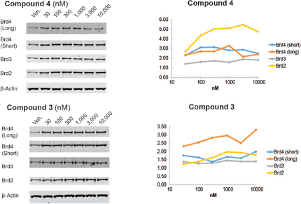
(+)-JQ-1 purchased from MedChemExpress. Usage Cited in: J Med Chem. 2018 Jan 25;61(2):504-513. [Abstract]
Protein degradation profile of VHL-based BET degraders. Sub-confluent HeLa cells are treated for 24 h with varying concentration of test compounds JQ1(Compound 3), I-BET726 (Compound 4). Protein extracts are separated by SDS-PAGE and then analyzed by Western blot. Proteins Brd4, Brd3, Brd2 and β-actin are probed for JQ1and I-BET726 with specific antibodies.
-
Anal Chem
Near-Infrared Triggered Self-Accelerating Nanozyme Camouflaged with a Cancer Cell Membrane for Precise Targeted Imaging and Enhanced Cancer Immunotherapy. [Abstract]2023 Sep 12;95(36):13575-13585. PMID: 37649359 -
Int J Nanomedicine
2024 Jul 3:19:6717-6730. PMID: 38979530 -
Elife
FAK activity sustains intrinsic and acquired ovarian cancer resistance to platinum chemotherapy. [Abstract]2019 Sep 3:8:e47327. PMID: 31478830 -
Oncoimmunology
2019 Mar 1;8(5):e1581529. PMID: 31069140 -
J Agric Food Chem
Super-Enhancer-Associated circ RHOQ Regulates Intramuscular Fat Deposition by Modulating the miR-5093/ IRF5 Axis. [Abstract]2025 Dec 3;73(48):30904-30921. PMID: 41252683 -
EMBO Rep
2024 Dec;25(12):5352-5382. PMID: 39420119 -
EMBO Rep
NONO enhances mRNA processing of super-enhancer-associated GATA2 and HAND2 genes in neuroblastoma. [Abstract]2023 Feb 6;24(2):e54977. PMID: 36416237 -
J Agric Food Chem
Resveratrol Reactivates Latent HIV through Increasing Histone Acetylation and Activating Heat Shock Factor 1. [Abstract]2017 Jun 7;65(22):4384-4394. PMID: 28471170 -
JCI Insight
2026 Feb 3:e194683. PMID: 41632526 -
-
JCI Insight
Inhibition of bromodomain extraterminal histone readers alleviates skin fibrosis in experimental models of scleroderma. [Abstract]2022 May 9;7(9):e150871. PMID: 35349485 -
Ecotoxicol Environ Saf
The epigenetic regulator BRD4 is involved in cadmium-triggered inflammatory response in rat kidney. [Abstract]2021 Aug 13:224:112620. PMID: 34392152 -
JCI Insight
BRD4 inhibition and FXR activation, individually beneficial in cholestasis, are antagonistic in combination. [Abstract]2020 Dec 8;6(1):e141640. PMID: 33290278 -
Cancer Cell Int
Trichostatin A downregulates bromodomain and extra-terminal proteins to suppress osimertinib resistant non-small cell lung carcinoma. [Abstract]2021 Apr 15;21(1):216. PMID: 33858423 -
Eur J Med Chem
Design, synthesis, and biological evaluation of novel 4,4'-bipyridine derivatives acting as CDK9-Cyclin T1 protein-protein interaction inhibitors against triple-negative breast cancer. [Abstract]2023 Dec 5:261:115858. PMID: 37837671 -
Front Immunol
BRD4 Inhibition Attenuates Inflammatory Pain by Ameliorating NLRP3 Inflammasome-Induced Pyroptosis. [Abstract]2022 Jan 26;13:837977. PMID: 35154163 -
Front Immunol
JQ1 as a BRD4 Inhibitor Blocks Inflammatory Pyroptosis-Related Acute Colon Injury Induced by LPS. [Abstract]2021 Feb 18:12:609319. PMID: 33679744 -
Biochem Pharmacol
Discovery of Bi-magnolignan as a novel BRD4 inhibitor inducing apoptosis and DNA damage for cancer therapy. [Abstract]2025 May:235:116843. PMID: 40024351 -
Biochem Pharmacol
Up-regulation of BRD4 contributes to gestational diabetes mellitus-induced cardiac hypertrophy in offspring by promoting mitochondria dysfunction in sex-independent manner. [Abstract]2024 Jun 27:116387. PMID: 38944397 -
Biochem Pharmacol
A synthetic resveratrol analog termed Q205 reactivates latent HIV-1 through activation of P-TEFb. [Abstract]2022 Mar:197:114901. PMID: 34971588 -
Biochem Pharmacol
Novel bivalent BET inhibitor N2817 exhibits potent anticancer activity and inhibits TAF1. [Abstract]2021 Mar:185:114435. PMID: 33539817 -
Biochem Pharmacol
Macrophages confer resistance to BET inhibition in triple-negative breast cancer by upregulating IKBKE. [Abstract]2020 Oct;180:114126. PMID: 32603665 -
Biochem Pharmacol
Resveratrol promotes HSV-2 replication by increasing histone acetylation and activating NF-κB. [Abstract]2020 Jan;171:113691. PMID: 31704236 -
Biochem Pharmacol
A novel bromodomain inhibitor, CPI-203, serves as an HIV-1 latency-reversing agent by activating positive transcription elongation factor b. [Abstract]2019 Jun:164:237-251. PMID: 30991051 -
Biochem Pharmacol
PR-957, a selective immunoproteasome inhibitor, reactivates latent HIV-1 through p-TEFb activation mediated by HSF-1. [Abstract]2018 Oct:156:511-523. PMID: 30170098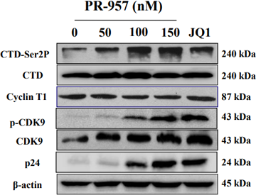
(+)-JQ-1 purchased from MedChemExpress. Usage Cited in: Biochem Pharmacol. 2018 Oct:156:511-523. [Abstract]
Western blot detection of the p-TEFb component CDK9, Cyclin T1 and CDK9 phosphorylation on Thr186, as well as its downstream RNA poly II CTD and phosphate CTD after J-Lat 10.6 cells are treated with PR-957 in dose-dependent manner.
-
ACS Biomater Sci Eng
Development of a Vessel-on-a-Chip as a Viral Infection Model and Antiviral Drug Screening Platform with Viral Mimics. [Abstract]2025 Jul 14;11(7):4381-4393. PMID: 40581966 -
Mol Cancer Ther
A Combination of BRD4 and HDAC3 Inhibitors Synergistically Suppresses Glioma Stem Cell Growth by Blocking GLI1/IL6/STAT3 Signaling Axis. [Abstract]2020 Dec;19(12):2542-2553. PMID: 32999044 -
Mol Cancer Ther
A Novel Combination Approach Targeting an Enhanced Protein Synthesis Pathway in MYC-driven (Group 3) Medulloblastoma. [Abstract]2020 Jun;19(6):1351-1362. PMID: 32371591 -
Genome Res
Altered hydroxymethylation is seen at regulatory regions in pancreatic cancer and regulates oncogenic pathways. [Abstract]2017 Nov;27(11):1830-1842. PMID: 28986391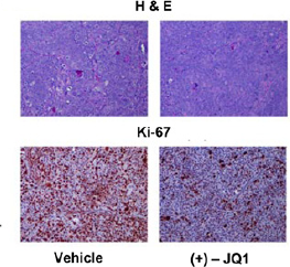
(+)-JQ-1 purchased from MedChemExpress. Usage Cited in: Genome Res. 2017 Nov;27(11):1830-1842. [Abstract]
Ki67 immunohistochemistry and H&E stained sections from representative xenograft per treatment arm. A marked decrease in the proliferation marker Ki67 is observed in xenografts treated with JQ1.
-
Mol Cancer Ther
Dual Targeting of Bromodomain and Extraterminal Domain Proteins, and WNT or MAPK Signaling, Inhibits c-MYC Expression and Proliferation of Colorectal Cancer Cells. [Abstract]2016 Jun;15(6):1217-26. PMID: 26983878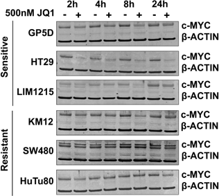
(+)-JQ-1 purchased from MedChemExpress. Usage Cited in: Mol Cancer Ther. 2016 Jun;15(6):1217-26. [Abstract]
JQ1-mediated c-MYC repression correlates with growth inhibition. BETi preferentially inhibit c-MYC protein expression in sensitive cell lines. JQ1-sensitive GP5D, HT29 and LIM1215 cells and JQ1-resistant KM12, SW480 and HuTu80 cells where treated with JQ1 (500 nM) for 2-24 hours and c-MYC protein expression determined by western blot.
-
-
Cancer Drug Resist
The BET inhibitor sensitivity is associated with the expression level of CDC25B in pancreatic cancer models. [Abstract]2024 Oct 18:7:40. PMID: 39534870 -
Drug Des Devel Ther
(+)-JQ1 Upregulates SIRT3 to Suppress cGAS/STING Pathway-Mediated Neuronal Inflammation and Ferroptosis After Hypoxic-Ischemic Encephalopathy. [Abstract]2026 Feb 25:20:578815. PMID: 41778146 -
Commun Biol
Pigment epithelium-derived factor promotes peritoneal dissemination of ovarian cancer through induction of immunosuppressive macrophages. [Abstract]2022 Sep 2;5(1):904. PMID: 36056141 -
J Clin Endocrinol Metab
Transcription Factor ASCL1 Acts as a Novel Potential Therapeutic Target for the Treatment of the Cushing's Disease. [Abstract]2022 Jul 14;107(8):2296-2306. PMID: 35521682 -
Commun Biol
Three-dimensional CRISPR screening reveals epigenetic interaction with anti-angiogenic therapy. [Abstract]2021 Jul 15;4(1):878. PMID: 34267311 -
Respir Res
2023 Jan 31;24(1):37. PMID: 36721187 -
Int J Mol Sci
The Chromatin Remodeler Chd8 Regulates Hematopoietic Stem and Progenitor Cell Survival and Differentiation During Zebrafish Embryogenesis. [Abstract]2025 Nov 6;26(21):10805. PMID: 41226840 -
Int J Mol Sci
BRD4 Mediates Transforming Growth Factor-β-Induced Smooth Muscle Cell Differentiation from Mesenchymal Progenitor Cells. [Abstract]2025 Aug 21;26(16):8074. PMID: 40869394 -
J Nutr Biochem
Identification of potential pathogenic hepatic super-enhancers regulatory network in high-fat diet induced hyperlipidemia. [Abstract]2024 Apr:126:109584. PMID: 38242178 -
Int J Mol Sci
Glucocorticoid Receptor and β-Catenin Interact in Prostate Cancer Cells and Their Co-Inhibition Attenuates Tumorsphere Formation, Stemness, and Docetaxel Resistance. [Abstract]2023 Apr 12;24(8):7130. PMID: 37108293 -
Int J Mol Sci
2022 Dec 2;23(23):15163. PMID: 36499487 -
PLoS Pathog
BRD4 inhibition exerts anti-viral activity through DNA damage-dependent innate immune responses. [Abstract]2020 Mar 24;16(3):e1008429. PMID: 32208449 -
Int J Oncol
Bromodomain inhibitor jq1 induces cell cycle arrest and apoptosis of glioma stem cells through the VEGF/PI3K/AKT signaling pathway. [Abstract]2019 Oct;55(4):879-895. PMID: 31485609 -
PLoS Pathog
An Epigenetic Compound Library Screen Identifies BET Inhibitors That Promote HSV-1 and -2 Replication by Bridging P-TEFb to Viral Gene Promoters through BRD4. [Abstract]2016 Oct 20;12(10):e1005950. PMID: 27764245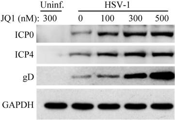
(+)-JQ-1 purchased from MedChemExpress. Usage Cited in: PLoS Pathog. 2016 Oct 20;12(10):e1005950. [Abstract]
Dose effect of JQ1 on HSV infection. Vero cells are treated with JQ1 at concentrations as indicated. HSV-1 or HSV-2 at 1 MOI are used. The samples are used for protein expression analysis by western blot.
-
Pharmaceuticals (Basel)
Bromodomain and Extra-Terminal Inhibitor BMS-986158 Reverses Latent HIV-1 Infection In Vitro and Ex Vivo by Increasing CDK9 Phosphorylation and Recruitment. [Abstract]2022 Mar 10;15(3):338. PMID: 35337136 -
Pharmaceuticals (Basel)
2021 Dec 15;14(12):1308. PMID: 34959707 -
Front Cell Infect Microbiol
CPI-637 as a Potential Bifunctional Latency-Reversing Agent That Targets Both the BRD4 and TIP60 Proteins. [Abstract]2021 Jul 19:11:686035. PMID: 34350133 -
Int Immunopharmacol
USP22/BRD4 mediated hedgehog pathway activation contributes to airway remodeling in asthma. [Abstract]2025 Mar 24:153:114538. PMID: 40132456 -
-
Eur J Pharmacol
2020 Oct 15;885:173500. PMID: 32861663 -
ACS Appl Bio Mater
Carrier-Free Small-Molecule Drug Nanoassembly Elicits Chemoimmunotherapy via Co-inhibition of PD-L1/mTOR. [Abstract]2020 Jul 20;3(7):4543-4555. PMID: 35025453 -
Mol Cancer Res
Intron 1-Mediated Regulation of EGFR Expression in EGFR-Dependent Malignancies Is Mediated by AP-1 and BET Proteins. [Abstract]2019 Nov;17(11):2208-2220. PMID: 31444232 -
Eur J Pharmacol
BRD4 promotes the stemness of gastric cancer cells via attenuating miR-216a-3p-mediated inhibition of Wnt/β-catenin signaling. [Abstract]2019 Jun 5:852:189-197. PMID: 30876979 -
J Mol Cell Cardiol
Inhibition of BRD4 attenuates transverse aortic constriction- and TGF-β-induced endothelial-mesenchymal transition and cardiac fibrosis. [Abstract]2019 Feb:127:83-96. PMID: 30529267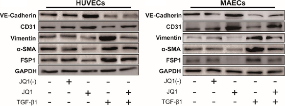
(+)-JQ-1 purchased from MedChemExpress. Usage Cited in: J Mol Cell Cardiol. 2019 Feb:127:83-96. [Abstract]
The expression of EndMT-related proteins, VE-cadherin, CD31, vimentin, α-SMA and FSP1, were compared among Ctrl, JQ1(-), JQ1, TGF-β1, and TGF-β1+JQ1 groups in both HUVECs and MAECs.
-
Cancer Biol Ther
BRD4 PROTAC degrader MZ1 exerts anticancer effects in acute myeloid leukemia by targeting c-Myc and ANP32B genes. [Abstract]2022 Dec 31;23(1):1-15. PMID: 36170346 -
Cancer Biol Ther
Targeting BRD4 proteins suppresses the growth of NSCLC through downregulation of eIF4E expression. [Abstract]2018 May 4;19(5):407-415. PMID: 29333921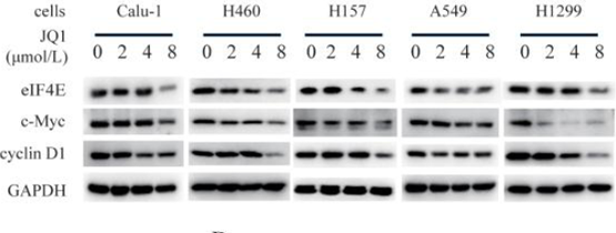
(+)-JQ-1 purchased from MedChemExpress. Usage Cited in: Cancer Biol Ther. 2018 May 4;19(5):407-415. [Abstract]
NSCLC cells are treated with 0-8 μM JQ1 for 24h, then the whole-cell lysates are prepared and subjected to western blot assay.
-
J Infect Dis
2025 Jul 16:jiaf373. PMID: 40668935 -
Antimicrob Agents Chemother
A New Small-Molecule Compound, Q308, Silences Latent HIV-1 Provirus by Suppressing Tat- and FACT-Mediated Transcription. [Abstract]2021 Nov 17;65(12):e0047021. PMID: 34491808 -
Cancers (Basel)
Enhanced Antitumoral Activity of Encapsulated BET Inhibitors When Combined with PARP Inhibitors for the Treatment of Triple-Negative Breast and Ovarian Cancers. [Abstract]2022 Sep 15;14(18):4474. PMID: 36139634 -
Transcription
Oxidative stress induces Ser 2 dephosphorylation of the RNA polymerase II CTD and premature transcription termination. [Abstract]2021 Oct;12(5):277-293. PMID: 34874799 -
Cancers (Basel)
Deacetylase Plus Bromodomain Inhibition Downregulates ERCC2 and Suppresses the Growth of Metastatic Colon Cancer Cells. [Abstract]2021 Mar 22;13(6):1438. PMID: 33809839 -
ACS Omega
XQ2, a Novel Derivative of Resveratrol, Reactivates Latent HIV‑1 via the Activation of Positive Transcription Elongation Factor B. [Abstract]2026 Feb 11;11(7):11739-11752. PMID: 41768657 -
Hum Genomics
Integrative analysis of a novel super-enhancer-associated lncRNA prognostic signature and identifying LINC00945 in aggravating glioma progression. [Abstract]2023 Mar 31;17(1):33. PMID: 37004060 -
Eur J Pharm Biopharm
Colorectal cancer inhibition by BET inhibitor JQ1 is MYC-independent and not improved by nanoencapsulation. [Abstract]2022 Feb:171:39-49. PMID: 34998911 -
Eur J Pharm Biopharm
Prevention of hepatic stellate cell activation using JQ1- and atorvastatin-loaded chitosan nanoparticles as a promising approach in therapy of liver fibrosis. [Abstract]2019 Jan:134:96-106. PMID: 30471341
(+)-JQ-1 purchased from MedChemExpress. Usage Cited in: Eur J Pharm Biopharm. 2019 Jan:134:96-106. [Abstract]
Western blot analysis of p21 and G2/M regulatory molecules, cyclin B1, CDC2, p-CDC2 (Tyr-15) and CDC25A in the treatment of NL101 and BMN673.
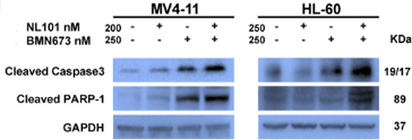
(+)-JQ-1 purchased from MedChemExpress. Usage Cited in: Eur J Pharm Biopharm. 2019 Jan:134:96-106. [Abstract]
Western blot of cleaved Caspase-3 and cleaved PARP-1 in AML cells with the treatment of NL101 and BMN673.
-
Cancer Sci
Isocitrate dehydrogenase 1 mutation sensitizes intrahepatic cholangiocarcinoma to the BET inhibitor JQ1. [Abstract]2018 Nov;109(11):3602-3610. PMID: 30156013 -
Drug Dev Res
Disruption of the FOXM1 Regulatory Region Inhibits Tumor Progression in Ovarian Cancer by CRISPR-Cas9. [Abstract]2025 Feb;86(1):e70049. PMID: 39829431 -
Stem Cell Rev Rep
Disruption of CTCF Boundary at HOXA Locus Promote BET Inhibitors' Therapeutic Sensitivity in Acute Myeloid Leukemia. [Abstract]2020 Dec;16(6):1280-1291. PMID: 33057942 -
iScience
Transcriptional synergy in human aortic endothelial cells is vulnerable to combination p300/CBP and BET bromodomain inhibition. [Abstract]2024 May 16;27(6):110011. PMID: 38868181 -
iScience
Deep genomic analysis of malignant peripheral nerve sheath tumor cell lines challenges current malignant peripheral nerve sheath tumor diagnosis. [Abstract]2023 Jan 31;26(2):106096. PMID: 36818284 -
Tissue Eng Regen Med
2021 Oct;18(5):841-850. PMID: 34003467 -
Sci Rep
BRD4 and MYB inhibition overcomes venetoclax resistance in EVI1-rearranged acute myeloid leukemia. [Abstract]2025 Oct 23;15(1):37099. PMID: 41131284 -
Sci Rep
BRD2 phase separation activates super-enhancer-driven ATG7 transcription to promote ferritinophagy in depression. [Abstract]2025 Oct 27;15(1):37493. PMID: 41145740 -
Sci Rep
TGFβ1 accelerated the progression of diabetic nephropathy via up-regulating BRD4/Notch1/YAP signaling induced fibrosis and proliferation in fibroblasts. [Abstract]2025 Jul 13;15(1):25338. PMID: 40653516 -
Sci Rep
Super-enhancer-driven SLCO4A1-AS1 is a new biomarker and a promising therapeutic target in glioblastoma. [Abstract]2025 Jan 6;15(1):954. PMID: 39762261 -
Oral Oncol
Development of a novel prognostic signature derived from super-enhancer-associated gene by machine learning in head and neck squamous cell carcinoma. [Abstract]2024 Sep 7:159:107016. PMID: 39244857 -
Sci Rep
The bromodomain inhibitor JQ1 up-regulates the long non-coding RNA MALAT1 in cultured human hepatic carcinoma cells. [Abstract]2022 May 11;12(1):7779. PMID: 35546353 -
J Biol Chem
Degradation and inhibition of epigenetic regulatory protein BRD4 exacerbate Alzheimer's disease-related neuropathology in cell models. [Abstract]2022 Apr;298(4):101794. PMID: 35248531 -
Oncol Rep
BET inhibitors combined with chemotherapy synergistically inhibit the growth of NSCLC cells. [Abstract]2021 May;45(5):70. PMID: 33760217 -
J Biol Chem
Benzolactam-related compounds promote apoptosis of HIV-infected human cells via protein kinase C-induced HIV latency reversal. [Abstract]2019 Jan 4;294(1):116-129. PMID: 30413535 -
Sci Rep
Enhancement of adenovirus infection and adenoviral vector-mediated gene delivery by bromodomain inhibitor JQ1. [Abstract]2018 Aug 1;8(1):11554. PMID: 30068949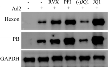
(+)-JQ-1 purchased from MedChemExpress. Usage Cited in: Sci Rep. 2018 Aug 1;8(1):11554. [Abstract]
A549 cells are mock-treated (0.1% DMSO in culture medium) or treated with RVX-208 at 500 nM, PFI-1 at 500 nM, JQ1 at 300 nM, or with 300 nM (-)-JQ1, an inactive enantiomer of JQ1. The cells are then infected with Ad2 at 1 PFU/cell for 24 h. Viral hexon and penton base (PB) protein is detected by immunoblotting analysis.
-
J Biol Chem
Bromodomain and Extraterminal Protein Inhibition Blocks Growth of Triple-negative Breast Cancers through the Suppression of Aurora Kinases. [Abstract]2016 Nov 4;291(45):23756-23768. PMID: 27650498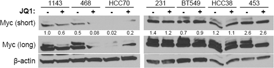
(+)-JQ-1 purchased from MedChemExpress. Usage Cited in: J Biol Chem. 2016 Nov 4;291(45):23756-23768. [Abstract]
BET inhibition blocks growth of TNBC cells without consistently downregulating MYC. Western blot analysis of MYC expression levels in TNBC cell lines treated for 24 hours with vehicle or 500 nM JQ1. Values on the western blot are relative to the vehicle-treated 1143 sample following normalization to β-actin.
-
Clin Transl Immunology
Integration of single-cell and bulk transcriptomics reveals the association of manganese metabolism-related genes with prognosis and immune infiltration in lung adenocarcinoma. [Abstract]2026 Mar 11;15(3):e70085. PMID: 42016144 -
ACS Infect Dis
Bcl-2 Antagonist Obatoclax Reactivates Latent HIV-1 via the NF-κB Pathway and Induces Latent Reservoir Cell Apoptosis in Latently Infected Cells. [Abstract]2023 Nov 10;9(11):2105-2118. PMID: 37796279 -
ACS Chem Biol
Cellular Resistance Mechanisms to Targeted Protein Degradation Converge Toward Impairment of the Engaged Ubiquitin Transfer Pathway. [Abstract]2019 Oct 18;14(10):2215-2223. PMID: 31553577 -
ACS Chem Biol
2015 Aug 21;10(8):1770-7. PMID: 26035625 -
Biochim Biophys Acta
2016 Dec;1859(12):1527-1537. PMID: 27717711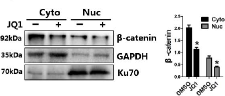
(+)-JQ-1 purchased from MedChemExpress. Usage Cited in: Biochim Biophys Acta. 2016 Dec;1859(12):1527-1537. [Abstract]
JQ1 treatment decreases β-catenin levels in HCT116 cells, but increases TAZ protein. When cells reach confluence, they are serum-starved overnight and pretreated with DMSO or JQ1 (500 nM) for 1 hour, followed by growth medium (containing 10% serum) stimulation for 24 hours.
-
J Proteome Res
Chemoproteomics Reveals FAM114A2 as a Functional Target of Wikstroelide E for Reversal of HIV-1 Latency. [Abstract]2025 Oct 23. PMID: 41131870 -
Heliyon
The landscape of super-enhancer regulates remote target gene transcription through loop domains in adipose tissue of pig. [Abstract]2024 Feb 9;10(4):e25725. PMID: 38390098 -
Heliyon
Co-overexpression of BRD4 and CDK7 promotes cell proliferation and predicts poor prognosis in HCC. [Abstract]2024 Jan 9;10(2):e24389. PMID: 38293462 -
Parasit Vectors
Comparative analysis of Microtus fortis and murine hosts reveals a correlation between BRD4 and hepatic inflammation during Schistosoma japonicum infection. [Abstract]2025 Jul 4;18(1):259. PMID: 40616163 -
Med Oncol
Combination of PSMA targeting alpha-emitting radioligand [212Pb]Pb-AB001 with BET bromodomain inhibitors in in vitro prostate cancer models. [Abstract]2025 Jul 22;42(8):362. PMID: 40694184 -
Exp Cell Res
2023 Sep 15;430(2):113718. PMID: 37468057 -
Toxicol Appl Pharmacol
Aloperine mitigates cigarette smoke-induced inflammation and pyroptosis by inhibiting the BRD4/NLRP3/GSDMD pathway in chronic obstructive pulmonary disease. [Abstract]2026 Feb:507:117704. PMID: 41485499 -
Cell Cycle
2019 Aug;18(15):1784-1797. PMID: 31223053 -
Cancer Res Commun
Targeting the Clear Cell Sarcoma Oncogenic Driver Fusion Gene EWSR1::ATF1 by HDAC Inhibition. [Abstract]2023 Jul 3;3(7):1152-1165. PMID: 37405123 -
Front Oncol
A Super-Enhancer Driven by FOSL1 Controls miR-21-5p Expression in Head and Neck Squamous Cell Carcinoma. [Abstract]2021 Apr 16:11:656628. PMID: 33937067 -
Front Neurosci
Inhibition of BET Protein Function Suppressed the Overactivation of the Canonical NF-κB Signaling Pathway in 6-OHDA-Lesioned Rat Model of Levodopa-Induced Dyskinesia. [Abstract]2022 Jun 21;16:896322. PMID: 35801173 -
Funct Integr Genomics
CRISPR-Cas9-mediated deletion enhancer of MECOM play a tumor suppressor role in ovarian cancer. [Abstract]2024 Jul 12;24(4):125. PMID: 38995475 -
Cell Biol Int
Targeting BET proteins inhibited the growth of non-small cell lung carcinoma through downregulation of Met expression. [Abstract]2023 Mar;47(3):622-633. PMID: 36448366 -
Noncoding RNA
Bromodomain and Extra-Terminal Family Proteins BRD2, BRD3, and BRD4 Contribute to H19-Dependent Transcriptional Regulation of Cell Adhesion Molecules, Modulating Metastatic Dissemination Program in Prostate Cancer. [Abstract]2025 Apr 29;11(3):33. PMID: 40407591 -
Bioorg Med Chem
Enhancing therapeutic efficacy in homologous recombination-proficient pancreatic cancer via the combination of PARP1-PROTAC and a BRD4 inhibitor. [Abstract]2024 Oct 26:115:117970. PMID: 39476572 -
Discov Oncol
2025 Oct 29;16(1):1985. PMID: 41160272 -
Discov Oncol
Acetylation increases expression, interaction with TRAPPC4 and surface localization of PD-L1. [Abstract]2023 Aug 21;14(1):152. PMID: 37603071 -
-
Am J Cancer Res
The novel BET degrader, QCA570, is highly active against the growth of human NSCLC cells and synergizes with osimertinib in suppressing osimertinib-resistant EGFR-mutant NSCLC cells. [Abstract]2022 Feb 15;12(2):779-792. PMID: 35261801 -
Mol Biol Rep
BRD4 inhibition by JQ1 protects against LPS-induced cardiac dysfunction by inhibiting activation of NLRP3 inflammasomes. [Abstract]2022 Sep;49(9):8197-8207. PMID: 35854052 -
J Cell Biochem
The BET Bromodomain Inhibitor JQ1 Suppresses Chondrosarcoma Cell Growth via Regulation of YAP/p21/c-Myc Signaling. [Abstract]2017 Aug;118(8):2182-2192. PMID: 28059436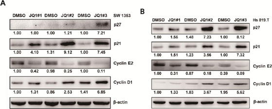
(+)-JQ-1 purchased from MedChemExpress. Usage Cited in: J Cell Biochem. 2017 Aug;118(8):2182-2192. [Abstract]
JQ1 induces G0/G1 cell cycle arrest and apoptosis of chondrosarcoma cells via regulating p21, p27, Cyclin E2, and Cyclin D1 expression. (A and B) JQ1 regulates the expression of cell cycle regulators such as p21, p27, Cyclin E2, and Cyclin D1. SW 1353 and Hs 819.T cells are seeded into a 6-well plate for 72 h, then the cells are treated with different concentrations of JQ1 (JQ1#1: 200 nM; JQ1#2: 2 μM; JQ1#3: 20 μM) for another 24 h. Total cell lysate is used for the analysis of protein expressio
-
Invest New Drugs
Targeting Myc through BET-PROTAC elicits potent anti-lymphoma activity in diffuse large B cell lymphoma. [Abstract]2025 Jun;43(3):621-633. PMID: 40307411 -
Mol Biol Cell
2024 Jun 1;35(6):ar88. PMID: 38656803 -
SLAS Discov
Development of a high-throughput screening platform to identify new therapeutic agents for Medulloblastoma Group 3. [Abstract]2024 Feb 12;29(2):100147. PMID: 38355016 -
Exp Eye Res
JQ1, a selective inhibitor of BRD4, suppresses retinoblastoma cell growth by inducing cell cycle arrest and apoptosis. [Abstract]2021 Jan;202:108304. PMID: 33080301 -
Invest New Drugs
The bromodomain and extra-terminal domain inhibitor JQ1 synergistically sensitizes human colorectal cancer cells to topoisomerase I inhibitors through repression of Mre11-mediated DNA repair pathway. [Abstract]2021 Apr;39(2):362-376. PMID: 32981006 -
Brain Res
Inhibition of BRD4 decreases fibrous scarring after ischemic stroke in rats by inhibiting the phosphorylation of Smad2/3. [Abstract]2022 Dec 15:1797:148126. PMID: 36244457 -
-
Int J Stem Cells
Strategic Application of Epigenetic Regulators for Efficient Neuronal Reprogramming of Human Fibroblasts. [Abstract]2023 May 30;16(2):156-167. PMID: 36823979 -
Eur J Neurosci
Impact of JQ1 treatment on seizures, hippocampal gene expression, and gliosis in a mouse model of temporal lobe epilepsy. [Abstract]2024 Sep;60(6):5266-5283. PMID: 39149798 -
Oncol Lett
BET inhibitor JQ1 induces apoptosis of ovarian and endometrial endometrioid carcinoma cells by downregulating c‑Myc. [Abstract]2024 Dec 19;29(3):106. PMID: 39736925 -
Biochem Biophys Res Commun
Brd4-dependent CDK9 expression induction upon sustained pharmacological inhibition of P-TEFb kinase activity. [Abstract]2023 Sep 3:671:75-79. PMID: 37295357 -
Biochem Biophys Res Commun
The CDK4/6-UCHL5-BRD4 axis confers resistance to BET inhibitors in MLL-rearranged leukemia cells by suppressing BRD4 protein degradation. [Abstract]2022 Jan 15:588:147-153. PMID: 34954522 -
Biochem Biophys Res Commun
ATF2 inhibits ani-tumor effects of BET inhibitor in a negative feedback manner by attenuating ferroptosis. [Abstract]2021 Jun 18:558:216-223. PMID: 33008584 -
Biochem Bioph Res Co
Suppressing BRD4 exhibits protective effects against vincristine-induced peripheral neuropathy by alleviating inflammation and oxidative stress. [Abstract]2020 Nov 5;532(2):271-279. PMID: 32868081 -
Biochem Biophys Res Commun
2019 Dec 3;520(2):250-256. PMID: 31594641 -
Exp Hematol
JQ-1/Bortezomib combination strongly impairs MM and PEL survival by inhibiting c-Myc and mTOR despite the activation of pro-survival mechanisms. [Abstract]2023 Mar-Apr:119-120:28-41. PMID: 36623719 -
Neurosci Lett
Inhibition of BRD4 expression attenuates the inflammatory response and apoptosis by downregulating the HMGB-1/NF-κB signaling pathway following traumatic brain injury in rats. [Abstract]2023 Aug 24:812:137385. PMID: 37423465 -
Hematology
BRD4 PROTAC degrader MZ1 exhibits anti-B-cell acute lymphoblastic leukemia effects via targeting CCND3. [Abstract]2023 Dec;28(1):2247253. PMID: 37594294 -
Biosci Biotechnol Biochem
Synergistic effect of cerium chloride and calcium chloride alters calcium signaling in keratinocytes to promote epidermal differentiation. [Abstract]2024 Nov 22;88(12):1432-1441. PMID: 39333009 -
Bio Protoc
2017 Jul 5;7(13):e2368. PMID: 34541110 -
-
-
-
-
bioRxiv
Spatially-Resolved Multiomic Atlas of Leiomyosarcoma Identifies Two Clinically Relevant Epigenetically-Driven Cell States. [Abstract]2026 May 26:2026.05.22.726988. PMID: 42244620 -
bioRxiv
Radiation synergizes with BET inhibition to stimulate durable, systemic anti-tumor immunity in murine cancer models. [Abstract]2026 Feb 18:2026.02.16.706212. PMID: 41757027 -
Royal College of Surgeons in Ireland
-
bioRxiv
A human sensory neuron model for varicella-zoster virus latency and reactivation in vitro. [Abstract]2025 Dec 12:2025.12.12.693952. PMID: 41415445 -
-
bioRxiv
2025 Sep 18:2025.09.17.676776. PMID: 41000743 -
-
bioRxiv
2025 Sep 3:2025.08.30.673282. PMID: 40949948 -
-
bioRxiv
Herpes simplex virus 1 strain 17+ with R2 mutation in UL37 has residual retrograde transport. [Abstract]2025 Jun 26:2025.06.25.661543. PMID: 40667132 -
bioRxiv
2025 May 4:2025.04.29.651320. PMID: 40654780 -
bioRxiv
Resistance to the KRASG12D Inhibitor MRTX1133 is Associated with Increased Sensitivity to BET Inhibition. [Abstract]2025 May 13:2025.05.10.653074. PMID: 40463163 -
-
-
-
Blood Neoplasia
2025 Jan 20;2(2):100069. PMID: 40453140 -
bioRxiv
2024 Sep 27:2024.09.25.615094. PMID: 39386645 -
-
-
-
-
bioRxiv
Exploration of the Tunability of BRD4 Degradation by DCAF16 Trans-labelling Covalent Glues. [Abstract]2023 Oct 10:2023.10.07.561308. PMID: 37873358 -
-
-
bioRxiv
Bifunctional small molecules that induce nuclear localization and targeted transcriptional regulation. [Abstract]2023 Jul 7:2023.07.07.548101. PMID: 37461636 -
-
-
-
-
bioRxiv
Template-assisted covalent modification of DCAF16 underlies activity of BRD4 molecular glue degraders. [Abstract]2023 Feb 15:2023.02.14.528208. PMID: 36824856 -
-
-
bioRxiv
Regulated Induced Proximity Targeting Chimeras (RIPTACs): a Novel Heterobifunctional Small Molecule Therapeutic Strategy for Killing Cancer Cells Selectively. [Abstract]2023 Jan 2:2023.01.01.522436. PMID: 36711980 -
-
-
Oxid Med Cell Longev
DSC2 Suppresses the Metastasis of Gastric Cancer through Inhibiting the BRD4/Snail Signaling Pathway and the Transcriptional Activity of β-Catenin. [Abstract]2022 Sep 6;2022:4813571. PMID: 36120591 -
-
-
-
-
-
-
-
-
-
-
-
-
-
Oncotarget
Bromodomain protein BRD4 inhibitor JQ1 regulates potential prognostic molecules in advanced renal cell carcinoma. [Abstract]2018 May 1;9(33):23003-23017. PMID: 29796168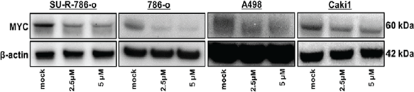
(+)-JQ-1 purchased from MedChemExpress. Usage Cited in: Oncotarget. 2018 May 1;9(33):23003-23017. [Abstract]
The expression of MYC protein is markedly repressed in JQ1-treated ccRCC cells (2.5 and 5 μM) in comparison with that in mock cells. β-Actin is used as a loading control. Densitometry analyses using ImageJ software are performed.
-
-
Oncotarget
Molecular-genetic profiling and high-throughput in vitro drug screening in NUT midline carcinoma-an aggressive and fatal disease. [Abstract]2017 Dec 2;8(68):112313-112329. PMID: 29348827


Solvent & Solubility
DMSO : 100 mg/mL (218.82 mM; Need ultrasonic; Hygroscopic DMSO has a significant impact on the solubility of product, please use newly opened DMSO)
Please refer to the solubility information to select the appropriate solvent. Once prepared, please aliquot and store the solution to prevent product inactivation from repeated freeze-thaw cycles.
Storage method and period of stock solution: -80°C, 1 year; -20°C, 6 months. When stored at -80°C, please use it within 1 year. When stored at -20°C, please use it within 6 months.
Concentration (start) × Volume (start) = Concentration (final) × Volume (final)
Select the appropriate dissolution method based on your experimental animal and administration route.
- For the following dissolution methods, please ensure to first prepare a clear stock solution using an In Vitro approach and then sequentially add co-solvents:
- To ensure reliable experimental results, the clarified stock solution can be appropriately stored based on storage conditions. As for the working solution for In Vivo experiments, it is recommended to prepare freshly and use it on the same day.
- The percentages shown for the solvents indicate their volumetric ratio in the final prepared solution. If precipitation or phase separation occurs during preparation, heat and/or sonication can be used to aid dissolution.
Add each solvent one by one: 10% DMSO 40% PEG300 5% Tween-80 45% Saline
Solubility: ≥ 2.5 mg/mL (5.47 mM); Clear solution
This protocol yields a clear solution of ≥ 2.5 mg/mL (saturation unknown).
Taking 1 mL working solution as an example, add 100 μL DMSO stock solution (25.0 mg/mL) to 400 μL PEG300, and mix evenly; then add 50 μL Tween-80 and mix evenly; then add 450 μL Saline to adjust the volume to 1 mL.
Preparation of Saline: Dissolve 0.9 g sodium chloride in ddH₂O and dilute to 100 mL to obtain a clear Saline solution.
Add each solvent one by one: 10% DMSO 90% (20% SBE-β-CD in Saline)
Solubility: ≥ 2.5 mg/mL (5.47 mM); Clear solution
This protocol yields a clear solution of ≥ 2.5 mg/mL (saturation unknown).
Taking 1 mL working solution as an example, add 100 μL DMSO stock solution (25.0 mg/mL) to 900 μL 20% SBE-β-CD in Saline, and mix evenly.
Preparation of 20% SBE-β-CD in Saline (4°C, storage for one week): 2 g SBE-β-CD powder is dissolved in 10 mL Saline, completely dissolve until clear.
For the following dissolution methods, please prepare the working solution directly:
It is recommended to prepare fresh solutions and use them promptly within a short period of time.
The percentages shown for the solvents indicate their volumetric ratio in the final prepared solution. If precipitation or phase separation occurs during preparation, heat and/or sonication can be used to aid dissolution.
Add each solvent one by one: 20% HP-β-CD in Saline
Solubility: 10 mg/mL (21.88 mM); Suspended solution; Need ultrasonic
Add each solvent one by one: 15% Cremophor EL 85% Saline
Solubility: 10 mg/mL (21.88 mM); Clear solution; Need ultrasonic
Please enter the basic information of animal experiments:
-
-
-
-
Recommended: Prepare an additional quantity of animals to account for potential losses during experiments.
Please enter your animal formula composition:
-
%DMSO +
Recommended: Keep the proportion of DMSO in working solution below 2% if your animal is weak.
-
%+
-
+%Tween-80 + +
-
%Saline +
The co-solvents required include: DMSO, . All of co-solvents are available by MedChemExpress (MCE). , Tween 80. All of co-solvents are available by MedChemExpress (MCE).
Working solution concentration: 0.22 mg/mL
Method for preparing stock solution: mg drug dissolved in μL DMSO. Stock solution concentration: mg/mL.
1. Take μL DMSO stock solution;
2. Add μL .
μL , mix evenly;
3. Then add μL Tween 80, mix evenly;
4. Then add μL
Please ensure that the stock solution in the first step is dissolved to a clear state, and add co-solvents in sequence. You can use ultrasonic heating (ultrasonic cleaner, recommended frequency 20-40 kHz), vortexing, etc. to assist dissolution.

Protocol
NUT midline carcinoma patient cell lines (797 and 11060) are plated in T-25 flasks and grown in DMEM (797) or RPMI (11060) containing 10 % fetal bovine serum. Cells are treated with either 250 nM (+)-JQ1, 250 nM (-)-JQ1 or the equivalent volume of DMSO (0.025%). At the desired time point, 2×10⁶ cells are spun at 500× g for 5 minutes at 4°C and washed with PBS. Pellets are resuspended in 1 mL of cold PBS and added dropwise while gently vortexing to 9 mL 70 % ethanol in a 15 mL polypropylene centrifuge tube. Fixed cells are then frozen at -20°C overnight. The next day, cells are centrifuged at 500× g for 10 minutes at 4°C and washed with 3 mL of cold PBS. Cells are resuspended in 500 μL of propidium iodide staining solution (0.2 mg/mL RNAse A, 0.02 mg/mL propidium iodide, 0.1 % Triton-X in PBS) and incubated for 20 minutes at 37°C. Samples are then transferred to ice and analyzed on a BD FACS Canto II. Histograms are generated and cell cycle analysis is performed using FlowJo flow cytometry analysis software[1].
MedChemExpress (MCE) has not independently confirmed the accuracy of these methods. They are for reference only.
Mice[1]
Matched cohorts of mice with established tumors are randomized to treatment with (+)-JQ1 (50 mg/kg) or vehicle, administered by daily intraperitoneal injection. Male CD1 mice (24-29 g) are treated with a single dose of (+)-JQ1 at 5 mg/kg for intravenous tail vein injection studies and 10 mg/kg for oral gavage studies.
Rats[3]
Adult male Sprague-Dawley rats are treated with vehicle or (+)-JQ1 (10 mg/kg). Treatment is administered IP at 1/100 body mass. Rats are checked twice-daily for mortality and weighed on days 1, 3, 7, 14, and 21. The treatment regimen utilized 4 days of 50 mg/kg JQ1 administered daily which is decreased to 10 mg/kg twice daily for the remainder of the study due to the appearance of adverse effects in a subset of animals. For all animals completing 3 weeks of treatment, testis mass, sperm motility, and sperm counts are determined as described for mouse studies. In brief, testes are fixed in Bouin’s and prepared for histology. The other half is minced in warm M16 buffer and used for sperm counts and motility studies.
MedChemExpress (MCE) has not independently confirmed the accuracy of these methods. They are for reference only.


Purity & Documentation
-
Data Sheet (287 KB)
-
SDS (396 KB)
- English - EN (396 KB)
- Français - FR (396 KB)
- Deutsch - DE (396 KB)
- Norwegian - NO (396 KB)
- Español - ES (396 KB)
- Swedish - SV (396 KB)
- Italian - IT (396 KB)
- Korean - KR (396 KB)
- Portuguese - PT (396 KB)
-
Handling Instructions (2659 KB)
References
[1]. Filippakopoulos P, et al. Selective inhibition of BET bromodomains. Nature. 2010 Dec 23;468(7327):1067-73. [Content Brief]
[2]. Sakamaki JI, et al. Bromodomain Protein BRD4 Is a Transcriptional Repressor of Autophagy and LysosomalFunction. Mol Cell. 2017 May 18;66(4):517-532.e9. [Content Brief]
[3]. Matzuk MM, et al. Small-molecule inhibition of BRDT for male contraception. Cell. 2012 Aug 17;150(4):673-84. [Content Brief]
[4]. Zhong X, et al. JQ1 attenuates neuroinflammation by inhibiting the inflammasome-dependent canonical pyroptosis pathway in SAE. Brain Res Bull. 2022 Oct 15;189:174-183. [Content Brief]


Complete Stock Solution Preparation Table
Please refer to the solubility information to select the appropriate solvent. Once prepared, please aliquot and store the solution to prevent product inactivation from repeated freeze-thaw cycles.
Storage method and period of stock solution: -80°C, 1 year; -20°C, 6 months. When stored at -80°C, please use it within 1 year. When stored at -20°C, please use it within 6 months.
| Optional Solvent | Concentration Solvent Mass | 1 mg | 5 mg | 10 mg | 25 mg |
|---|---|---|---|---|---|
| DMSO | 1 mM | 2.1882 mL | 10.9412 mL | 21.8823 mL | 54.7058 mL |
| 5 mM | 0.4376 mL | 2.1882 mL | 4.3765 mL | 10.9412 mL | |
| 10 mM | 0.2188 mL | 1.0941 mL | 2.1882 mL | 5.4706 mL | |
| 15 mM | 0.1459 mL | 0.7294 mL | 1.4588 mL | 3.6471 mL | |
| 20 mM | 0.1094 mL | 0.5471 mL | 1.0941 mL | 2.7353 mL | |
| 25 mM | 0.0875 mL | 0.4376 mL | 0.8753 mL | 2.1882 mL | |
| 30 mM | 0.0729 mL | 0.3647 mL | 0.7294 mL | 1.8235 mL | |
| 40 mM | 0.0547 mL | 0.2735 mL | 0.5471 mL | 1.3676 mL | |
| 50 mM | 0.0438 mL | 0.2188 mL | 0.4376 mL | 1.0941 mL | |
| 60 mM | 0.0365 mL | 0.1824 mL | 0.3647 mL | 0.9118 mL | |
| 80 mM | 0.0274 mL | 0.1368 mL | 0.2735 mL | 0.6838 mL | |
| 100 mM | 0.0219 mL | 0.1094 mL | 0.2188 mL | 0.5471 mL |


 Powered by Bioz