



Bcl-XL Antibody (YA588)
(Synonyms: BCL2L, BCLX, BCL2L1, Bcl-2-like protein 1, Bcl2-L-1, Apoptosis regulator Bcl-X)Based on 1 publication(s) in Google Scholar
Bcl-XL Antibody (YA588) is a Rabbit-derived and non-conjugated IgG monoclonal antibody, targeting to Bcl-XL.
-
Host:
Rabbit
-
Isotype:
IgG
-
Application:
WB, IHC-P, ICC/IF, IP, FC, IF-Tissue
-
Reactivity :
Human
-
Formulation:
Supplied in PBS (pH7.4), 0.1% BSA, 40% Glycerol, 0.2% Proclin950.
-
Conjugation:
Non-conjugated


To place orders, for customer services and technical support, please contact: MedChemExpress USA
Tel: 609-228-6898 E-mail: [email protected] [email protected]



Publications Citing Use of MedChemExpress (MCE) Bcl-XL Antibody (YA588)
More

Applications
| Application |
WB

WB: Western Blot
|
ICC/IF
ICC/IF: Immunocytochemistry/
Immunofluorescence |
IF-Tissue
IF-Tissue: Immunofluorescence-Tissue
|
IHC-P
IHC-P: Immunohistochemistry-Paraffin
|
FC
FC: Flow Cytometry
|
IP
IP: Immunoprecipitation
|
|---|---|---|---|---|---|---|
| Dilution Ratio | 1:1000-5000 | 1:50-200 | 1:50-200 | 1:200-1000 | 1:1000 | Use at an assay dependent concentration. |


Product Details
Bcl-XL Antibody (YA588) is a Rabbit-derived and non-conjugated IgG monoclonal antibody, targeting to Bcl-XL.
-
Host Rabbit

-
Clonality Recombinant,Monoclonal

-
Species ReactivityHuman
-
Observed Molecular WeightObserved band size: 30 kDa
Note: Due to possible protein modifications or aggregation, the molecular weight should be confirmed by actual measurement, and the predicted value is for reference only.

-
Calculated Molecular Weight Predicted band size: 26 kDa

Synthetic peptide corresponding to Human Bcl-XL.AA range:37-86.
Endogenous
Protein A affinity purified.
Non-conjugated
Unmodified
IgG


Product Properties
-
Appearance
Solution
-
Formulation
Supplied in PBS (pH7.4), 0.1% BSA, 40% Glycerol, 0.2% Proclin950.
-
Concentration
Batch-dependent, Please check the COA for the concentration of each lot. Check Lot Concentration
-
Storage & Stability
Stored at -20°C for 1 year. Avoid repeated freeze / thaw cycles.
-
Shipping
Shipping with blue ice.


Publications (1)
-
Journal Impact Factor
-
Most Recent
-
Front Vet Sci
Unrevealing the therapeutic potential of artesunate against emerging zoonotic Babesia microti infection in the murine model. [Abstract]2024 May 9:11:1383291. PMID: 38784653


Verification Images
-
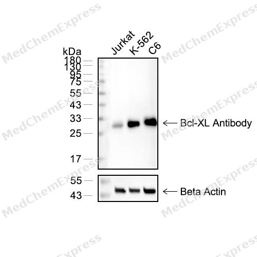 Western blot analysis of extracts from Jurkat(lane 2(20μg) , K-562(lane 3(20μg) and C6(lane 4(20μg) using Bcl-XL(HY-P80030) Rabbit mAb. Proteins were transferred to a PVDF membrane and blocked with 5% non-fat milk in TBST for 2 hour at room temperature. The primary antibody (1/1000) and Loading control antibody (Beta Actin, HY-P80438, 1/10000) was used in 5% non-fat milk in TBST at 4°C overnight. Goat Anti-Mouse/Rabbit IgG-HRP Secondary Antibody (1/10000) was used for 1 hour at room temperature.
 -
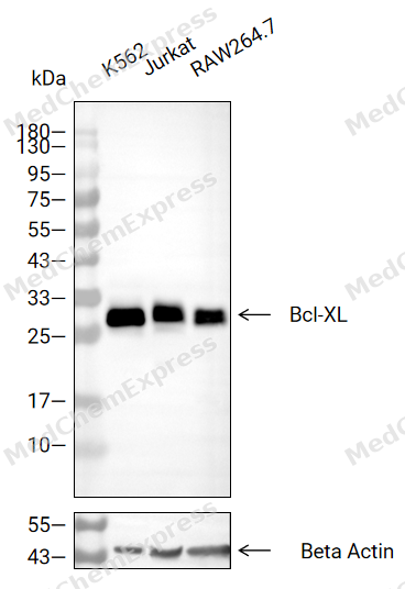 Western blot analysis of extracts from K-562 (lane 2(20μg), Jurkat (lane 3(20μg), RAW264.7 (lane 4(20μg),using Bcl-XL Antibody. Proteins were transferred to a PVDF membrane and blocked with 5% BSA in TBST for 2 hour at room temperature. The primary antibody and Loading control antibody (Beta Actin, HY-P80438, 1/3000) was used in 5% BSA in TBST at 4°C overnight. Goat Anti-Mouse/Rabbit IgG-HRP Secondary Antibody (HY-P8004/HY-P8001, 1/10,000) was used for 1 hour at room temperature.
 -
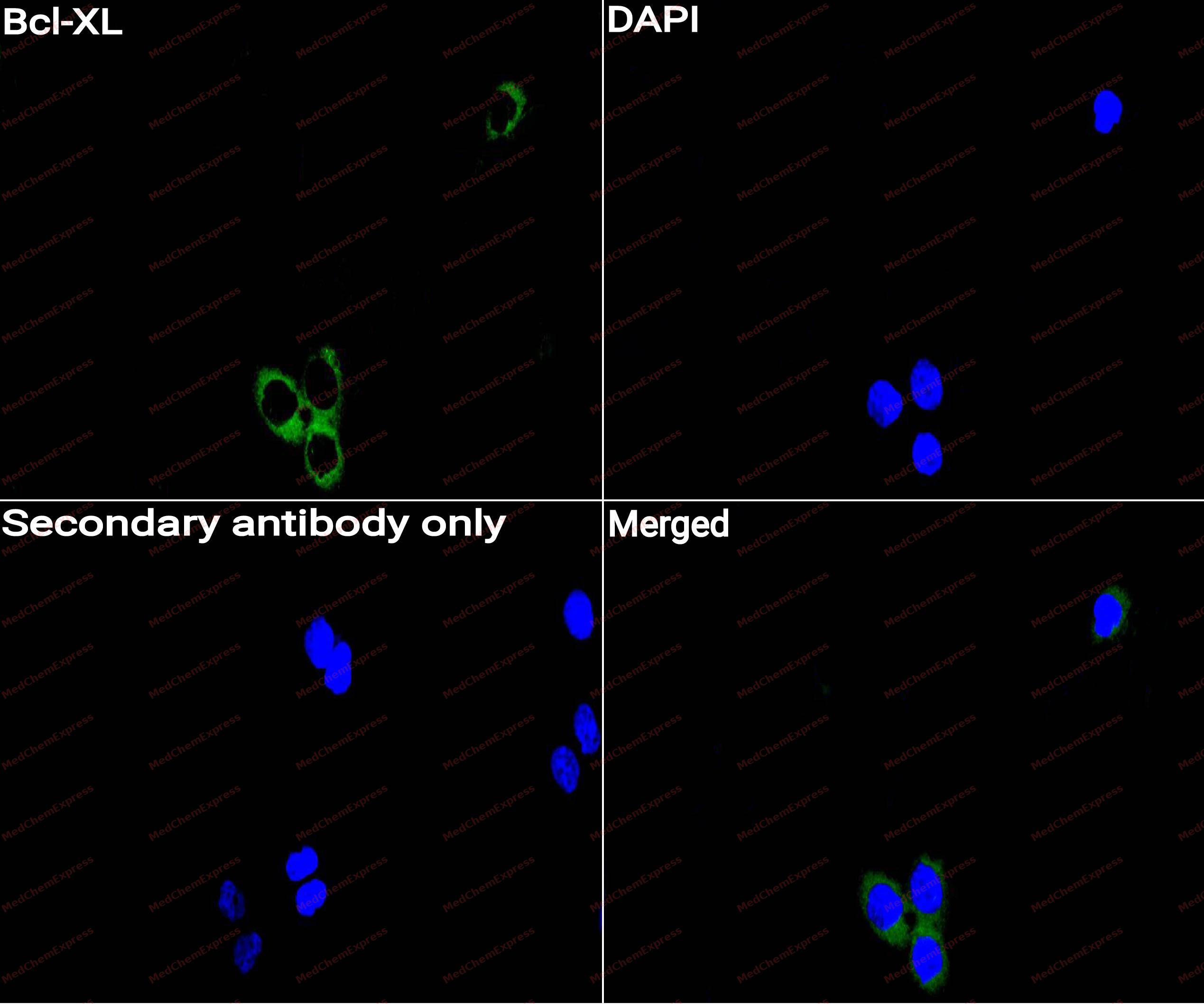 Immunocytochemistry analysis of Hela cells labeling Bcl-XL with Bcl-XL Antibody (HY-P80030) at 1/100 dilution. Cells were fixed in 4% paraformaldehyde for 15 minutes at room temperature, permeabilized with 0.1% Triton X-100 for 10 minutes at room temperature, then blocked with QuickBlock™ Blocking Buffer for Immunol Staining for 10 min at room temperature. Cells were then incubated with Bcl-XL Antibody (HY-P80030)at 1/100 dilution in QuickBlock™ Blocking Buffer for Immunol Staining at 4 ℃. Alexa Fluor® 488-conjugated AffiniPure Goat Anti-Rabbit IgG H&L(HY-P8002,Green) was used as the secondary antibody at 1/1,000 dilution. PBS instead of the primary antibody was used as the secondary antibody only control. The Nuclear counterstain was DAPI (Blue).
 -
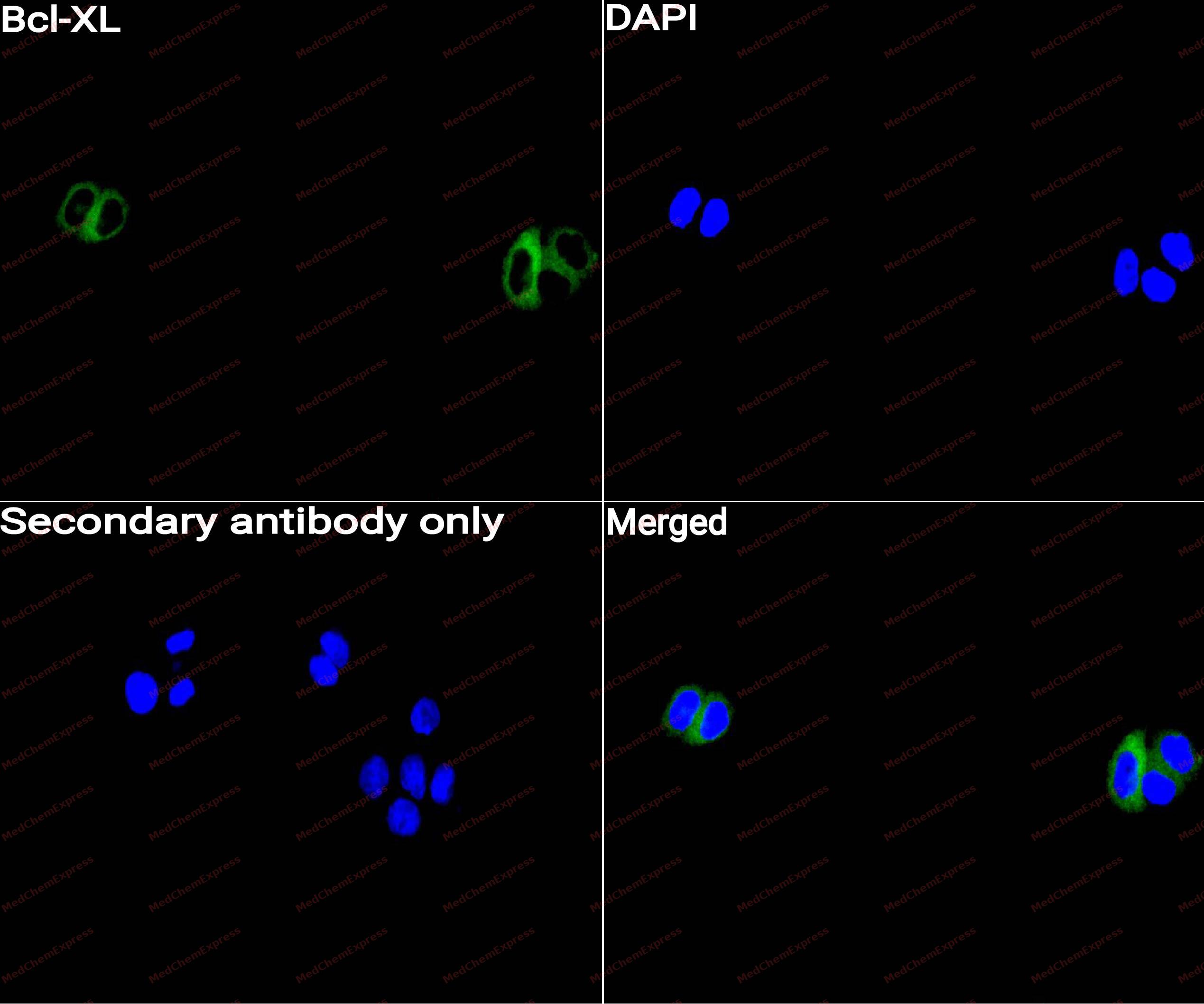 Immunocytochemistry analysis of Hela cells labeling Bcl-XL with Bcl-XL Antibody (HY-P80030) at 1/50 dilution. Cells were fixed in 4% paraformaldehyde for 15 minutes at room temperature, permeabilized with 0.1% Triton X-100 for 10 minutes at room temperature, then blocked with QuickBlock™ Blocking Buffer for Immunol Staining for 10 min at room temperature. Cells were then incubated with Bcl-XL Antibody (HY-P80030) at 1/50 dilution in QuickBlock™ Blocking Buffer for Immunol Staining at 4 ℃. Alexa Fluor® 488-conjugated AffiniPure Goat Anti-Rabbit IgG H&L(HY-P8002, Green) was used as the secondary antibody at 1/1,000 dilution. PBS instead of the primary antibody was used as the secondary antibody only control. The Nuclear counterstain was DAPI (Blue).
 -
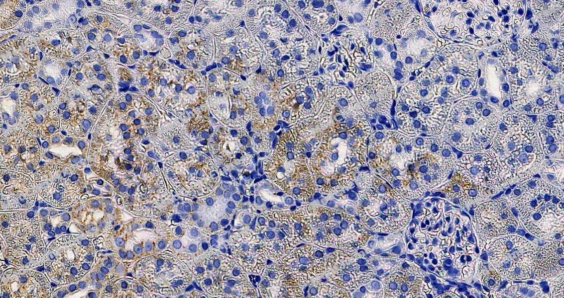 Immunohistochemical analysis of paraffin-embedded rat kidney tissue using Bcl-XL Antibody. The section was pre-treated using heat mediated antigen retrieval with Tris-EDTA buffer (pH 9.0) for 8 minutes. The tissues were blocked in QuickBlock for 20 minutes at room temperature, washed with ddH2O and PBS, and then probed with the primary antibody at 1/100 dilution in 4℃ overnight. The detection was performed using an HRP conjugated compact polymer system. DAB was used as the chromogen. Tissues were counterstained with hematoxylin and mounted with DPX.
 -
Immunohistochemical analysis of paraffin-embedded rat kidney tissue using Bcl-XL Antibody. The section was pre-treated using heat mediated antigen retrieval with Tris-EDTA buffer (pH 9.0) for 8 minutes. The tissues were blocked in QuickBlock for 20 minutes at room temperature, washed with ddH2O and PBS, and then probed with the primary antibody at 1/100 dilution in 4℃ overnight. The detection was performed using an HRP conjugated compact polymer system. DAB was used as the chromogen. Tissues were counterstained with hematoxylin and mounted with DPX.
-
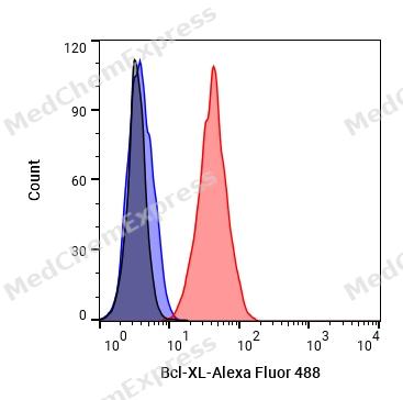 Flow cytometric analysis of 1X10^6 K562 cells labeling Bcl-XL Antibody (HY-P80030, red). Cells were fixed with 4% paraformaldehyde and permeabilised with 90% methanol. Then stained with the primary antibody at 1/1000 dilution for an hour at 4℃. Alexa Fluor® 488-conjugated AffiniPure Goat Anti-Rabbit IgG H&L (HY-P8002) was used as the secondary antibody at 1/1,000 dilution for 30 minutes at 4℃. Rabbit IgG Isotype Control (HY-P80879, blue) was used as the isotype control, cells without incubation with primary antibody were used as the unlabeled control (black).


Background
-
Function
Bcl-xL, encoded by BCL2L1, is a major anti-apoptotic member of the BCL-2 family that regulates mitochondrial apoptosis through interactions with pro-apoptotic proteins and preservation of mitochondrial integrity[1][2]. Mechanistically, Bcl-xL suppresses mitochondrial outer membrane permeabilization, limits BAX/BAK activation, and modulates cellular bioenergetics, calcium signaling, and autophagy, linking survival pathways with metabolic adaptation under cellular stress[1][3]. Beyond canonical apoptosis control, Bcl-xL interacts with Beclin-1 and intracellular calcium regulators, including IP3 receptors and VDAC1, thereby influencing autophagy, endoplasmic reticulum-mitochondrial communication, and stress responses[1][4]. In disease models, elevated Bcl-xL expression promotes tumor cell survival, therapeutic resistance, angiogenesis, and metastatic progression across hematologic malignancies and solid tumors, including melanoma and breast cancer[1][5]. Compared with related BCL2L1 splice isoforms, Bcl-xL functions predominantly as a pro-survival protein, whereas the alternatively spliced Bcl-xS isoform exhibits pro-apoptotic activity, highlighting the biological significance of isoform-specific regulation in apoptosis research[2][6]. For experimental applications, Bcl-xL is a validated target of BH3 mimetics and other apoptosis-directed strategies, and its inhibition is widely used to investigate mitochondrial apoptotic priming, drug resistance mechanisms, and cancer cell dependency on anti-apoptotic signaling networks[1][7].
-
Subcellular Localization
Mitochondrion inner membrane; Mitochondrion outer membrane; Mitochondrion matrix; Cytoplasmic vesicle, secretory vesicle, synaptic vesicle membrane; Cytoplasm, cytosol; Cytoplasm, cytoskeleton, microtubule organizing center, centrosome; Nucleus membrane; Single-pass membrane protein; Cytoplasmic side
-
Expression
Tissue_specificity:Bcl-X (S) is highly expressed in cells with a rapid cell turnover rate, such as developing lymphocytes. In contrast, Bcl-X (L) is found in tissues containing long-lived amitotic cells, such as adult brain tissue. -
Isoforms & Post-Translational Modification
Q07817 has 3 isomers: Q07817-1: 26049 Da (predicted); Q07817-2: 18894 Da (predicted); Q07817-3: 25290 Da (predicted).
Proteolytically cleaved by caspases during apoptosis. The cleaved protein, lacking the BH4 motif, has pro-apoptotic activity;Phosphorylated on Ser-62 by CDK1. This phosphorylation is partial in normal mitotic cells, but complete in G2-arrested cells upon DNA-damage, thus promoting subsequent apoptosis probably by triggering caspases-mediated proteolysis. Phosphorylated by PLK3, leading to regulate the G2 checkpoint and progression to cytokinesis during mitosis. Phosphorylation at Ser-49 appears during the S phase and G2, disappears rapidly in early mitosis during prometaphase, metaphase and early anaphase, and re-appears during telophase and cytokinesis;Ubiquitinated by RNF183 during prolonged ER stress, leading to degradation by the proteosome -
Subunit
Homodimer. Interacts with BCL2L11 (By similarity). Interacts with BAD. Interacts with PGAM5. Interacts with HEBP2. Interacts with p53/TP53 and BBC3; interaction with BBC3 disrupts the interaction with p53/TP53. Interacts with ATP5F1A and ATP5F1B; the interactions mediate the association of isoform Bcl-X(L) with the mitochondrial membrane ATP synthase F(1)F(0) ATP synthase. Interacts with VDAC1 (PubMed:25296756). Interacts with BCL2L11 (via BH3) (PubMed:27013495). Interacts with RNF183 (PubMed:29507230). Interacts with GIMAP3/IAN4 and GIMAP5/IAN5 (PubMed:16509771). Interacts with GIMAP5 and HSPA8/HSC70; the interaction between HSPA8 and BCL2L1 is impaired in the absence of GIMAP5 (By similarity). Interacts with isoform 4 of CLU; this interaction releases and activates BAX and promotes cell death (PubMed:21567405)
-
SwissProt ID
-
Synonyms
BCL2L, BCLX, BCL2L1, Bcl-2-like protein 1, Bcl2-L-1, Apoptosis regulator Bcl-X
-
Research Field
Cell Biology
Documentation
-
Data Sheet (262 KB)
-
SDS (251 KB)
- English - EN (251 KB)
- Français - FR (251 KB)
- Deutsch - DE (251 KB)
- Norwegian - NO (251 KB)
- Español - ES (251 KB)
- Swedish - SV (251 KB)
- Italian - IT (251 KB)
- Korean - KR (251 KB)
- Portuguese - PT (251 KB)
-
User Guide for Antibodies (1077 KB)
References
[2]. Warren CFA, et al. BCL-2 family isoforms in apoptosis and cancer. Cell Death Dis. 2019 Feb 21;10(3):177. [Content Brief]
[3]. Li M, et al. Bcl-XL: A multifunctional anti-apoptotic protein. Pharmacol Res. 2020;151:104547.
[4]. Bcl-xL gene information from NCBI.
[5]. Kim TJ, et al. Risk of Second Primary Malignancies among Patients with Early Gastric Cancer Exposed to Recurrent Computed Tomography Scans. Cancers (Basel). 2021 Mar 7;13(5):1144. [Content Brief]
[6]. Chen W, et al. Alternative splicing of BCL-X and implications for treating hematological malignancies. Oncol Lett. 2021;22(4):670. [Content Brief]
[7]. Campbell KJ, et al. Targeting BCL-2 regulated apoptosis in cancer. Open Biol. 2018;8(5):180002. [Content Brief]

 Powered by Bioz
