



Caspase-3 Antibody (YA557)
(Synonyms: CPP32, CASP3, Caspase-3, CASP-3, Apopain, Cysteine protease CPP32, Protein Yama, SREBP cleavage activity 1, CPP-32, SCA-1)Based on 6 publication(s) in Google Scholar
Caspase-3 Antibody (YA557) is a Rabbit-derived and non-conjugated IgG monoclonal antibody, targeting to Caspase-3.
-
Host:
Rabbit
-
Isotype:
IgG
-
Application:
WB, ICC/IF, IHC-P, IP, FC
-
Reactivity :
Human
-
Formulation:
Supplied in 1*TBS (pH7.4), 0.05% BSA and 40% Glycerol. Preservative: 0.05% Sodium Azide.
-
Conjugation:
Non-conjugated


To place orders, for customer services and technical support, please contact: MedChemExpress USA
Tel: 609-228-6898 E-mail: [email protected] [email protected]



-

HY-P80028
Bax Antibody (YA591) -
HY-P80566
Bcl-2 Antibody (YA590) -
HY-P80623
Cleaved-Caspase 3 p12 Antibody (YA499) -
HY-P80050
Caspase-9 Antibody (YA553) -
HY-P80029
Bcl-2 Antibody (YA589) -
HY-P80049
Caspase-8 Antibody (YA554) -
HY-P80027
Bax Antibody (YA825) -
HY-P80102
Cytochrome C Antibody (YA479) -
HY-P8001
HRP-conjugated Goat Anti-Rabbit IgG H&L -
HY-P80137
GAPDH Antibody (YA418) -
HY-P86370
Cleaved Caspase-3 (Asp175) Antibody (YA6062) -
HY-K0010
Protease Inhibitor Cocktail (EDTA-Free, 100× in DMSO)
Publications Citing Use of MedChemExpress (MCE) Caspase-3 Antibody (YA557)
More

Applications
| Application |
WB

WB: Western Blot
|
ICC/IF
ICC/IF: Immunocytochemistry/
Immunofluorescence |
IHC-P
IHC-P: Immunohistochemistry-Paraffin
|
FC
FC: Flow Cytometry
|
IP
IP: Immunoprecipitation
|
|---|---|---|---|---|---|
| Dilution Ratio | 1:500 | 1:50 | 1:50 | 1:20-1:50 | Use at an assay dependent concentration. |


Product Details
Caspase-3 Antibody (YA557) is a Rabbit-derived and non-conjugated IgG monoclonal antibody, targeting to Caspase-3.
-
Host Rabbit

-
Clonality Recombinant,Monoclonal

-
Species ReactivityHuman
-
Observed Molecular WeightObserved band size: 17 kDa
Note: Due to possible protein modifications or aggregation, the molecular weight should be confirmed by actual measurement, and the predicted value is for reference only.

-
Calculated Molecular Weight Predicted band size: 32 kDa

Synthetic peptide corresponding to Human Caspase-3.AA range:60-100.
Endogenous
Protein A affinity purified.
Non-conjugated
Unmodified
IgG


Product Properties
-
Appearance
Solution
-
Formulation
Supplied in 1*TBS (pH7.4), 0.05% BSA and 40% Glycerol. Preservative: 0.05% Sodium Azide.
-
Concentration
Batch-dependent, Please check the COA for the concentration of each lot. Check Lot Concentration
-
Storage & Stability
Stored at -20°C for 1 year. Avoid repeated freeze / thaw cycles.
-
Shipping
Shipping with blue ice.


Publications (6)
-
Journal Impact Factor
-
Most Recent
-
J Transl Med
Long non-coding RNA RP11-197K6.1 as ceRNA promotes colorectal cancer progression via miR-135a-5p/DLX5 axis. [Abstract]2024 May 17;22(1):469. PMID: 38760791 -
Sci Rep
Sodium propionate protects against bronchopulmonary dysplasia by inhibiting IL-17-mediated apoptosis of alveolar epithelial cells. [Abstract]2025 Apr 5;15(1):11722. PMID: 40188136


Verification Images
-
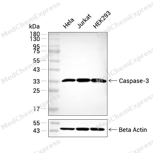 Western blot analysis of extracts from Hela (lane 2(20μg), Jurkat (lane 3(20μg) and HEK293 (lane 4(20μg) using Caspase-3(HY-P80046) Rabbit mAb. Proteins were transferred to a PVDF membrane and blocked with 5% non-fat milk in TBST for 2 hour at room temperature. The primary antibody (1/1000) and Loading control antibody (Beta Actin, HY-P80438, 1/10000) was used in 5% non-fat milk in TBST at 4°C overnight. Goat Anti-Mouse IgG-HRP Secondary Antibody (1/10000) was used for 1 hour at room temperature.
 -
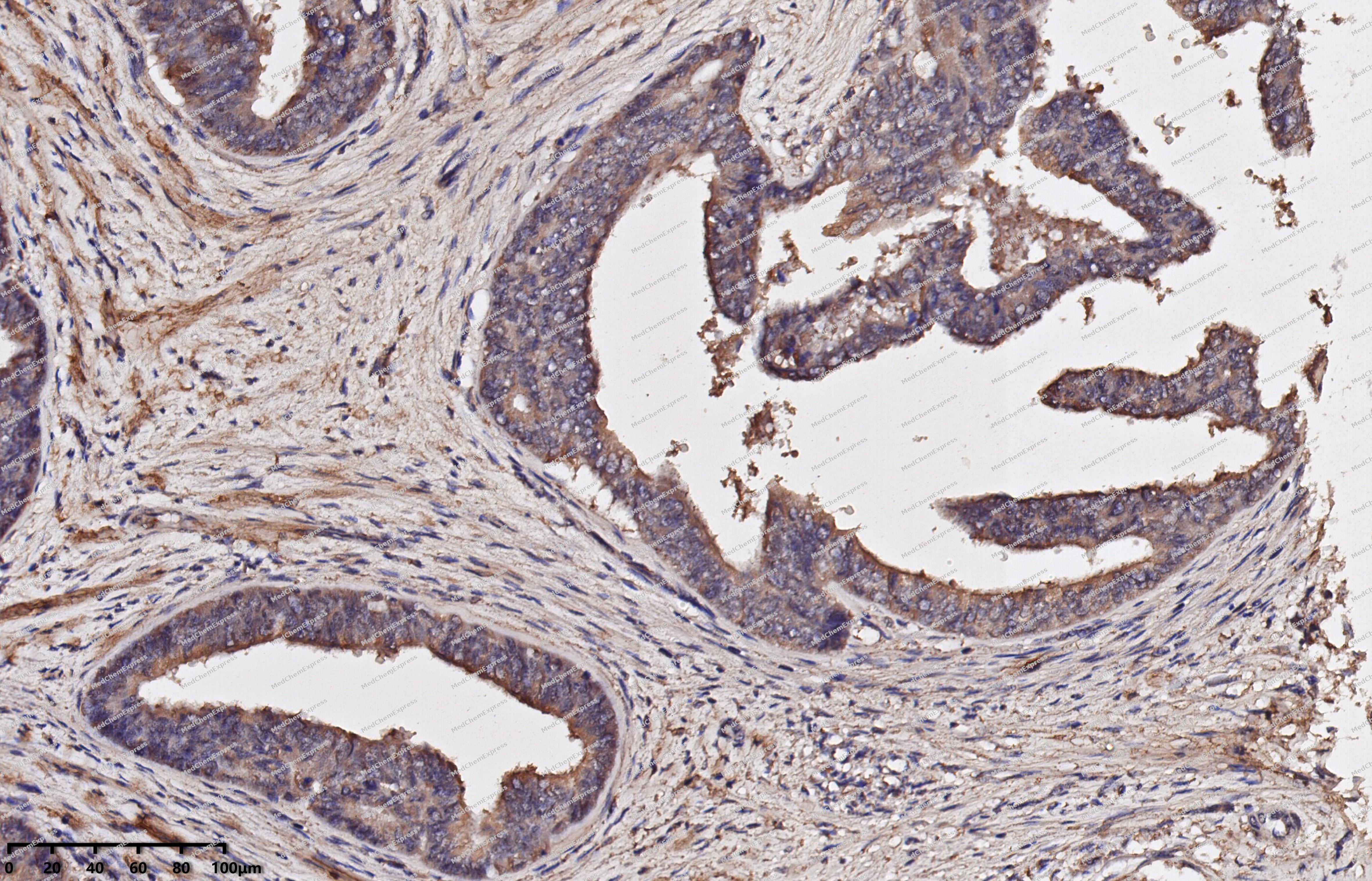 Immunohistochemical analysis of paraffin-embedded human cervical cancer using Caspase-3 antibody. The section was pre-treated using heat mediated antigen retrieval with EDTA (pH 9.0) for 14 minutes. The tissues were blocked in 5% BSA for 30 minutes at room temperature, washed with TBST, and then probed with the primary antibody (HY-P80046, 1/50) for 30 minutes at room temperature. The detection was performed using Polymer HRP-conjugated Goat Anti-Mouse/Rabbit lgG(H&L) secondary antibody (HY-P83652). DAB was used as the chromogen. Tissues were counterstained with hematoxylin and mounted with DPX.
 -
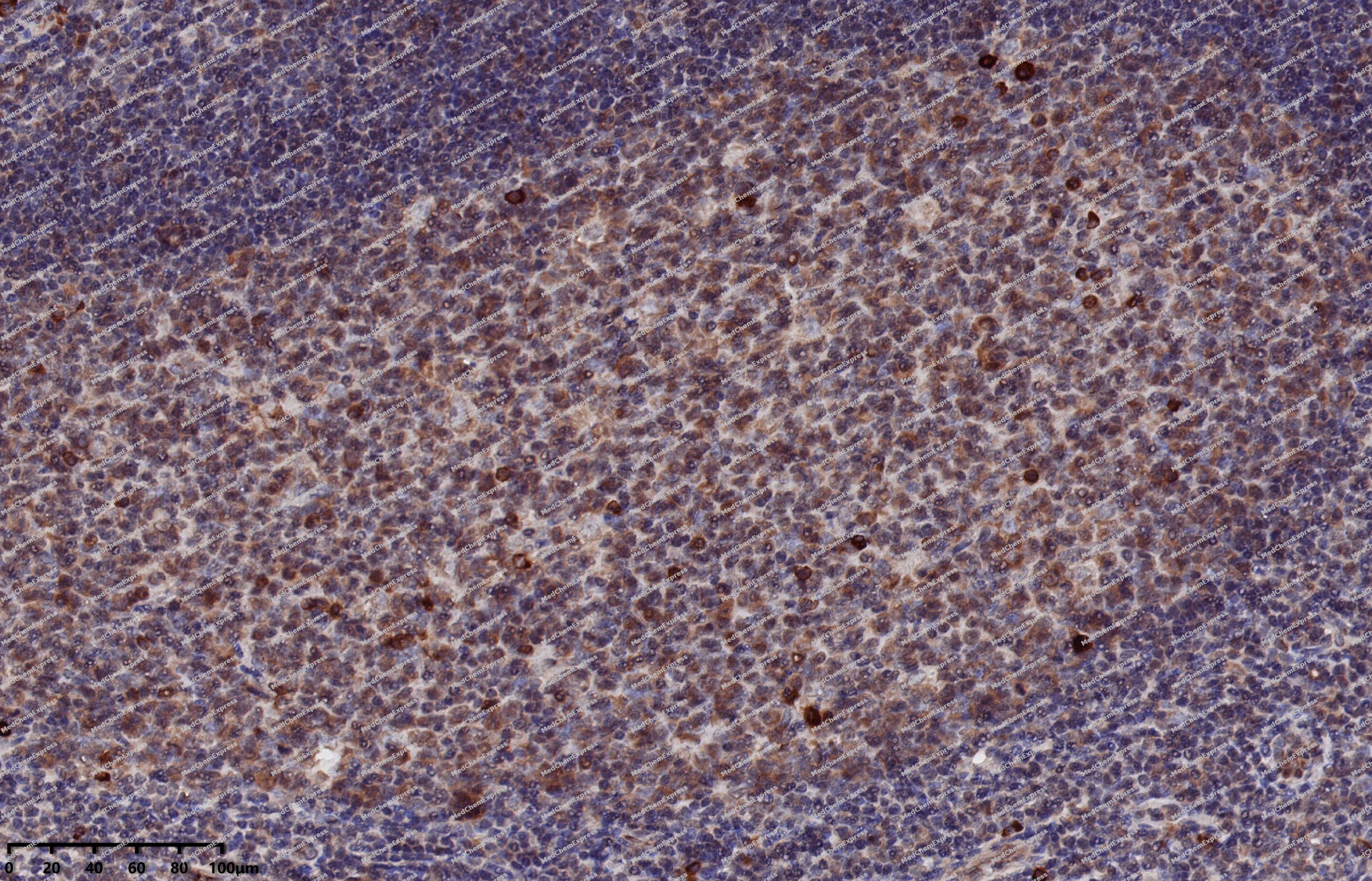 Immunohistochemical analysis of paraffin-embedded human tonsil using Caspase-3 antibody. The section was pre-treated using heat mediated antigen retrieval with EDTA (pH 9.0) for 14 minutes. The tissues were blocked in 5% BSA for 30 minutes at room temperature, washed with TBST, and then probed with the primary antibody (HY-P80046, 1/50) for 30 minutes at room temperature. The detection was performed using Polymer HRP-conjugated Goat Anti-Mouse/Rabbit lgG(H&L) secondary antibody (HY-P83652). DAB was used as the chromogen. Tissues were counterstained with hematoxylin and mounted with DPX.
 -
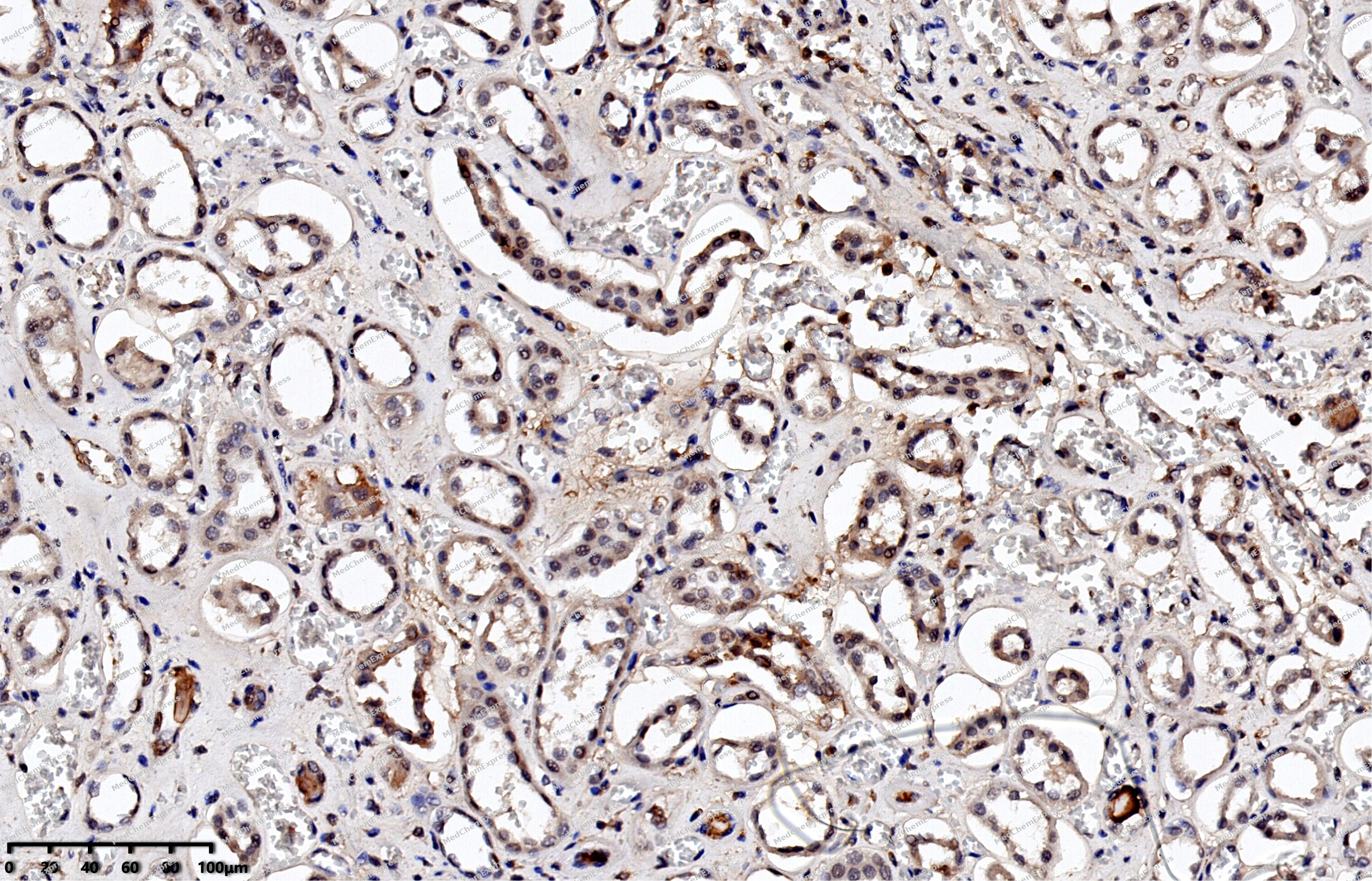 Immunohistochemical analysis of paraffin-embedded human kidney using Caspase-3 antibody. The section was pre-treated using heat mediated antigen retrieval with EDTA (pH 9.0) for 14 minutes. The tissues were blocked in 5% BSA for 30 minutes at room temperature, washed with TBST, and then probed with the primary antibody (HY-P80046, 1/50) for 30 minutes at room temperature. The detection was performed using Polymer HRP-conjugated Goat Anti-Mouse/Rabbit lgG(H&L) secondary antibody (HY-P83652). DAB was used as the chromogen. Tissues were counterstained with hematoxylin and mounted with DPX.
 -
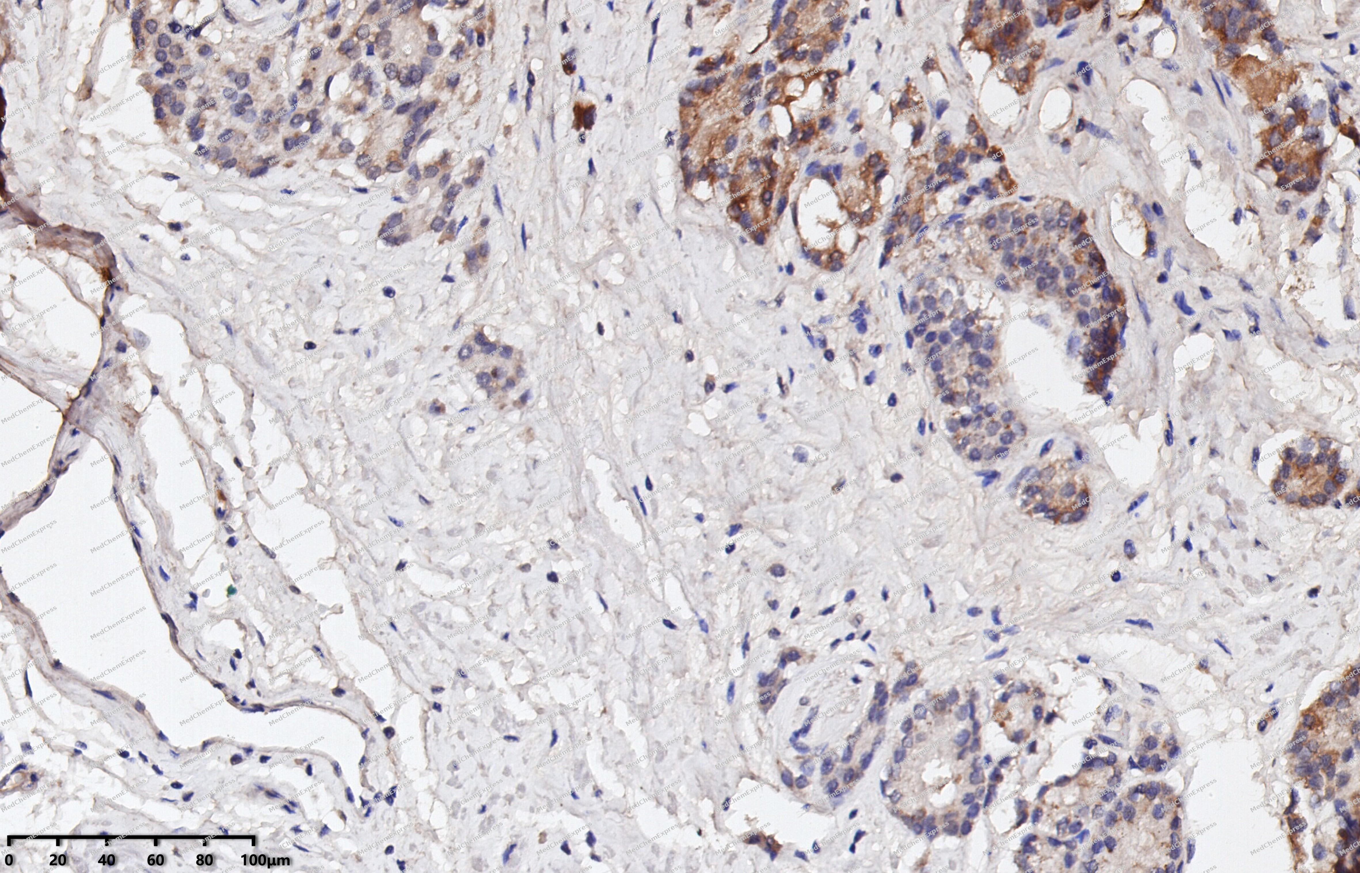 Immunohistochemical analysis of paraffin-embedded human prostate cancer using Caspase-3 antibody. The section was pre-treated using heat mediated antigen retrieval with EDTA (pH 9.0) for 14 minutes. The tissues were blocked in 5% BSA for 30 minutes at room temperature, washed with TBST, and then probed with the primary antibody (HY-P80046, 1/50) for 30 minutes at room temperature. The detection was performed using Polymer HRP-conjugated Goat Anti-Mouse/Rabbit lgG(H&L) secondary antibody (HY-P83652). DAB was used as the chromogen. Tissues were counterstained with hematoxylin and mounted with DPX.
 -
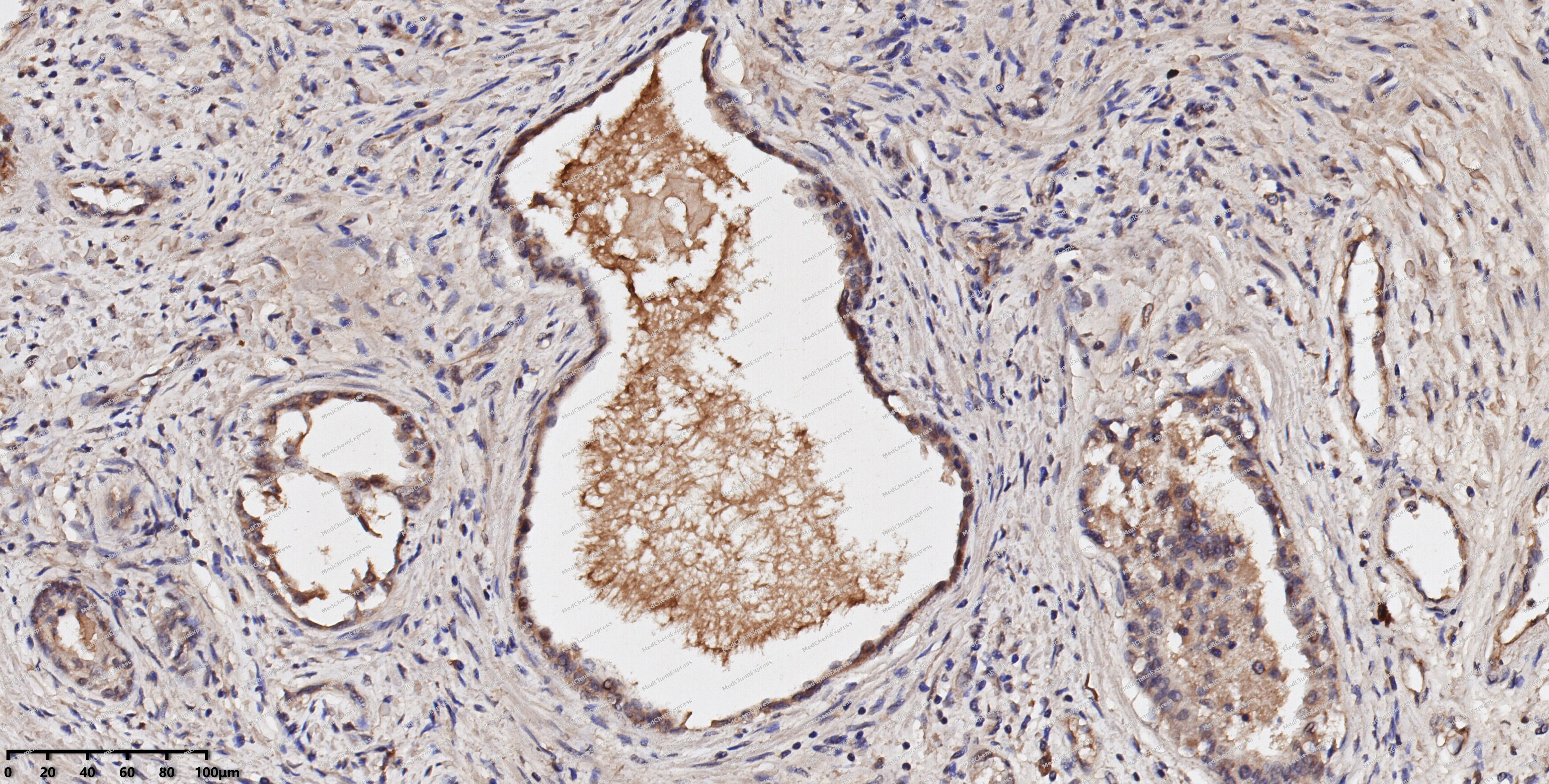 Immunohistochemical analysis of paraffin-embedded human prostate using Caspase-3 antibody. The section was pre-treated using heat mediated antigen retrieval with EDTA (pH 9.0) for 14 minutes. The tissues were blocked in 5% BSA for 30 minutes at room temperature, washed with TBST, and then probed with the primary antibody (HY-P80046, 1/50) for 30 minutes at room temperature. The detection was performed using Polymer HRP-conjugated Goat Anti-Mouse/Rabbit lgG(H&L) secondary antibody (HY-P83652). DAB was used as the chromogen. Tissues were counterstained with hematoxylin and mounted with DPX.
 -
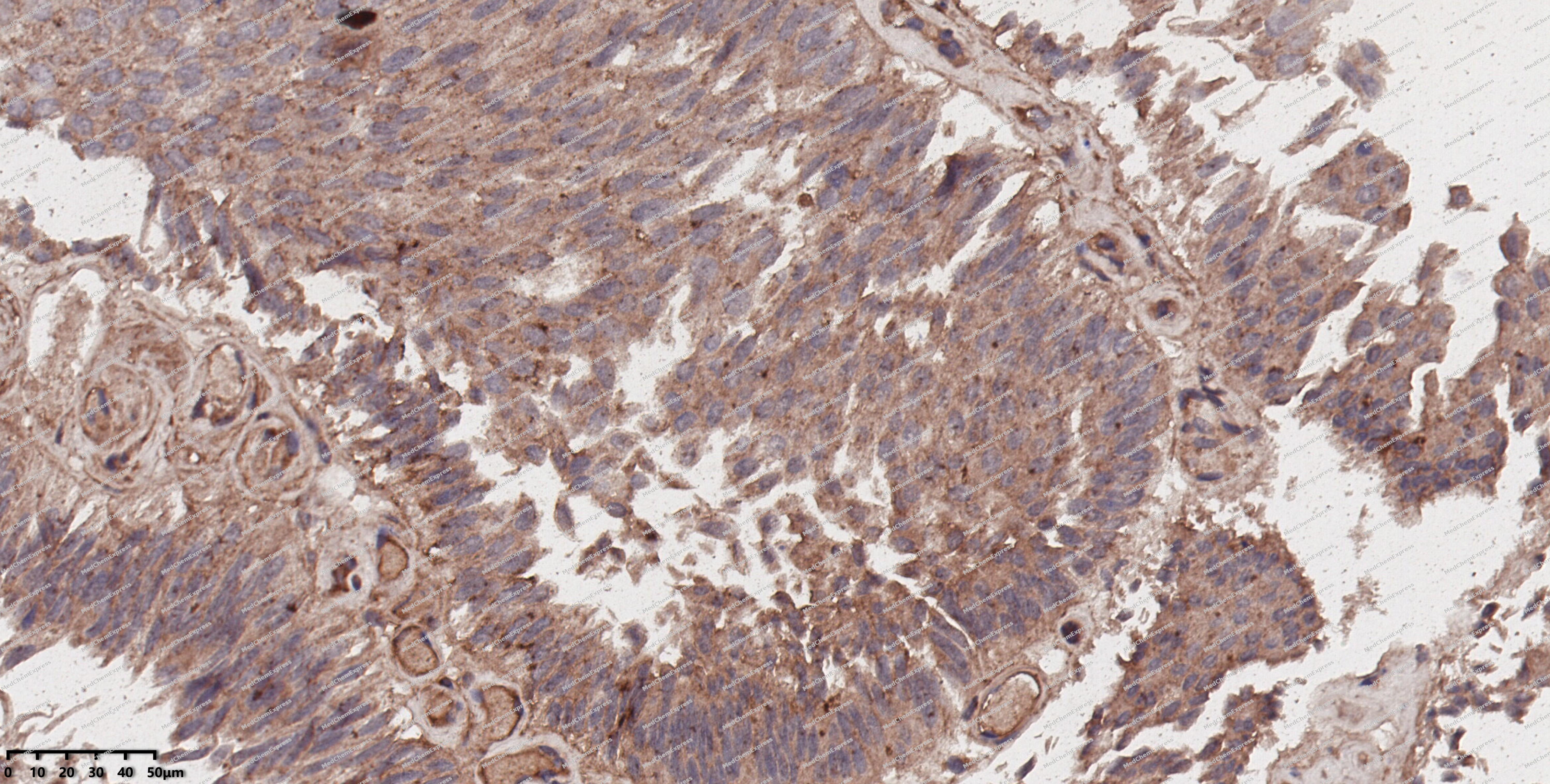 Immunohistochemical analysis of paraffin-embedded human colon cancer using Caspase-3 antibody. The section was pre-treated using heat mediated antigen retrieval with EDTA (pH 9.0) for 14 minutes. The tissues were blocked in 5% BSA for 30 minutes at room temperature, washed with TBST, and then probed with the primary antibody (HY-P80046, 1/50) for 30 minutes at room temperature. The detection was performed using Polymer HRP-conjugated Goat Anti-Mouse/Rabbit lgG(H&L) secondary antibody (HY-P83652). DAB was used as the chromogen. Tissues were counterstained with hematoxylin and mounted with DPX.
 -
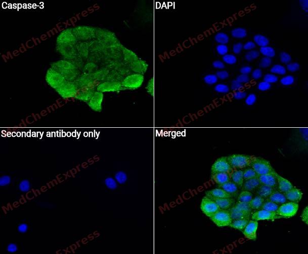 Immunocytochemistry analysis of Hela cells labeling Caspase-3 with Caspase-3 Antibody (HY-P80046)at 1/50 dilution. Cells were fixed in 4% paraformaldehyde for 15 minutes at room temperature, permeabilized with 0.1% Triton X-100 for 10 minutes at room temperature, then blocked with QuickBlock™ Blocking Buffer for Immunol Staining for 10 min at room temperature. Cells were then incubated with Caspase-3 Antibody (HY-P80046) at 1/50 dilution in QuickBlock™ Blocking Buffer for Immunol Staining at 4 ℃. Alexa Fluor® 488-conjugated AffiniPure Goat Anti-Rabbit IgG H&L(HY-P8002, Green) was used as the secondary antibody at 1/1,000 dilution. PBS instead of the primary antibody was used as the secondary antibody only control. The Nuclear counterstain was DAPI (Blue).
 -
 Flow cytometric analysis of 1X10⁶ Ramos cells labeling Caspase-3 Antibody(HY-P80046, red). Cells were fixed with 4% paraformaldehyde and permeabilised with 90% methanol. Then stained with the primary antibody at 1/50 dilution for an hour at 4℃. AF488-conjugated Goat Anti-Rabbit IgG H&L (HY-P8002) was used as the secondary antibody at 1/1,000 dilution for 30 minutes at 4℃. Rabbit IgG Isotype Control (HY-P80879, blue) was used as the isotype control, cells without incubation with primary antibody were used as the unlabeled control (black).


Background
-
Function
Caspase 3 is a central executioner caspase in apoptosis, where initiator caspase 9 activates executioner caspases 3 and 7 by proteolysis[1]. Mechanistically, caspase 3 cleaves caspase 9 at Asp330 and enhances apoptosis by relieving XIAP-mediated inhibition, rather than directly activating caspase 9[1]. This places caspase 3 within apoptosome-linked feedback control and inhibitor-of-apoptosis regulation, while preserving a distinct functional profile from caspase 7[1][2]. Compared with caspase 7, caspase 3 can cleave caspase 9 under physiologically relevant conditions, and engineered activation studies show that caspase 3 or caspase 7, but not caspase 6 alone, is sufficient to induce apoptosis[1][2]. In disease models, caspase 3 and caspase 7 deletion did not reduce infarct size or ventricular remodeling after transient myocardial ischemia-reperfusion, limiting assumptions about canonical caspase-dependent apoptosis in adult cardiomyocytes[3]. In prostate cancer samples, procaspase 3 and cleaved caspase 3 predicted biochemical progression after radical prostatectomy, supporting their use as apoptosis-related tumor markers[4]. For experimental applications, caspase 3 research benefits from isoform-aware probes because common DEVD-based peptide inhibitors and substrates cannot reliably distinguish caspase 3 from caspase 7[5].
-
Subcellular Localization
Cytoplasm
-
Expression
Tissue_specificity:It is highly expressed in the lungs, spleen, heart, liver, and kidneys. It is moderately expressed in the brain and skeletal muscle, and lowly expressed in the testes. It is also present in many cell lines, with the highest expression in immune system cells. -
Subunit
Heterotetramer that consists of two anti-parallel arranged heterodimers, each one formed by a 17 kDa (p17) and a 12 kDa (p12) subunit. Interacts with BIRC6/bruce
-
SwissProt ID
-
Synonyms
CPP32, CASP3, Caspase-3, CASP-3, Apopain, Cysteine protease CPP32, Protein Yama, SREBP cleavage activity 1, CPP-32, SCA-1
-
Research Field
Cell Biology
Documentation
-
Data Sheet (263 KB)
-
SDS (252 KB)
- English - EN (252 KB)
- Français - FR (252 KB)
- Deutsch - DE (252 KB)
- Norwegian - NO (252 KB)
- Español - ES (252 KB)
- Swedish - SV (252 KB)
- Italian - IT (252 KB)
- Korean - KR (252 KB)
- Portuguese - PT (252 KB)
-
User Guide for Antibodies (1077 KB)
References
[1]. Denault JB, et al. Caspase 3 attenuates XIAP (X-linked inhibitor of apoptosis protein)-mediated inhibition of caspase 9. Biochem J. 2007 Jul 1;405(1):11-9. [Content Brief]
[2]. Gray DC, et al. Activation of specific apoptotic caspases with an engineered small-molecule-activated protease. Cell. 2010 Aug 20;142(4):637-46. [Content Brief]
[3]. Inserte J, et al. Studies on the role of apoptosis after transient myocardial ischemia: genetic deletion of the executioner caspases-3 and -7 does not limit infarct size and ventricular remodeling. Basic Res Cardiol. 2016 Mar;111(2):18. [Content Brief]
[4]. Rodríguez-Berriguete G, et al. Prognostic value of inhibitors of apoptosis proteins (IAPs) and caspases in prostate cancer: caspase-3 forms and XIAP predict biochemical progression after radical prostatectomy. BMC Cancer. 2015 Oct 27;15:809. [Content Brief]
[5]. Batool A, et al. Eukaryotic initiation factor 4E is a novel effector of mTORC1 signaling pathway in cross talk with Mnk1. Mol Cell Biochem. 2020 Feb;465(1-2):13-26. [Content Brief]

 Powered by Bioz
