



EBP1 Antibody (YA2509)
(Synonyms: PA2G4; EBP1; Proliferation-associated protein 2G4; Cell cycle protein p38-2G4 homolog; hG4-1; ErbB3-binding protein 1)
Based on 1 Customer Validation
EBP1 Antibody (YA2509) is a Rabbit-derived and non-conjugated IgG monoclonal antibody, targeting to EBP1.
-
Host:
Rabbit
-
Isotype:
IgG
-
Application:
WB, IHC-P, ICC/IF, FC, IP
-
Reactivity :
Human, Mouse, Rat
-
Formulation:
Supplied in rabbit IgG in 50mM Tris-Glycine(pH 7.4), 0.15M NaCl, 40%Glycerol, 0.01% sodium azide and 0.05% BSA.
-
Conjugation:
Non-conjugated


To place orders, for customer services and technical support, please contact: MedChemExpress USA
Tel: 609-228-6898 E-mail: [email protected] [email protected]





Applications
| Application |
WB

WB: Western Blot
|
IHC-P
IHC-P: Immunohistochemistry-Paraffin
|
ICC/IF
ICC/IF: Immunocytochemistry/
Immunofluorescence |
FC
FC: Flow Cytometry
|
IP
IP: Immunoprecipitation
|
|---|---|---|---|---|---|
| Dilution Ratio | 1:1000-1:2000 | 1:100-1:200 | 1:50-1:200 | 1:50-1:100 | 1:40 |


Product Details
EBP1 Antibody (YA2509) is a Rabbit-derived and non-conjugated IgG monoclonal antibody, targeting to EBP1.
-
Host Rabbit

-
Clonality Recombinant,Monoclonal

-
Species ReactivityHuman, Mouse, Rat
-
Observed Molecular WeightObserved band size: 48 kDa
Note: Due to possible protein modifications or aggregation, the molecular weight should be confirmed by actual measurement, and the predicted value is for reference only.

-
Calculated Molecular Weight Predicted band size: 44 kDa

Entrez Gene: 5036 Human ; 18813 Mouse ; 288778 Rat
SwissProt: Q9UQ80 Human ; P50580 Mouse ;
OMIM: 602145 Human
A synthesized peptide derived from human EBP1 / PA2G4 aa300-394/394.
Endogenous
Affinity Purified
Non-conjugated
Unmodified
IgG


Product Properties
-
Appearance
Solution
-
Formulation
Supplied in rabbit IgG in 50mM Tris-Glycine(pH 7.4), 0.15M NaCl, 40%Glycerol, 0.01% sodium azide and 0.05% BSA.
-
Concentration
Batch-dependent, Please check the COA for the concentration of each lot. Check Lot Concentration
-
Storage & Stability
Stored at -20°C for 1 year. Avoid repeated freeze / thaw cycles.
-
Shipping
Shipping with blue ice.


Verification Images
-
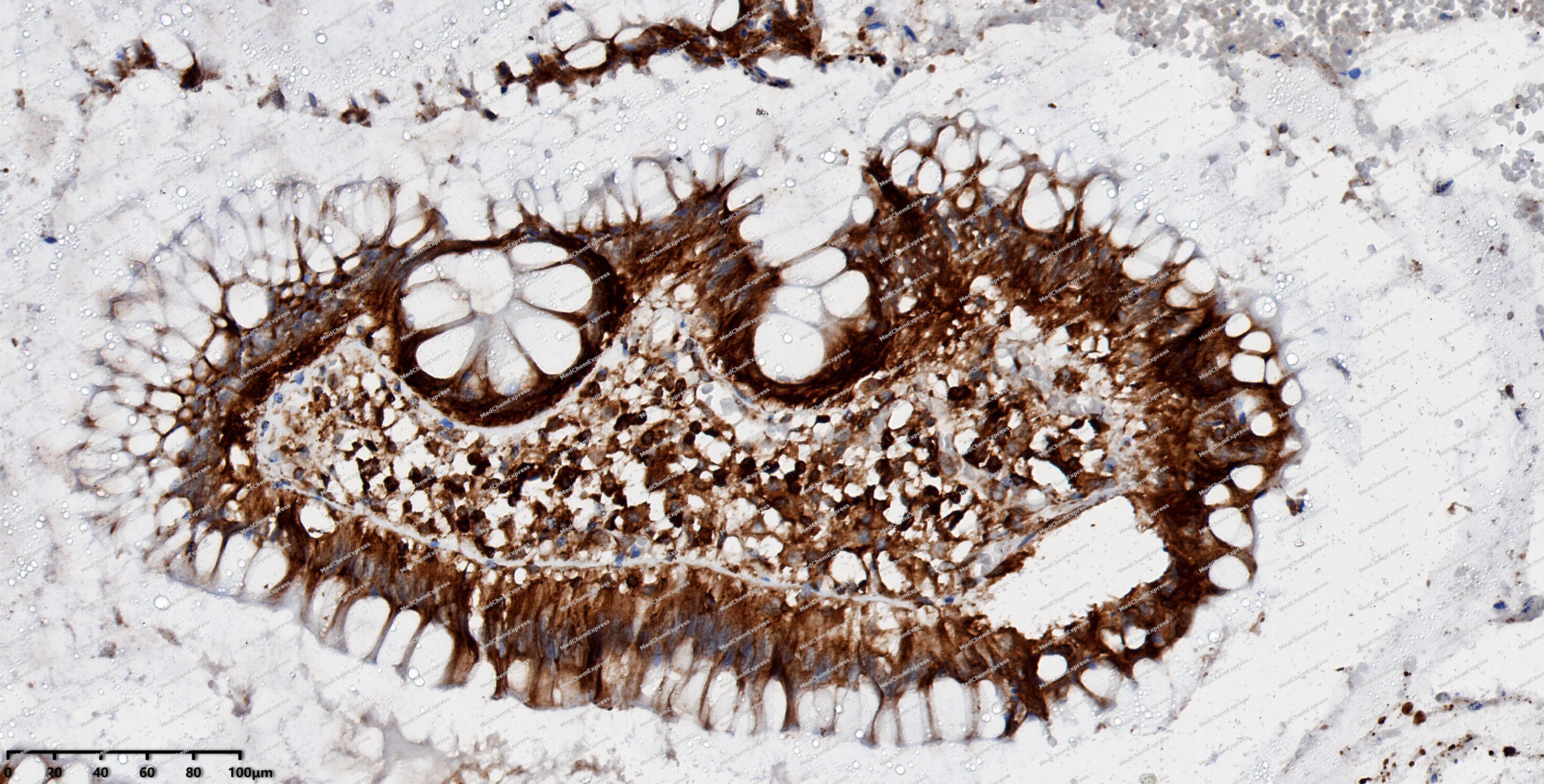 Immunohistochemical analysis of paraffin-embedded human colon cancer using EBP1 antibody. The section was pre-treated using heat mediated antigen retrieval with EDTA (pH 9.0) for 14 minutes. The tissues were blocked in 5% BSA for 30 minutes at room temperature, washed with TBST, and then probed with the primary antibody (HY-P82764, 1/200) for 30 minutes at room temperature. The detection was performed using Polymer HRP-conjugated Goat Anti-Mouse/Rabbit lgG(H&L) secondary antibody (HY-P83652). DAB was used as the chromogen. Tissues were counterstained with hematoxylin and mounted with DPX.
 -
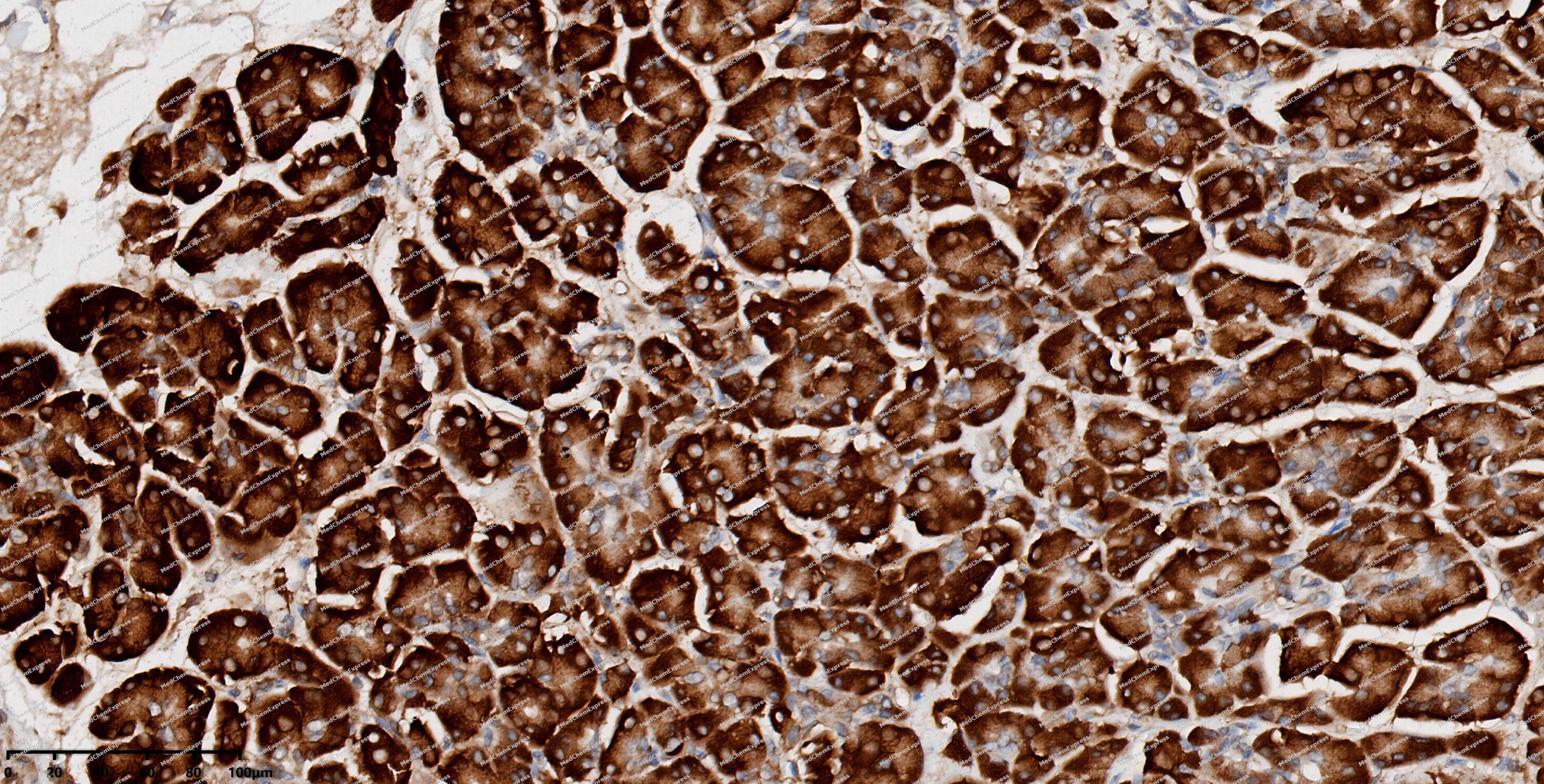 Immunohistochemical analysis of paraffin-embedded human pancreas using EBP1 antibody. The section was pre-treated using heat mediated antigen retrieval with EDTA (pH 9.0) for 14 minutes. The tissues were blocked in 5% BSA for 30 minutes at room temperature, washed with TBST, and then probed with the primary antibody (HY-P82764, 1/200) for 30 minutes at room temperature. The detection was performed using Polymer HRP-conjugated Goat Anti-Mouse/Rabbit lgG(H&L) secondary antibody (HY-P83652). DAB was used as the chromogen. Tissues were counterstained with hematoxylin and mounted with DPX.
 -
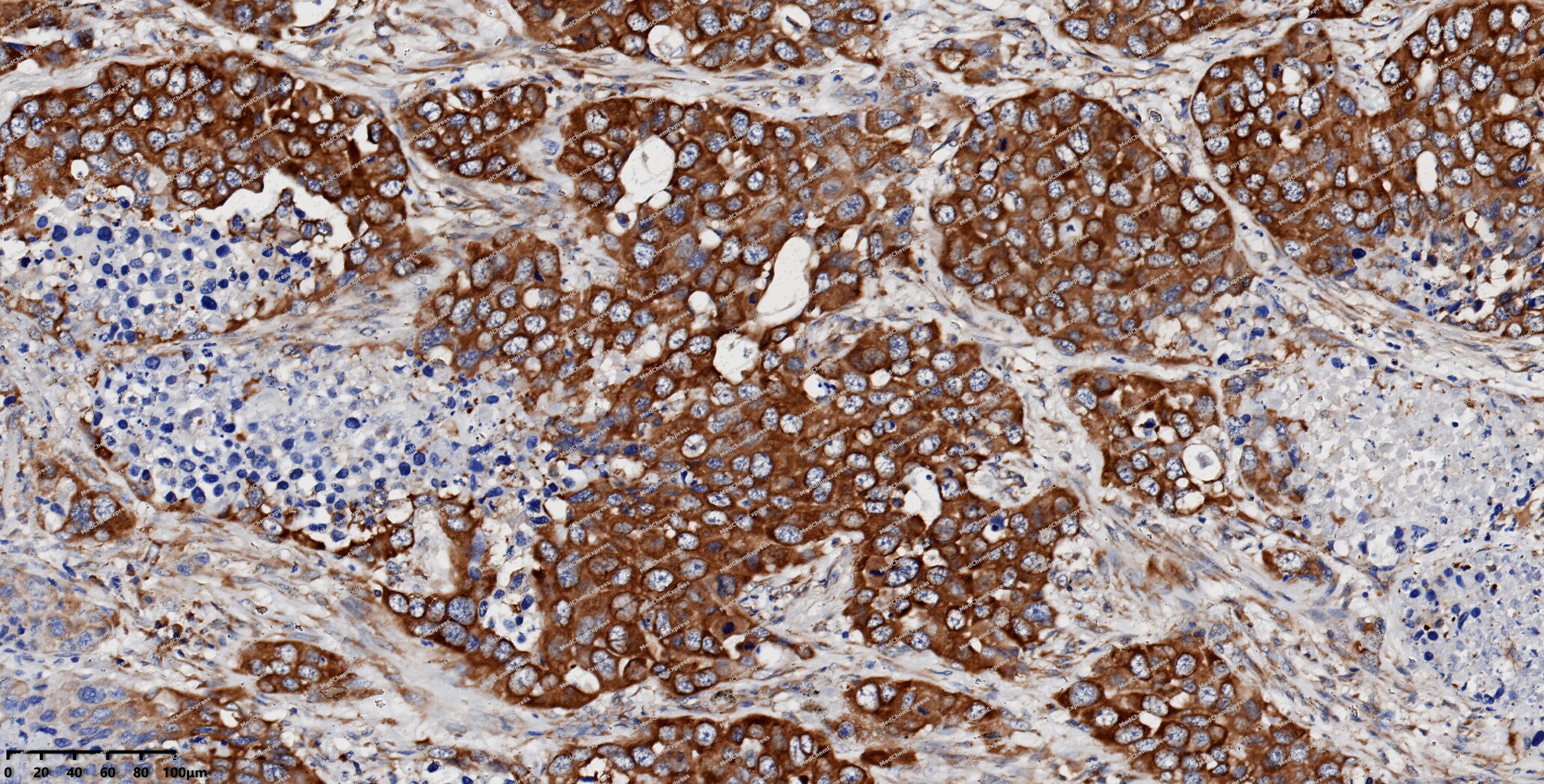 Immunohistochemical analysis of paraffin-embedded human lung cancer using EBP1 antibody. The section was pre-treated using heat mediated antigen retrieval with EDTA (pH 9.0) for 14 minutes. The tissues were blocked in 5% BSA for 30 minutes at room temperature, washed with TBST, and then probed with the primary antibody (HY-P82764, 1/200) for 30 minutes at room temperature. The detection was performed using Polymer HRP-conjugated Goat Anti-Mouse/Rabbit lgG(H&L) secondary antibody (HY-P83652). DAB was used as the chromogen. Tissues were counterstained with hematoxylin and mounted with DPX.
 -
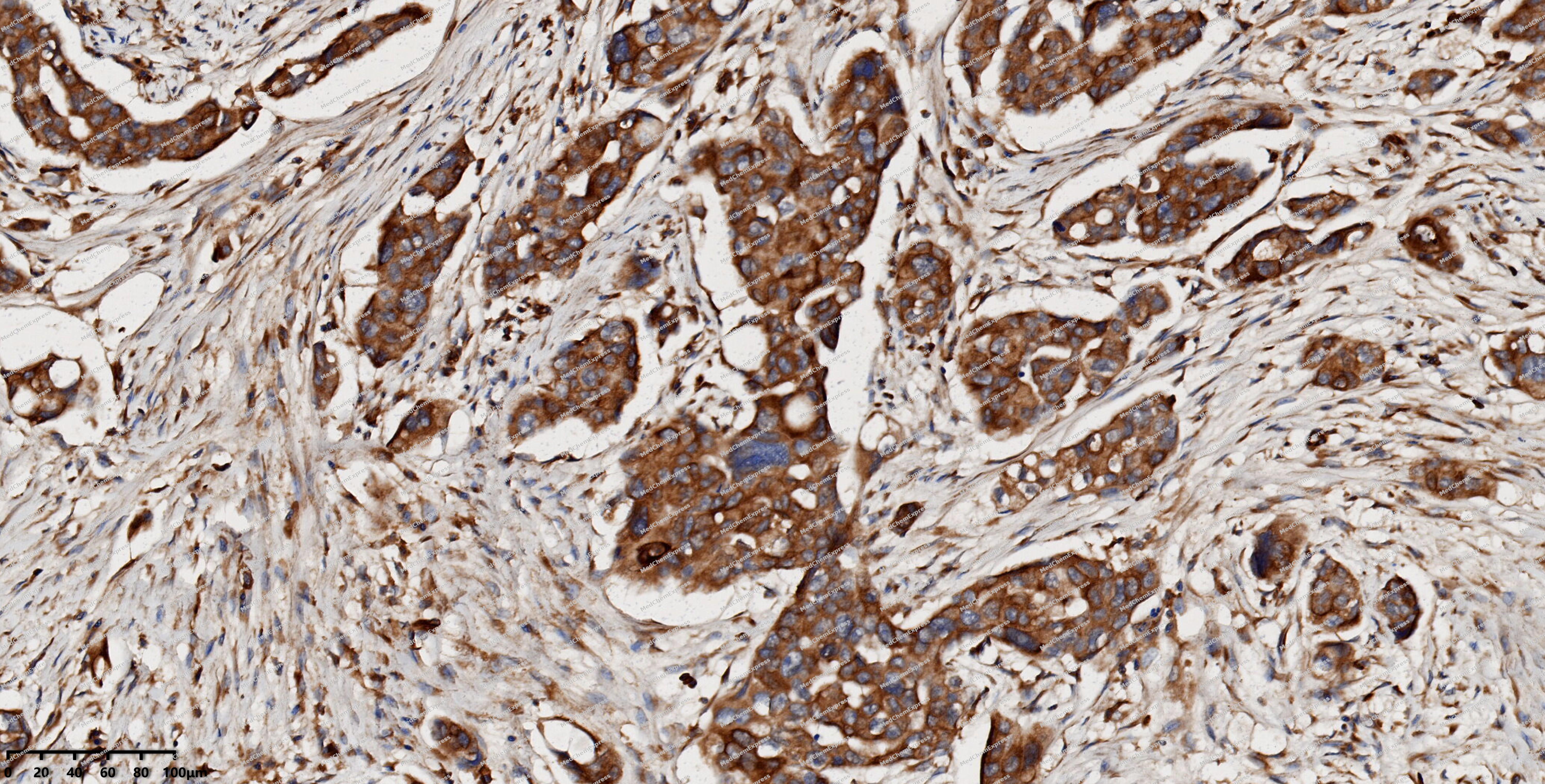 Immunohistochemical analysis of paraffin-embedded human ovarian cancer using EBP1 antibody. The section was pre-treated using heat mediated antigen retrieval with EDTA (pH 9.0) for 14 minutes. The tissues were blocked in 5% BSA for 30 minutes at room temperature, washed with TBST, and then probed with the primary antibody (HY-P82764, 1/200) for 30 minutes at room temperature. The detection was performed using Polymer HRP-conjugated Goat Anti-Mouse/Rabbit lgG(H&L) secondary antibody (HY-P83652). DAB was used as the chromogen. Tissues were counterstained with hematoxylin and mounted with DPX.
 -
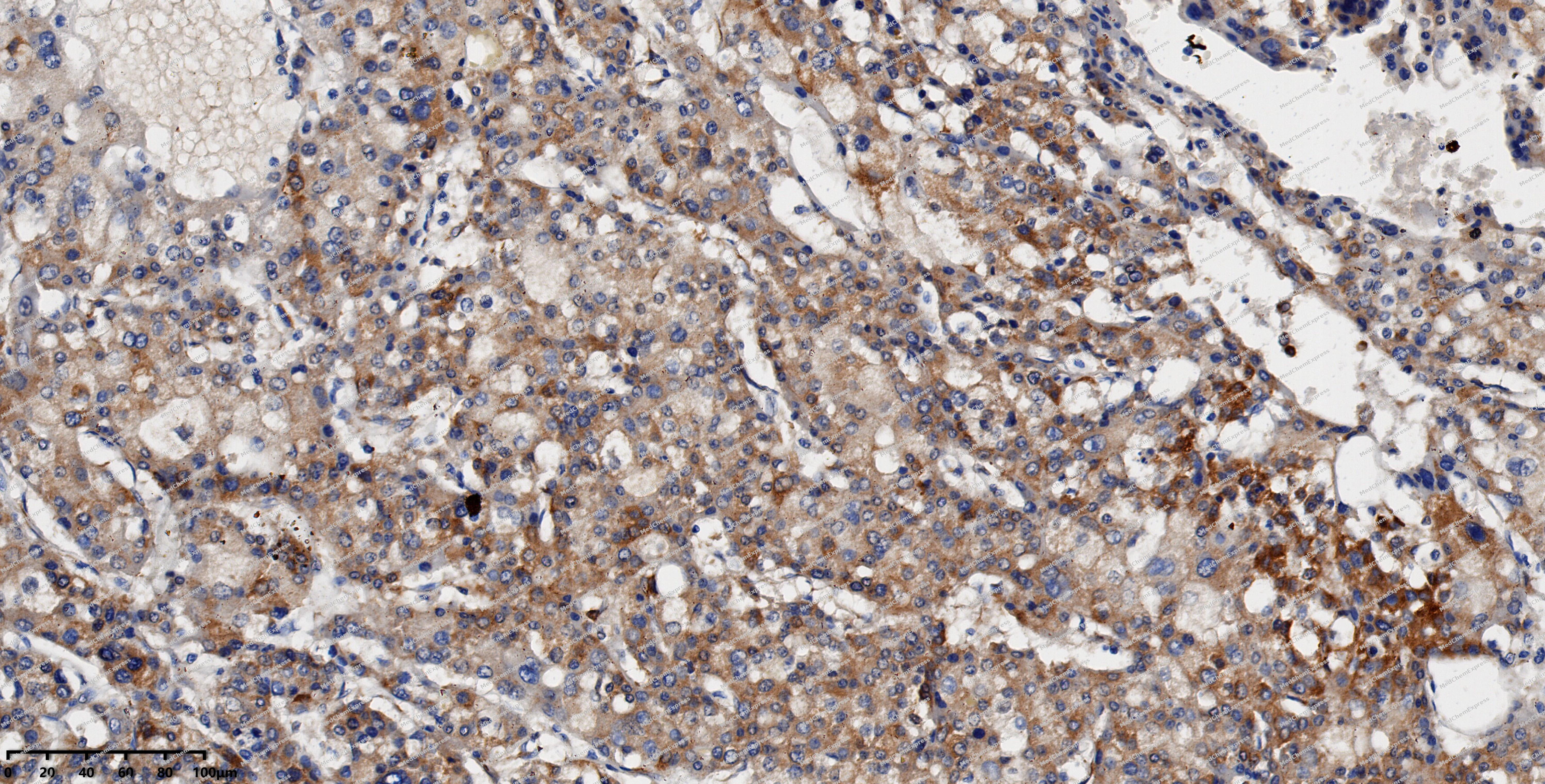 Immunohistochemical analysis of paraffin-embedded human liver cancer using EBP1 antibody. The section was pre-treated using heat mediated antigen retrieval with EDTA (pH 9.0) for 14 minutes. The tissues were blocked in 5% BSA for 30 minutes at room temperature, washed with TBST, and then probed with the primary antibody (HY-P82764, 1/200) for 30 minutes at room temperature. The detection was performed using Polymer HRP-conjugated Goat Anti-Mouse/Rabbit lgG(H&L) secondary antibody (HY-P83652). DAB was used as the chromogen. Tissues were counterstained with hematoxylin and mounted with DPX.
 -
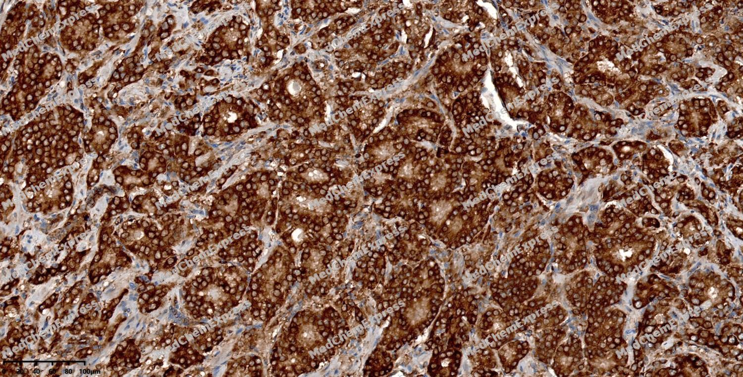 Immunohistochemical analysis of paraffin-embedded human prostate cancer using EBP1 antibody. The section was pre-treated using heat mediated antigen retrieval with EDTA (pH 9.0) for 14 minutes. The tissues were blocked in 5% BSA for 30 minutes at room temperature, washed with TBST, and then probed with the primary antibody (HY-P82764, 1/200) for 30 minutes at room temperature. The detection was performed using Polymer HRP-conjugated Goat Anti-Mouse/Rabbit lgG(H&L) secondary antibody (HY-P83652). DAB was used as the chromogen. Tissues were counterstained with hematoxylin and mounted with DPX.
 -
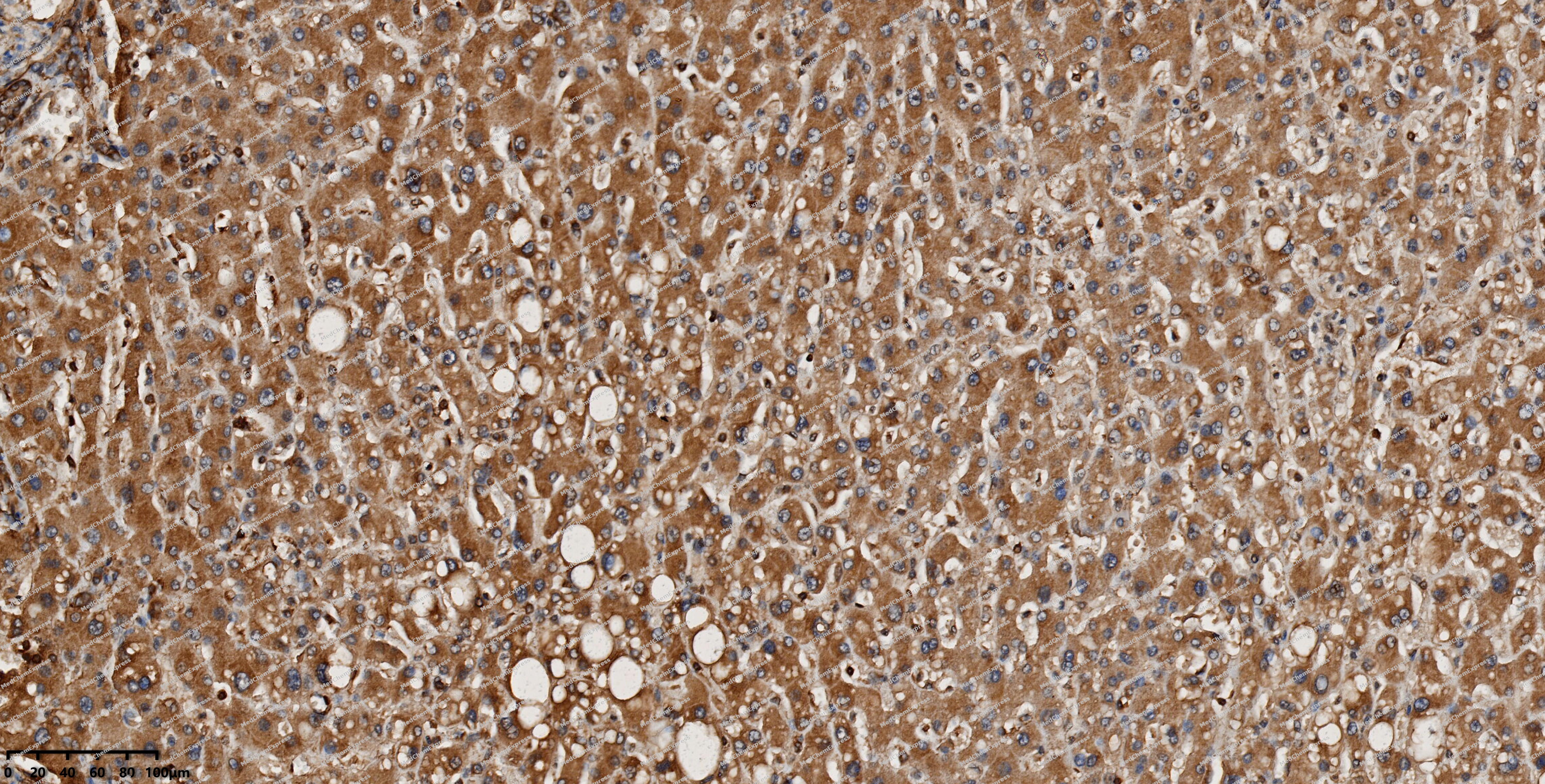 Immunohistochemical analysis of paraffin-embedded human liver using EBP1 antibody. The section was pre-treated using heat mediated antigen retrieval with EDTA (pH 9.0) for 14 minutes. The tissues were blocked in 5% BSA for 30 minutes at room temperature, washed with TBST, and then probed with the primary antibody (HY-P82764, 1/200) for 30 minutes at room temperature. The detection was performed using Polymer HRP-conjugated Goat Anti-Mouse/Rabbit lgG(H&L) secondary antibody (HY-P83652). DAB was used as the chromogen. Tissues were counterstained with hematoxylin and mounted with DPX.
 -
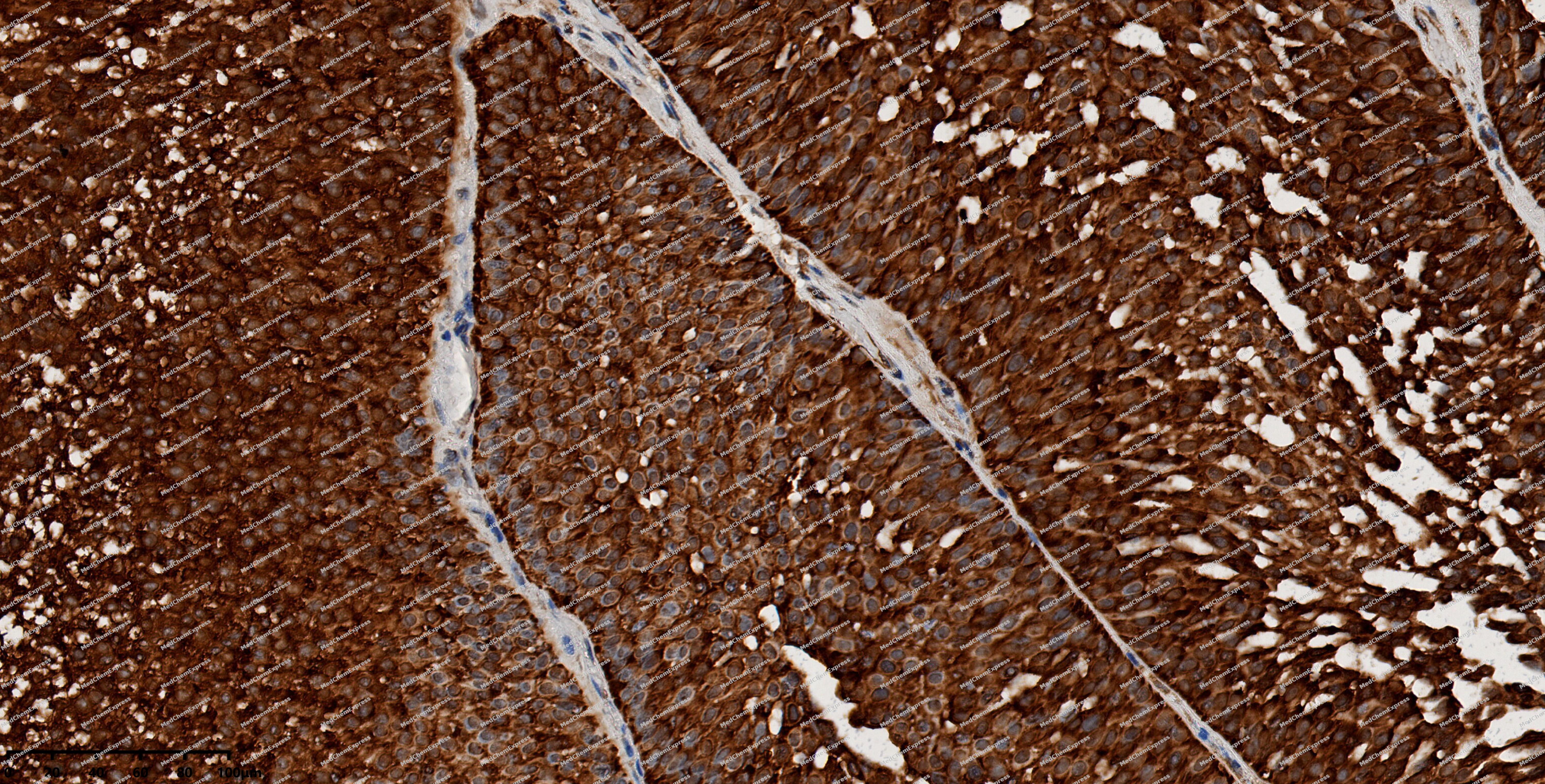 Immunohistochemical analysis of paraffin-embedded human bladder cancer using EBP1 antibody. The section was pre-treated using heat mediated antigen retrieval with EDTA (pH 9.0) for 14 minutes. The tissues were blocked in 5% BSA for 30 minutes at room temperature, washed with TBST, and then probed with the primary antibody (HY-P82764, 1/200) for 30 minutes at room temperature. The detection was performed using Polymer HRP-conjugated Goat Anti-Mouse/Rabbit lgG(H&L) secondary antibody (HY-P83652). DAB was used as the chromogen. Tissues were counterstained with hematoxylin and mounted with DPX.


Background
-
Function
EBP1 may play a role in a ERBB3-regulated signal transduction pathway. Seems be involved in growth regulation. Acts a corepressor of the androgen receptor (AR) and is regulated by the ERBB3 ligand neuregulin-1/heregulin (HRG). Inhibits transcription of some E2F1-regulated promoters, probably by recruiting histone acetylase (HAT) activity. Binds RNA. Associates with 28S, 18S and 5.8S mature rRNAs, several rRNA precursors and probably U3 small nucleolar RNA. May be involved in regulation of intermediate and late steps of rRNA processing. May be involved in ribosome assembly. Mediates cap-independent translation of specific viral IRESs (internal ribosomal entry site). Regulates cell proliferation, differentiation, and survival. Isoform 1 suppresses apoptosis whereas isoform 2 promotes cell differentiation
-
Subcellular Localization
Cytoplasm; Nucleus, nucleolus; Cytoplasm
-
Expression
Tissue_specificity:Isoform 2 is undetectable whereas isoform 1 is strongly expressed in cancer cells (at protein level) . Isoform 1 and isoform 2 are widely expressed, including heart, brain, lung, pancreas, skeletal muscle, kidney, placenta and liver -
Isoforms & Post-Translational Modification
Q9UQ80 has 2 isomers: Q9UQ80-1: 43787 Da (predicted); Q9UQ80-2: 38059 Da (predicted).
Phosphorylated on serine and threonine residues. Phosphorylation is enhanced by HRG treatment. Basal phosphorylation is PKC-dependent and HRG-induced phosphorylation is predominantly PKC-independent. Phosphorylation at Ser-361 by PKC/PRKCD regulates its nucleolar localization;In cancer cells, isoform 2 is polyubiquitinated leading to its proteasomal degradation and phosphorylation by PKC/PRKCD enhances polyubiquitination -
Subunit
Isoform 2 interacts with the cytoplasmic domain of non-phosphorylated ERBB3; the interaction requires PKC activity. Interacts with AR. Treatment with HRG leads to dissociation from ERBB3 and increases association with AR. Interacts with NCL/nucleolin. Component of a ribonucleoprotein complex containing at least PA2G4, NCL, TOP1, PABPC2, RPLP0, acetylated histone H1 (HIST1H1A or H1F1), histone H1 2/4, RPL4, RPL8, RPL15, RPL18, RPL18A, RPL21, RPL11, RPL12, RPL28, RPL27, RPLP2 and RPL24. Interacts with HDAC2. Interacts with RB1; the interaction is enhanced upon PA2G4 dephosphorylation. Interacts with AKT1 (By similarity). Isoform 1 and isoform 2 interact with RNF20. Isoform 2 interacts with HUWE1. Interacts with DNAJC21 (PubMed:27346687)
-
SwissProt ID
-
Synonyms
PA2G4; EBP1; Proliferation-associated protein 2G4; Cell cycle protein p38-2G4 homolog; hG4-1; ErbB3-binding protein 1
-
Research Field
Epigenetics and Nuclear Signaling


Documentation

 Powered by Bioz
