



HIPK2 Antibody (YA1913)
(Synonyms: hHIPk2; Hipk2; Nbak1; Stank)
Based on 1 Customer Validation
HIPK2 Antibody (YA1913) is a Rabbit-derived and non-conjugated IgG monoclonal antibody, targeting to HIPK2.
-
Host:
Rabbit
-
Isotype:
IgG
-
Application:
WB, ICC/IF, IP, FC
-
Reactivity :
Human, Mouse
-
Formulation:
Supplied in 50mM Tris-Glycine(pH 7.4), 0.15M NaCl, 40%Glycerol, 0.01% sodium azide and 0.05% BSA.
-
Conjugation:
Non-conjugated


To place orders, for customer services and technical support, please contact: MedChemExpress USA
Tel: 609-228-6898 E-mail: [email protected] [email protected]





Applications
| Application |
WB

WB: Western Blot
|
ICC/IF
ICC/IF: Immunocytochemistry/
Immunofluorescence |
IP
IP: Immunoprecipitation
|
FC
FC: Flow Cytometry
|
|---|---|---|---|---|
| Dilution Ratio | 1:500-1:1000 | 1:50-1:200 | 1:30 | 1:50-1:100 |


Product Details
HIPK2 Antibody (YA1913) is a Rabbit-derived and non-conjugated IgG monoclonal antibody, targeting to HIPK2.
-
Host Rabbit

-
Clonality Recombinant,Monoclonal

-
Species ReactivityHuman, Mouse
-
Observed Molecular WeightObserved band size: 130,100 kDa
Note: Due to possible protein modifications or aggregation, the molecular weight should be confirmed by actual measurement, and the predicted value is for reference only.

-
Calculated Molecular Weight Predicted band size: 131 kDa

Entrez Gene: 28996 Human ; 15258 Mouse ;
SwissProt: Q9H2X6 Human ; Q9QZR5 Mouse ;
OMIM: 606868 Human
A synthesized peptide derived from human HIPK2 aa150-200/1198.
Endogenous
Affinity Chromatography
Non-conjugated
Unmodified
IgG


Product Properties
-
Appearance
Liquid
-
Formulation
Supplied in 50mM Tris-Glycine(pH 7.4), 0.15M NaCl, 40%Glycerol, 0.01% sodium azide and 0.05% BSA.
-
Concentration
Batch-dependent, Please check the COA for the concentration of each lot. Check Lot Concentration
-
Storage & Stability
Stored at -20°C for 1 year. Avoid repeated freeze / thaw cycles.
-
Shipping
Shipping with blue ice.


Verification Images
-
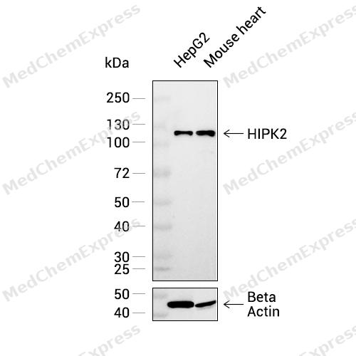
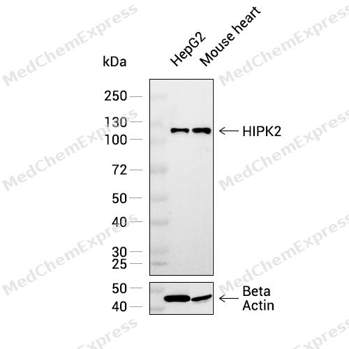 Western blot analysis was performed on extracts from HepG2 (lane 1, 15 μg), and Mouse heart (lane 2, 15 μg) using HIPK2 Rabbit mAb.Proteins were transferred to a PVDF membrane and blocked with 5% non - fat milk in TBST at 4°C overnight.The primary antibody (1:1000 dilution) and the loading control antibody (beta-Actin, HY-P83730, 1:20000 dilution) were incubated in 5% non-fat milk in TBST for 1 hour at 37°C.Goat Anti - Rabbit IgG - HRP Secondary Antibody (1:20000 dilution) was then applied for 40 minutes at 37°C.
 -
 Flow cytometric analysis of 1X10⁶ Raji cells labeling HIPK2 Antibody (HY-P82168, red). Cells were fixed with 4% paraformaldehyde and permeabilised with 90% methanol. Then stained with the primary antibody at 1/100 dilution for an hour at 4℃. AF488-conjugated Goat Anti-Rabbit IgG H&L (HY-P8002) was used as the secondary antibody at 1/1,000 dilution for 30 minutes at 4℃. Rabbit IgG Isotype Control (HY-P80879, blue) was used as the isotype control, cells without incubation with primary antibody were used as the unlabeled control (black).


Background
-
Function
HIPK2 is a Serine/threonine-protein kinase involved in transcription regulation, p53/TP53-mediated cellular apoptosis and regulation of the cell cycle. Acts as a corepressor of several transcription factors, including SMAD1 and POU4F1/Brn3a and probably NK homeodomain transcription factors. Phosphorylates PDX1, ATF1, PML, p53/TP53, CREB1, CTBP1, CBX4, RUNX1, EP300, CTNNB1, HMGA1, ZBTB4 and DAZAP2. Inhibits cell growth and promotes apoptosis through the activation of p53/TP53 both at the transcription level and at the protein level (by phosphorylation and indirect acetylation). The phosphorylation of p53/TP53 may be mediated by a p53/TP53-HIPK2-AXIN1 complex. Involved in the response to hypoxia by acting as a transcriptional co-suppressor of HIF1A. Mediates transcriptional activation of TP73. In response to TGFB, cooperates with DAXX to activate JNK. Negative regulator through phosphorylation and subsequent proteasomal degradation of CTNNB1 and the antiapoptotic factor CTBP1. In the Wnt/beta-catenin signaling pathway acts as an intermediate kinase between MAP3K7/TAK1 and NLK to promote the proteasomal degradation of MYB. Phosphorylates CBX4 upon DNA damage and promotes its E3 SUMO-protein ligase activity. Activates CREB1 and ATF1 transcription factors by phosphorylation in response to genotoxic stress. In response to DNA damage, stabilizes PML by phosphorylation. PML, HIPK2 and FBXO3 may act synergically to activate p53/TP53-dependent transactivation. Promotes angiogenesis, and is involved in erythroid differentiation, especially during fetal liver erythropoiesis. Phosphorylation of RUNX1 and EP300 stimulates EP300 transcription regulation activity. Triggers ZBTB4 protein degradation in response to DNA damage. In response to DNA damage, phosphorylates DAZAP2 which localizes DAZAP2 to the nucleus, reduces interaction of DAZAP2 with HIPK2 and prevents DAZAP2-dependent ubiquitination of HIPK2 by E3 ubiquitin-protein ligase SIAH1 and subsequent proteasomal degradation. Modulates HMGA1 DNA-binding affinity. In response to high glucose, triggers phosphorylation-mediated subnuclear localization shifting of PDX1. Involved in the regulation of eye size, lens formation and retinal lamination during late embryogenesis[1].
-
Subcellular Localization
Nucleus, PML body; Cytoplasm; Cytoplasm, Stress granule
-
Expression
Tissue_specificity:Highly expressed in heart, muscle and kidney. Weakly expressed in a ubiquitous way. Down-regulated in several thyroid and breast tumors
Induction:Unstable in unstressed cells but stabilized upon DNA damage. Induced by UV irradiation and other genotoxic agents (adriamycin ADR, cisplatin CDDP, etoposide, IR, roscovitin) , thus triggering p53/TP53 apoptotic response. Consistutively negatively regulated by SIAH1 and WSB1 through proteasomal degradation. This negative regulation is impaired upon genotoxic stress. Repressed upon hypoxia (often associated with tumors) , through MDM2- (an E3 ubiquitin ligases) mediated proteasomal degradation, thus inactivating p53/TP53 apoptotic response. This hypoxia repression is reversed by zinc. The stabilization mediated by DNA damage requires the damage checkpoint kinases ATM and ATR -
Isoforms & Post-Translational Modification
Q9H2X6 has 3 isomers: Q9H2X6-1: 130966 Da (predicted); Q9H2X6-2: 101013 Da (predicted); Q9H2X6-3: 128158 Da (predicted).
Autophosphorylation at Tyr-361 in the activation loop activates the kinase and promotes nuclear localization;Sumoylated. When conjugated it is directed to nuclear speckles. Desumoylated by SENP1 (By similarity). Sumoylation on Lys-32 is promoted by the E3 SUMO-protein ligase CBX4;Ubiquitinated by FBXO3, WSB1 and SIAH1, leading to rapid proteasome-dependent degradation. The degradation mediated by FBXO3, but not ubiquitination, is prevented in the presence of PML. The degradation mediated by WSB1 and SIAH1 is reversibly reduced upon DNA damage;Cleaved at Asp-923 and Asp-984 by CASP6 in a p53/TP53-dependent manner. The cleaved form lacks the autoinhibitory C-terminal domain (AID), resulting in a hyperactive kinase, which potentiates p53/TP53 Ser-46 phosphorylation and subsequent activation of the cell death machinery -
Subunit
Interacts with CREB1, SIAH1, WSB1, CBX4, TRADD, p53/TP53, TP73, TP63, CREBBP, DAXX, P53DINP1, SKI, SMAD1, SMAD2 and SMAD3, but not SMAD4. Interacts with ATF1, PML, RUNX1, EP300, NKX1-2, NKX2-5, UBE2I, HMGA1, CTBP1, AXIN1, NLK, MYB, POU4F1, POU4F2, POU4F3, UBE2I, UBL1 and ZBTB4. Probably part of a complex consisting of p53/TP53, HIPK2 and AXIN1. Interacts with SP100; positively regulates TP53-dependent transcription. Interacts with SUMO1P1/SUMO5 (PubMed:27211601). Interacts with DAZAP2; the interaction results in phosphorylation of DAZAP2 which causes localization of DAZAP2 to the nucleus, reduces interaction of DAZAP2 with HIPK2 and prevents DAZAP2-dependent degradation of HIPK2 (PubMed:33591310). Interacts with SIAH1; the interaction is promoted by DAZAP2 and results in SIAH1-mediated ubiquitination and subsequent proteasomal degradation of HIPK2 (PubMed:33591310)
-
SwissProt ID
-
Synonyms
hHIPk2; Hipk2; Nbak1; Stank
-
Research Field
Cell Biology


Documentation
-
Data Sheet (259 KB)
-
SDS (251 KB)
- English - EN (251 KB)
- Français - FR (251 KB)
- Deutsch - DE (251 KB)
- Norwegian - NO (251 KB)
- Español - ES (251 KB)
- Swedish - SV (251 KB)
- Italian - IT (251 KB)
- Korean - KR (251 KB)
- Portuguese - PT (251 KB)
-
User Guide for Antibodies (1077 KB)

 Powered by Bioz
