



PDLIM1 Antibody (YA1565)
(Synonyms: PDLIM1; CLIM1; CLP36; PDZ and LIM domain protein 1; C-terminal LIM domain protein 1; Elfin; LIM domain protein CLP-36)
Based on 1 Customer Validation
PDLIM1 Antibody (YA1565) is a Rabbit-derived and non-conjugated IgG monoclonal antibody, targeting to PDLIM1.
-
Host:
Rabbit
-
Isotype:
IgG
-
Application:
WB, ICC/IF, IP
-
Reactivity :
Human, Mouse, Rat
-
Formulation:
Supplied in 50mM Tris-Glycine(pH 7.4), 0.15M NaCl, 40% Glycerol, 0.01% Sodium azide and 0.05% BSA
-
Conjugation:
Non-conjugated


To place orders, for customer services and technical support, please contact: MedChemExpress USA
Tel: 609-228-6898 E-mail: [email protected] [email protected]





Applications
| Application |
WB

WB: Western Blot
|
ICC/IF
ICC/IF: Immunocytochemistry/
Immunofluorescence |
IP
IP: Immunoprecipitation
|
|---|---|---|---|
| Dilution Ratio | 1:500-1:1000 | 1:50-1:200 | 1:20 |


Product Details
PDLIM1 Antibody (YA1565) is a Rabbit-derived and non-conjugated IgG monoclonal antibody, targeting to PDLIM1.
-
Host Rabbit

-
Clonality Recombinant,Monoclonal

-
Species ReactivityHuman, Mouse, Rat
-
Observed Molecular WeightObserved band size: 36 kDa
Note: Due to possible protein modifications or aggregation, the molecular weight should be confirmed by actual measurement, and the predicted value is for reference only.

-
Calculated Molecular Weight Predicted band size: 36 kDa

Entrez Gene: 9124 Human ; 54132 Mouse ;
SwissProt: O00151 Human ; O70400 Mouse ; Q3TZ17 Mouse ;
OMIM: 605900 Human
A synthetic peptide of human PDLIM1
Endogenous
Affinity Purified
Non-conjugated
Unmodified
IgG


Product Properties
-
Appearance
Solution
-
Formulation
Supplied in 50mM Tris-Glycine(pH 7.4), 0.15M NaCl, 40% Glycerol, 0.01% Sodium azide and 0.05% BSA
-
Concentration
Batch-dependent, Please check the COA for the concentration of each lot. Check Lot Concentration
-
Storage & Stability
Stored at -20°C for 1 year. Avoid repeated freeze / thaw cycles.
-
Shipping
Shipping with blue ice.


Verification Images
-
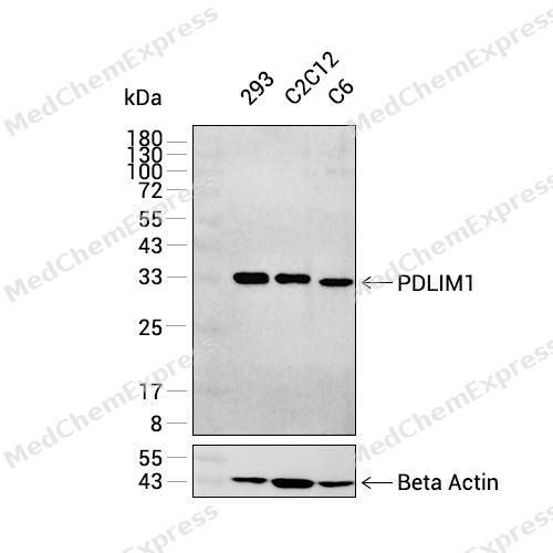 Western blot analysis was performed on extracts from 293 (lane 1, 15 μg), C2C12 (lane 2, 15 μg), and C6 (lane 3, 15 μg) using PDLIM1 Rabbit mAb.Proteins were transferred to a PVDF membrane and blocked with 5% non-fat milk in TBST at 4°C overnight.The primary antibody (1:1000 dilution) and the loading control antibody (beta-Actin, HY-P83730, 1:30000 dilution) were incubated in 5% non-fat milk in TBST for 1 hour at 37°C.Goat Anti-Rabbit IgG-HRP Secondary Antibody (1:20000 dilution) was then applied for 40 minutes at 37°C.
 -
 Western blot analysis of extracts from K562(lane 2(20ug) , C6(lane 3(20ug) and NIH/3T3(lane 4(40ug) using PDLIM1 Antibody (HY-P81820) Rabbit mAb. Proteins were transferred to a PVDF membrane and blocked with 5% non-fat milk in TBST for 2 hour at room temperature. The primary antibody (1/1000) and Loading control antibody (Beta Actin, HY-P83730, 1/10000) was used in 5% non-fat milk in TBST at 4°C overnight. Goat Anti-Mouse/Rabbit IgG-HRP Secondary Antibody (1/10000) was used for 1 hour at room temperature.
 -
 Immunocytochemistry analysis of 293T cells labeling PDLIM1 Antibody (HY-P81820) at 1/50 dilution. Cells were fixed in 4% paraformaldehyde for 15 minutes at room temperature, permeabilized with 0.1% Triton X-100 for 10 minutes at room temperature, then blocked with BSA for Immunol Staining for 10 min at room temperature. Cells were then incubated with PDLIM1 Antibody (HY-P81820) dilution in BSA for Immunol Staining at 4 ℃overnight. AF488-conjugated AffiniPure Goat Anti-Rabbit IgG H&L(HY-P8002, Green) was used as the secondary antibody at 1/1,000 dilution. PBS instead of the primary antibody was used as the secondary antibody only control. The Nuclear counterstain was DAPI (Blue).
 -
 Immunocytochemistry analysis of 293T cells labeling PDLIM1 Antibody (HY-P81820) at 1/200dilution. Cells were fixed in 4% paraformaldehyde for 15 minutes at room temperature, permeabilized with 0.1% Triton X-100 for 10 minutes at room temperature, then blocked with BSA for Immunol Staining for 10 min at room temperature. Cells were then incubated with PDLIM1 Antibody (HY-P81820) dilution in BSA for Immunol Staining at 4 ℃overnight. AF488-conjugated AffiniPure Goat Anti-Rabbit IgG H&L(HY-P8002, Green) was used as the secondary antibody at 1/1,000 dilution. PBS instead of the primary antibody was used as the secondary antibody only control. The Nuclear counterstain was DAPI (Blue).


Background
-
Function
PDLIM1 is a Cytoskeletal protein that may act as an adapter that brings other proteins (like kinases) to the cytoskeleton. Involved in assembly, disassembly and directioning of stress fibers in fibroblasts. Required for the localization of ACTN1 and PALLD to stress fibers. Required for cell migration and in maintaining cell polarity of fibroblasts[1].
-
Subcellular Localization
Cytoplasm; Cytoplasm, cytoskeleton; Cytoplasm, myofibril, sarcomere, Z line
-
Expression
Tissue_specificity:Strongly expressed in the heart and skeletal muscle, moderately expressed in the spleen, small intestine, colon, placenta, and lung. A lower level expression is seen in liver, thymus, kidney, prostate and pancreas and is not found in the brain, testis, ovary, and peripheral blood leukocytes -
Subunit
Interacts with ACTN1, ACTN2 and ACTN4
-
SwissProt ID
-
Synonyms
PDLIM1; CLIM1; CLP36; PDZ and LIM domain protein 1; C-terminal LIM domain protein 1; Elfin; LIM domain protein CLP-36
-
Research Field
Epigenetics and Nuclear Signaling


Documentation
-
Data Sheet (259 KB)
-
SDS (251 KB)
- English - EN (251 KB)
- Français - FR (251 KB)
- Deutsch - DE (251 KB)
- Norwegian - NO (251 KB)
- Español - ES (251 KB)
- Swedish - SV (251 KB)
- Italian - IT (251 KB)
- Korean - KR (251 KB)
- Portuguese - PT (251 KB)
-
User Guide for Antibodies (1077 KB)
References

 Powered by Bioz
