



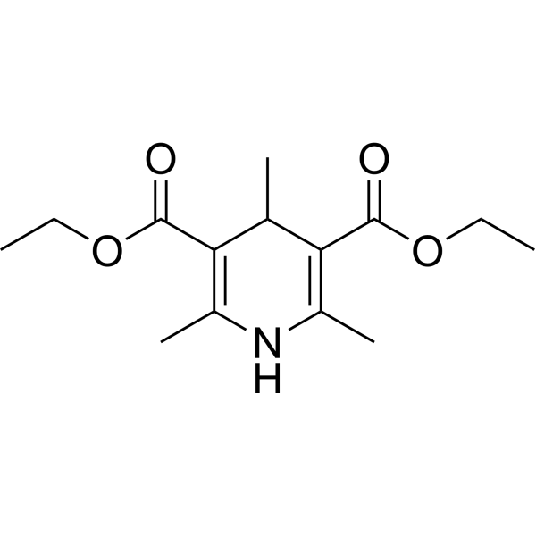
Diethyl 2,4,6-trimethyl-1,4-dihydropyridine-3,5-dicarboxylate

Based on 1 Customer Validation
Diethyl 2,4,6-trimethyl-1,4-dihydropyridine-3,5-dicarboxylate is an orally active porphyrin inducer and ferrochelatase inhibitor. Diethyl 2,4,6-trimethyl-1,4-dihydropyridine-3,5-dicarboxylate can induce small bile duct obstruction in mice, resulting in blocked bile excretion and causing cholestasis. Long-term use of Diethyl 2,4,6-trimethyl-1,4-dihydropyridine-3,5-dicarboxylate can cause damage to bile duct epithelial cells, inflammatory responses, and liver fibrosis. Diethyl 2,4,6-trimethyl-1,4-dihydropyridine-3,5-dicarboxylate can be used to simulate the pathological features of cholestatic liver diseases such as sclerosing cholangitis (PSC).
For research use only. We do not sell to patients.
- Purity : 99.83%
- CAS No.: 632-93-9
- Formula: C14H21NO4
- Molecular Weight:267.32
-
Storage:Powder -20°C, 3 years , 4°C, 2 years ; In solvent -80°C, 6 months , -20°C, 1 month

To place orders, for customer services and technical support, please contact: MedChemExpress USA
Tel: 609-228-6898 E-mail: [email protected] [email protected]


-
 Biological Activity
-
Chemical Information
-
Solvent & Solubility
- Protocols
- Purity & Documentation
- References
-
 Help & FAQs


-
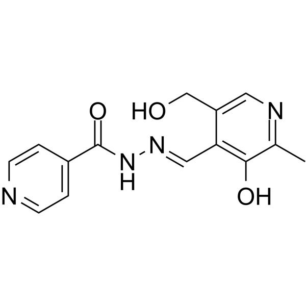
HY-114758
Pyridoxal isonicotinoyl hydrazoneFe-chelating Agent -

HY-118594R
Diethyl 2,4,6-trimethyl-1,4-dihydropyridine-3,5-dicarboxylate (Standard)Standard of Diethyl 2,4,6-trimethyl-1,4-dihydropyridine-3,5-dicarboxylate -
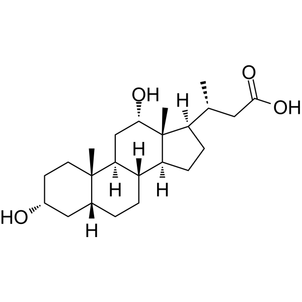
HY-N8268
Nordeoxycholic acidHydrophobic Bile Acid Metabolite -
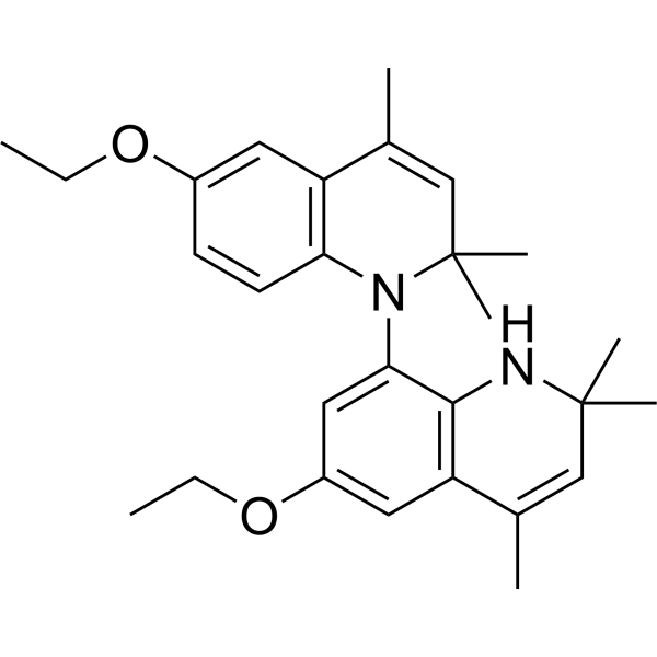
HY-138291
Ethoxyquin dimerAntioxidant -

HY-N8268R
Nordeoxycholic acid (Standard)Reference Standard -

HY-137922
SHS4121705Mitochondrial uncoupling Agent


Biological Activity
Description
In Vivo
-
Animal Model:1-month-old male C3H mice[1].
-
Dosage:0.17%
-
Administration:Fed continuously for 10 weeks
-
Result:Induced MDB formation in the livers of mice.
-
Animal Model:Male weanling C57BL/6 mice were fed with selenium-deficient diet (less than 0.02 mg selenium/kg) or selenium-replete (control) diet (supplemented with 0.25 mg selenium/kg as sodium selenite)[2].
-
Dosage:20 mg/100 g body weight
-
Administration:Administered intraperitoneally once, 4 h before phenobarbital, phorone, or Hemin (HY-19424) administration
-
Result:Blocked the induction of HO activity and HO-1 protein by phenobarbital in selenium-deficient mice.
Inhibited the rise in serum iron induced by phenobarbital.
Inhibited the induction of HO-1 by phorone treatment in selenium-replete mice.
Did not block the induction of HO-1 by hemin in either selenium - deficient or selenium-replete mice.
Induced Disease Models
Please do not refer to only one article to determine the experimental conditions. It is recommended to determine the optimal experimental conditions (animal strain, age, dosage, frequency and cycle, detection time and indicators, etc.) through preliminary experiments before the formal experiment.
Diethyl 2,4,6-trimethyl-1,4-dihydropyridine-3,5-dicarboxylate (0.17%; fed; continuously for 10 weeks) in 1-month-old male C3H mice induces the formation of Mallory Denk bodies (MDB). Refeeding Diethyl 2,4,6-trimethyl-1,4-dihydropyridine-3,5-dicarboxylate after drug withdrawal increases the proportion of FAT10-positive hepatocytes. Diethyl 2,4,6-trimethyl-1,4-dihydropyridine-3,5-dicarboxylate also alters the expression of tumor-related genes, and liver tumors form in mice after long-term drug withdrawal[1].
Diethyl 1,4-dihydro-2,4,6-trimethyl-3,5-pyridinedicarboxylate (20 mg/100 g body weight; intraperitoneal injection; once, 4 h before Phenobarbital, Phorone, or Hemin (HY-19424) administration) blocks heme synthesis, prevents the induction of HO-1 by oxidative stress, and inhibits the increase in serum iron induced by phenobarbital in C57BL/6 mice with selenium deficiency or sufficiency[2].
Administration: 0.1% (wt/wt) evenly mixed into standard rat diet • Free access • for 8, 14, 17 or 28 days (depending on the degree of injury)
or Caerulein (50 μg/kg, 7 hourly, total 7 times) plus LPS (10 mg/kg, once) • i.p.
(2) Diethyl 1,4-dihydro-2,4,6-trimethyl-3,5-pyridinedicarboxylate model is robust, reproducible, and has little impact on animal welfare; different strains of mice can produce similar phenotypes.
(3) The exposure time and whether to use the recovery period for Diethyl 1,4-dihydro-2,4,6-trimethyl-3,5-pyridinedicarboxylate can be adjusted according to the actual situation.
Histology analysis: The liver turned dark brown (indicating protoporphyrin pigment accumulation), and H&E staining showed that the damage was mainly concentrated in the portal vein area, accompanied by bile duct reaction, inflammation, and fibrosis.
MedChemExpress (MCE) has not independently confirmed the accuracy of these methods. They are for reference only.


Chemical Information
-
CAS No. 632-93-9
-
Appearance Solid
-
Molecular Weight 267.32
-
Formula C14H21NO4
-
Color White to off-white
-
SMILES
CC1C(C(OCC)=O)=C(NC(C)=C1C(OCC)=O)C
-
Shipping
Room temperature in continental US; may vary elsewhere.
-
Storage
Powder -20°C 3 years 4°C 2 years In solvent -80°C 6 months -20°C 1 month


Solvent & Solubility
In Vitro:
DMSO : 100 mg/mL (374.08 mM; Need ultrasonic; Hygroscopic DMSO has a significant impact on the solubility of product, please use newly opened DMSO)
Please refer to the solubility information to select the appropriate solvent. Once prepared, please aliquot and store the solution to prevent product inactivation from repeated freeze-thaw cycles.
Storage method and period of stock solution: -80°C, 6 months; -20°C, 1 month. When stored at -80°C, please use it within 6 months. When stored at -20°C, please use it within 1 month.
Please refer to the solubility information to select the appropriate solvent. Once prepared, please aliquot and store the solution to prevent product inactivation from repeated freeze-thaw cycles.
Storage method and period of stock solution: -80°C, 6 months; -20°C, 1 month. When stored at -80°C, please use it within 6 months. When stored at -20°C, please use it within 1 month.
Concentration (start) × Volume (start) = Concentration (final) × Volume (final)
In Vivo:
Select the appropriate dissolution method based on your experimental animal and administration route.
- For the following dissolution methods, please ensure to first prepare a clear stock solution using an In Vitro approach and then sequentially add co-solvents:
- To ensure reliable experimental results, the clarified stock solution can be appropriately stored based on storage conditions. As for the working solution for In Vivo experiments, it is recommended to prepare freshly and use it on the same day.
- The percentages shown for the solvents indicate their volumetric ratio in the final prepared solution. If precipitation or phase separation occurs during preparation, heat and/or sonication can be used to aid dissolution.
In Vivo Dissolution Calculator
Please enter the basic information of animal experiments:
-
-
-
-
Recommended: Prepare an additional quantity of animals to account for potential losses during experiments.
Please enter your animal formula composition:
-
%DMSO +
Recommended: Keep the proportion of DMSO in working solution below 2% if your animal is weak.
-
%+
-
+%Tween-80 + +
-
%Saline +
The co-solvents required include: DMSO, . All of co-solvents are available by MedChemExpress (MCE). , Tween 80. All of co-solvents are available by MedChemExpress (MCE).
Working solution concentration: 0.22 mg/mL
Method for preparing stock solution: mg drug dissolved in μL DMSO. Stock solution concentration: mg/mL.
1. Take μL DMSO stock solution;
2. Add μL .
μL , mix evenly;
3. Then add μL Tween 80, mix evenly;
4. Then add μL
Please ensure that the stock solution in the first step is dissolved to a clear state, and add co-solvents in sequence. You can use ultrasonic heating (ultrasonic cleaner, recommended frequency 20-40 kHz), vortexing, etc. to assist dissolution.

Protocols
-
Fibrosis/Collagen Morphometry
Fibrosis and collagen morphometry is based on the quantitative visualization of fibrillar collagen deposition in tissue sections using histochemical stains such as Sirius Red (Picrosirius Red) or Masson's trichrome, followed by image-based or polarization-enhanced analysis to estimate collagen proportional area as a surrogate of extracellular matrix accumulation during fibrotic remodeling. Sirius Red combined with polarized light microscopy enhances detection of collagen fibers due to birefringence properties, enabling more specific visualization of collagen type I and III fibrils compared to conventional bright-field histology, while whole-section or region-restricted digital morphometry reduces field-selection bias in fibrosis assessment. Alternative quantitative approaches include second harmonic generation (SHG) and two-photon excited fluorescence microscopy, which enable label-free detection of fibrillar collagen and have been validated against histological staining and biochemica
-
Collagen: Sirius Red Staining
Sirius Red or picrosirius red staining is a histochemical method for visualizing collagen-rich extracellular matrix in tissue sections, and collagen fibers are detected as red-stained structures under bright-field microscopy with enhanced birefringence under polarized light. Picrosirius red is useful for assessing total collagen organization, distribution, and fibrosis burden, but polarized color should not be interpreted as a definitive collagen type I versus type III readout because color is affected by fiber orientation, thickness, and packing.
-
Connective Tissue: Masson's Trichrome/Collagen Trichrome Staining
Masson’s Trichrome (collagen/trichrome staining) is a histological technique that differentially stains tissue compartments using sequential acidic dyes to distinguish collagen from muscle and cytoplasmic components based on dye affinity and tissue permeability differences, enabling visualization of fibrosis and connective tissue architecture in histological sections. The classical formulation typically uses Weigert's iron hematoxylin for nuclear staining, Biebrich scarlet-acid fuchsin for cytoplasm and muscle, and aniline blue (or light green variants) for collagen, producing a characteristic blue/green collagen signal contrasted against red cytoplasm and dark nuclei. The staining principle relies on selective displacement of smaller dye molecules by larger anionic dyes in collagen-rich regions under controlled acidified conditions, which enhances collagen-specific dye retention. This property makes the method widely used for fibrosis assessment in organs such as heart, liver, lung, a
-
Liver Histomorphometry
Liver histomorphometry is a quantitative histological approach used to measure structural alterations in hepatic tissue, including parenchymal loss, steatosis, fibrosis, and vascular remodeling, by combining stained tissue section analysis with stereological or computerized image-based measurements. Classical morphometric frameworks quantify volume fractions of liver compartments and fibrotic regions using systematic sampling and image analysis, enabling objective comparison of pathological changes across experimental groups. These approaches are widely applied in liver cirrhosis and fibrosis studies to reduce subjectivity in histological scoring and improve reproducibility of tissue evaluation. Recent methodological advances integrate automated image analysis and radiomics-based extraction of histological features from standard liver stains (e. g. , H&E and fibrotic stains), enabling quantitative correlation between morphometric features and fibrosis stages in non-alcoholic fatty live


Purity & Documentation
-
Data Sheet (279 KB)
-
SDS (393 KB)
- English - EN (393 KB)
- Français - FR (393 KB)
- Deutsch - DE (393 KB)
- Norwegian - NO (393 KB)
- Español - ES (393 KB)
- Swedish - SV (393 KB)
- Italian - IT (393 KB)
- Korean - KR (393 KB)
- Portuguese - PT (393 KB)
-
Handling Instructions (2659 KB)
References
[1]. Oliva J, et al. Fat10 is an epigenetic marker for liver preneoplasia in a drug-primed mouse model of tumorigenesis. Exp Mol Pathol. 2008 Apr;84(2):102-12. [Content Brief]
[2]. Mostert V, et al. Serum iron increases with acute induction of hepatic heme oxygenase-1 in mice. Drug Metab Rev. 2007;39(2-3):619-26. [Content Brief]
[4]. Wu B, et al. A spatiotemporal atlas of cholestatic injury and repair in mice. Nat Genet. 2024 May;56(5):938-952. [Content Brief]
[5]. Zhang J, et al. P4HA2 induces hepatic ductular reaction and biliary fibrosis in chronic cholestatic liver diseases. Hepatology. 2023 Jul 1;78(1):10-25. [Content Brief]
[6]. Kudira R, et al. Bile acids engage the SIPR-STAT3 signaling axis to modulate regulatory T cell responses in fibrosing cholangiopathies. J Hepatol. 2025 Nov;83(5):1128-1141 [Content Brief]


Complete Stock Solution Preparation Table
Please refer to the solubility information to select the appropriate solvent. Once prepared, please aliquot and store the solution to prevent product inactivation from repeated freeze-thaw cycles.
Storage method and period of stock solution: -80°C, 6 months; -20°C, 1 month. When stored at -80°C, please use it within 6 months. When stored at -20°C, please use it within 1 month.
| Optional Solvent | Concentration Solvent Mass | 1 mg | 5 mg | 10 mg | 25 mg |
|---|---|---|---|---|---|
| DMSO | 1 mM | 3.7408 mL | 18.7042 mL | 37.4083 mL | 93.5209 mL |
| 5 mM | 0.7482 mL | 3.7408 mL | 7.4817 mL | 18.7042 mL | |
| 10 mM | 0.3741 mL | 1.8704 mL | 3.7408 mL | 9.3521 mL | |
| 15 mM | 0.2494 mL | 1.2469 mL | 2.4939 mL | 6.2347 mL | |
| 20 mM | 0.1870 mL | 0.9352 mL | 1.8704 mL | 4.6760 mL | |
| 25 mM | 0.1496 mL | 0.7482 mL | 1.4963 mL | 3.7408 mL | |
| 30 mM | 0.1247 mL | 0.6235 mL | 1.2469 mL | 3.1174 mL | |
| 40 mM | 0.0935 mL | 0.4676 mL | 0.9352 mL | 2.3380 mL | |
| 50 mM | 0.0748 mL | 0.3741 mL | 0.7482 mL | 1.8704 mL | |
| 60 mM | 0.0623 mL | 0.3117 mL | 0.6235 mL | 1.5587 mL | |
| 80 mM | 0.0468 mL | 0.2338 mL | 0.4676 mL | 1.1690 mL | |
| 100 mM | 0.0374 mL | 0.1870 mL | 0.3741 mL | 0.9352 mL |


Diethyl 2,4,6-trimethyl-1,4-dihydropyridine-3,5-dicarboxylate Related Classifications
HY-118594 Related Classifications
 Powered by Bioz
Keywords