



Cathepsin S Protein, Mouse (HEK293, His)

Based on 1 Customer Validation
Cathepsin S Protein, Mouse (HEK293, His) is potent cysteine protease which can promote degradation of damaged or unwanted proteins in the endo-lysosomal pathway.
- Species: Mouse
- Source: HEK293
-
Storage:Stored at -20°C for 2 years from date of receipt. After reconstitution, it is stable at 4°C for 1 week or -20°C for longer (with carrier protein). It is recommended to freeze aliquots at -20°C or -80°C for extended storage.


To place orders, for customer services and technical support, please contact: MedChemExpress USA
Tel: 609-228-6898 E-mail: [email protected] [email protected]


-
 Biological Activity
-
Technical Parameters
-
Product Properties
- Documentation
- References
- Help & FAQs




Biological Activity
Description
Cathepsin S Protein, Mouse (HEK293, His) is potent cysteine protease which can promote degradation of damaged or unwanted proteins in the endo-lysosomal pathway.
Background
Cathepsin S is a member of the cysteine cathepsin protease family. Cathepsin S has specific roles such as MHC class II antigen presentation, where it is important in the degradation of the invariant chain. Cathepsin S is involved in a variety of pathological processes including arthritis, cancer, and cardiovascular disease, where it becomes secreted and can act on extracellular substrates. Cathepsin S has uniquely restricted tissue expression and is more stable at a neutral pH. Cathepsin S is unique amongst the cysteine cathepsin family due to restricted tissue expression, associated with antigen presenting cells localised in lymph and spleen, as well as other immune cells such as macrophages. Cathepsin S is thought to be a particularly potent cysteine protease cleaving elastin and generating bioactive elastin peptides, leading to the promotion of cardiovascular inflammation and calcification. Cathepsin S is also released by smooth muscle cells and macrophages as a systemic response to inflammation in a continuous recursive feedback loop[1][2].
Verified Bioactivity
Measured by its ability to cleave the fluorogenic peptide substrate Mca-RPKPVE-Nval-WRK(Dnp)-NH2 (HY-P2185A) incubated at room temperature in kinetic mode for 5 minutes. Read at excitation and emission wavelengths of 320 nm and 405 nm. The specific activity is >300 pmol/min/µg.(Activation description: The enzyme needs to be activated in acid buffer for an activated form.)
MCE Validation Data
-
Purity - SDS-PAGE
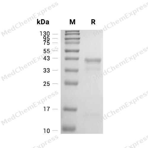
Purity - SDS-PAGE
-
Bioactivity - Enzymatic Activity Assay
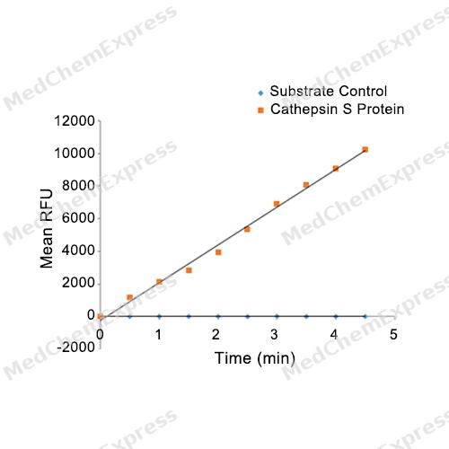
Bioactivity - Enzymatic Activity Assay

Assay Procedure
Materials
Assay buffer: 50 mM NaOAc,5 mM DTT, 250 mM NaCl, pH 4.5
Test protein: Cathepsin S Protein, Mouse (HEK293, His) (HY-P7757)
Substrate: NFF-3 TFA (HY-P2185A)
Standard: MCA-Pro-Leu-OH
Procedure
1. Dilute the standard with the assay buffer to the following concentrations: 0, 0.5, 1, 2, 3.9, 7.8, 15.6, 31.25, 62.5, 125, 250, and 500 μmol/L. Add 100 μL of each concentration to the wells to initiate the reaction, with three replicate wells for each concentration. Read the reaction in kinetic mode for 5 minutes at excitation and emission wavelengths of 320 nm and 405 nm, respectively. Plot the standard curve with the standard concentration as the x-axis and the measured RFU as the y-axis to obtain the standard curve equation.
2. First, reconstitute the protein to a concentration of 200 μg/mL with sterile water, then dilute Mouse Cathepsin S to 10 μg/mL with the assay buffer.
3. Incubate at room temperature for 2 hours.
4. Dilute Mouse Cathepsin S to 0.5 ng/μL with the assay buffer.
5. Dilute the substrate to 20 μM in assay buffer.
6. Add 50 μL of 0.5 ng/μL Mouse Cathepsin S and 50 μL of 20 μM substrate into a 96-well black plate. Include a substrate blank containing 50 μL of assay buffer and 50 μL of 20 μM substrate without any Mouse Cathepsin S. Perform each concentration in triplicate.
7. Read the RFU values in kinetic mode at excitation and emission wavelengths of 320 nm and 405 nm, respectively, for 5 minutes.
8. Calculate the specific activity:
Calculate the specific activity:
| Specific Activity (pmol/min/μg) = |
Adjusted Vmax (RFU/min) × Conversion Factor (pmol/RFU)
amount of enzyme (μg)
|
*Adjusted for Control
**Derived using calibration standard


Technical Parameters
-
Species Mouse
-
Source HEK293
-
Tag C-6*His
-
Accession
O70370 (V18-I340)
-
Molecular Construction
-
N-term
-
Cathepsin S (V18-I340)
Accession # O70370 -
6*His
-
C-term
-
-
Protein Length
Full Length of Mature Protein (with Propeptide)
-
Synonyms
CTSS; Cathepsin S
-
AA Sequence
VCSVAMEQLQRDPTLDYHWDLWKKTHEKEYKDKNEEEVRRLIWEKNLKFIMIHNLEYSMGMHTYQVGMNDMGDMTNEEILCRMGALRIPRQSPKTVTFRSYSNRTLPDTVDWREKGCVTEVKYQGSCGACWAFSAVGALEGQLKLKTGKLISLSAQNLVDCSNEEKYGNKGCGGGYMTEAFQYIIDNGGIEADASYPYKATDEKCHYNSKNRAATCSRYIQLPFGDEDALKEAVATKGPVSVGIDASHSSFFFYKSGVYDDPSCTGNVNHGVLVVGYGTLDGKDYWLVKNSWGLNFGDQGYIRMARNNKNHCGIASYCSYPEI
-
Molecular Weight
Approximately 40 kDa & 27-35 kDa & 8-17 kDa, based on SDS-PAGE under reducing conditions.
-
Glycosylation
Yes
-
Purity
≥ 95%, as determined by reducing SDS-PAGE.


Product Properties
Lyophilized powder
1.Lyophilized from a 0.22 μm filtered solution of 20 mM PB, 150 mM NaCl, pH 7.4.
2.Lyophilized from a 0.22 μm filtered solution of PBS, pH 7.4.
Please refer to the lot-specific COA for specific buffer information.
<1 EU/μg, determined by LAL method.
It is not recommended to reconstitute to a concentration less than 100 μg/mL in ddH2O. For long term storage it is recommended to add a carrier protein (0.1% BSA, 5% HSA, 10% FBS or 5% Trehalose).
Stored at -20°C for 2 years from date of receipt. After reconstitution, it is stable at 4°C for 1 week or -20°C for longer (with carrier protein). It is recommended to freeze aliquots at -20°C or -80°C for extended storage.
Room temperature in continental US; may vary elsewhere.


Documentation
-
Data Sheet (264 KB)
-
SDS (252 KB)
- English - EN (252 KB)
- Français - FR (252 KB)
- Deutsch - DE (252 KB)
- Norwegian - NO (252 KB)
- Español - ES (252 KB)
- Swedish - SV (252 KB)
- Italian - IT (252 KB)
- Korean - KR (252 KB)
- Portuguese - PT (252 KB)
-
Handling Instructions (2659 KB)


References
[1]. Richard D A Wilkinson, et al. Cathepsin S: therapeutic, diagnostic, and prognostic potential. Biol Chem. 2015 Aug;396(8):867-82. [Content Brief]
[2]. Brena F Sena, et al. Cathepsin S As an Inhibitor of Cardiovascular Inflammation and Calcification in Chronic Kidney Disease. Front Cardiovasc Med. 2018 Jan 5;4:88. [Content Brief]

Calculators
Concentration (start) × Volume (start) = Concentration (final) × Volume (final)


 Powered by Bioz

