
10 Results for "

apoptotic bodies

" in MedChemExpress (MCE) Product Catalog:
Products (10)

10 Results for "apoptotic bodies" in MCE Product Catalog:

Cat. No.: HY-Y1366
CAS No.: 116-09-6
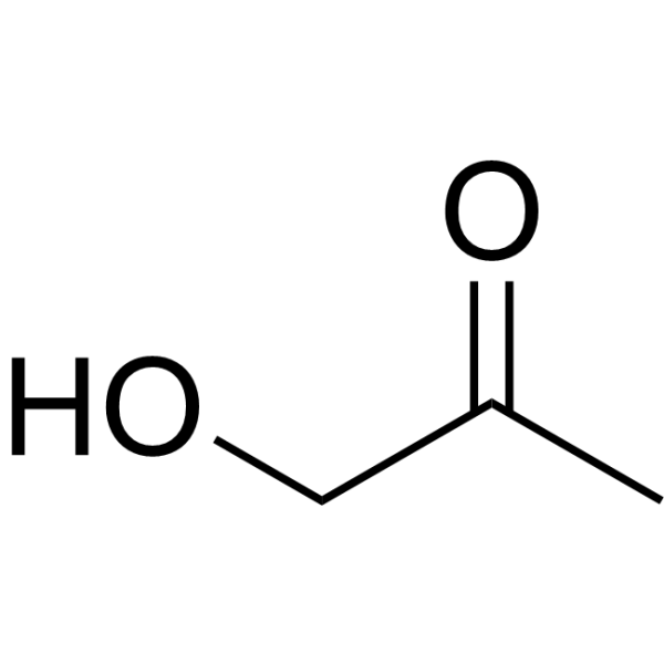
Hydroxyacetone is a toxic compound. Hydroxyacetone can be isolated from e-cigarette aerosols. Hydroxyacetone reduces the activity of cellular Mitochondrial reductase (with an IC50 of 5.53 mg/mL for mitochondrial reductase in BEAS-2B cells) and increases ROS levels. Hydroxyacetone induces mitochondrial stress and oxidative damage. Hydroxyacetone induces destabilization of F-actin. At high concentrations, Hydroxyacetone promotes cell rounding and Apoptotic body formation. Hydroxyacetone exerts toxic effects on cells including airway epithelial cells and possesses respiratory toxicity potential .\n


loading...
    loading...
Cat. No.: HY-P10943
CAS No.: 3051946-11-0
Target:  

Fluorescent Dye

Research Areas:  

Inflammation/Immunology Cancer

APO-15 is a phosphatidylserine-binding fluorescent probe and apoptosis imaging reagent. APO-15 exhibits high chemical stability under proteolytic and oxidative conditions, enables quantification and imaging of drug-induced apoptosis in preclinical mouse models, and is applicable to fixed tissue samples and multiple in vivo administration routes (Ex = 488 nm; Em = 525 nm). APO-15 can be used in studies related to acute lung injury and breast cancer .
loading...
    loading...
Cat. No.: HY-P1380A
Research Areas:  

Cancer

Difopein TFA is a 14-3-3 protein inhibitor. Difopein TFA acts as an apoptosis inducer, regulates apoptosis-related proteins, downregulates Bcl-2, upregulates Bax, activates caspase-9 and caspase-3, and induces nuclear fragmentation, membrane-enclosed apoptotic bodies and DNA ladder formation. Difopein TFA serves as a tumor growth inhibitor, which inhibits the proliferation of glioma cells and induces their apoptosis in in vivo nude mouse models. Difopein TFA is applicable to glioma-related research .
loading...
    loading...
Cat. No.: HY-W075770
CAS No.: 1313-99-1
Synonyms: Nickel monoxide
Nickel(II) oxide (nickel monoxide) is a chemical warfare agent that can enter the body through the respiratory tract and other routes, distributing to organs such as the lungs and testes. The nanoparticle form of nickel(II) oxide (NiO NPs) exhibits antibacterial, anti-leishmanial, anti-diabetic, and anti-cancer activities. NiO NPs can be activated by ultraviolet and visible light, generating reactive oxygen species (ROS). Nickel(II) oxide induces oxidative stress by generating reactive oxygen species, activating the TGF-β1-mediated MAPK and PI3K/AKT pathways, disrupting the MMPs/TIMPs balance, and upregulating the expression of inflammatory factors (IL-1β, IL-6) and apoptosis-related molecules (Bax, caspase-3, p53), while inhibiting the activity of the anti-apoptotic molecule Bcl-2. Nickel(II) oxide induces cytotoxicity, promotes fibrosis, triggers inflammatory responses, and causes apoptosis. Nickel(II) oxide can be applied in research on the safety assessment of nanomaterials, such as in the context of pulmonary fibrosis and reproductive system toxicity .
loading...
    loading...
Cat. No.: HY-N6674
CAS No.: 733035-26-2
Synonyms: ECO-4601; TLN-4601; BU 4664L
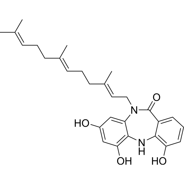
Diazepinomicin (ECO-4601) is an anticancer and antibacterial agent. Diazepinomicin can be produced by a Micromonospora strain. Diazepinomicin induces Apoptosis. Diazepinomicin inhibits the proteases Rhodesain and Cathepsin L at an IC50 of 70-90 μM. Diazepinomicin possesses anti-inflammatory and anti-tumor activity. Diazepinomicin has demonstrated activity against hepatocellular carcinoma. Diazepinomicin shows antiparasitic activity against trypomastigote forms of Trypanosoma brucei with an IC50 of 13.5 μM. Diazepinomicin exhibits moderate antibacterial activity against specific Gram-positive bacteria, with an MIC of approximately 32 μg/mL .
loading...
    loading...
Cat. No.: HY-168720
Target:  

Apoptosis

Research Areas:  

Cancer

Apoptosis inducer 32 (Compound 7g) is an apoptosis inducer with a KD of 42 μM, showing anti-tumor activity. Apoptosis inducer 32 caused significant cellular morphological changes in MDA-MB-231 cells, including membrane bubbling, nuclear fragmentation, and apoptotic body formation. The IC50 of Apoptosis inducer 32 in MCF-7, MDA-MB-231, and HEK cells is 4.77, 6.56 and 337.8 μM respectively .
loading...
    loading...
Cat. No.: HY-123647
CAS No.: 53126-64-0
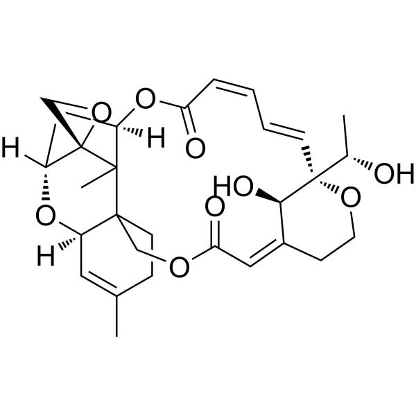
Satratoxin H is a toxic metabolite of Stachybotrys atra. Satratoxin H induces caspase-3 and PARP cleavage via p38 MAPK and JNK pathways, stimulates JNK, ERK, and p38 MAPK phosphorylation, and activates JNK and p38 MAPK in a glutathione-sensitive manner. Satratoxin H induces DNA double-stranded breaks, apoptotic body formation, intracellular reactive oxygen species generation, and endoplasmic reticulum stress via ATF6, PERK, and IRE1 pathways. Satratoxin H can be used for the research of central nervous system disorders and melanoma .
loading...
    loading...
Cat. No.: HY-W550859
CAS No.: 34023-49-9
Synonyms: 4-Sulfo-L-phenylalanine
Target:  

Apoptosis

Research Areas:  

Cancer

H-Phe(4-SO3H)-OH (4-Sulfo-L-phenylalanine) is a L-phenylalanine (HY-N0215) derivative. H-Phe(4-SO3H)-OH induces apoptosis, chromatin margination, and inhibits cell growth in human colon cancer cells. H-Phe(4-SO3H)-OH can be used for the research of colon cancer .
loading...
    loading...
Cat. No.: HY-P1380
CAS No.: 396834-58-5
Research Areas:  

Cancer

Difopein is a 14-3-3 protein inhibitor. Difopein acts as an apoptosis inducer, regulates apoptosis-related proteins, downregulates Bcl-2, upregulates Bax, activates caspase-9 and caspase-3, and induces nuclear fragmentation, membrane-enclosed apoptotic bodies and DNA ladder formation. Difopein serves as a tumor growth inhibitor, which inhibits the proliferation of glioma cells and induces their apoptosis in in vivo nude mouse models. Difopein is applicable to glioma-related research .
loading...
    loading...
Cat. No.: HY-L168
707 compounds

Extracellular vesicles (EVs) are small membrane binding structures that are released from cells into the surrounding environment and play a crucial role in mediating and regulating intercellular communication related to physiological and pathological processes. EVs are lipid membrane vesicles composed of proteins, lipids, and nucleic acids. EVs can be divided into several types based on their source, such as extracellular vesicles, microcapsules, and apoptotic vesicles. The size range of exosomes is 30-150nm, which are endocrine in multi vesicular endosomes (MVEs); microvesicles (50-1000nm) are secreted directly through extracellular interactions, thereby releasing plasma membrane vesicles. In contrast, apoptotic bodies are usually larger, ranging in size from 1 to 5 μ m. This is generated during programmed cell death. EV plays a crucial role in transmitting information between cells and influencing the behavior and function of receptor cells.

MCE designs a unique collection of 707 small molecules related to extracellular vesicles (EVs). It is a good tool to be used for research on metabolize, cancer and other diseases.

