1. Cell Cycle/DNA Damage PI3K/Akt/mTOR Autophagy
  2. ATM/ATR Autophagy
  3. KU-55933

KU-55933 is a potent ATM inhibitor with an IC50 and Ki of 12.9 and 2.2 nM, respectively, and is highly selective for ATM as compared to DNA-PK, PI3K/PI4K, ATR and mTOR.

For research use only. We do not sell to patients.

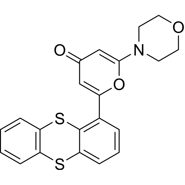
KU-55933 Chemical Structure

KU-55933 Chemical Structure

CAS No. : 587871-26-9

Size Price Stock Quantity
Solid + Solvent (Highly Recommended)
10 mM * 1 mL in DMSO
ready for reconstitution
USD 61 In-stock
Solution
10 mM * 1 mL in DMSO USD 61 In-stock
Solid
1 mg USD 28 In-stock
5 mg USD 55 In-stock
10 mg USD 77 In-stock
50 mg USD 220 In-stock
100 mg USD 385 In-stock
200 mg   Get quote  
500 mg   Get quote  

* Please select Quantity before adding items.

This product is a controlled substance and not for sale in your territory.

Customer Review

Based on 38 publication(s) in Google Scholar

Top Publications Citing Use of Products

36 Publications Citing Use of MCE KU-55933

Proliferation Assay
RT-PCR

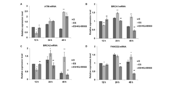
    KU-55933 purchased from MedChemExpress. Usage Cited in: Environ Pollut. 2018 Jul;238:1048-1055.  [Abstract]

    Time-effects of endosulfan on DNA damage and repair related genes. (AeD) Relative mRNA expression levels of ATM (A), BRCA1 (B), BRCA2 (C) and FANCD2 (D) are measured by qRT-PCR after endosulfan exposure (75 μM, for 12, 24 and 48 h) in the absence and presence of KU-55933.

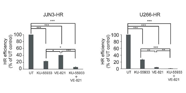
    KU-55933 purchased from MedChemExpress. Usage Cited in: Front Oncol. 2017 May 19;7:98.  [Abstract]

    ATM inhibition by treatment with KU-55933 (10 µM) strongly reduces HR efficiency in JJN3-HR and U266-HR, although cells are still able to perform HR to some extent.

    View All ATM/ATR Isoform Specific Products:

    • Biological Activity

    • Protocol

    • Purity & Documentation

    • References

    • Customer Review

    Description

    KU-55933 is a potent ATM inhibitor with an IC50 and Ki of 12.9 and 2.2 nM, respectively, and is highly selective for ATM as compared to DNA-PK, PI3K/PI4K, ATR and mTOR.

    IC50 & Target[1]

    ATM

    12.9 nM (IC50)

    DNA-PK

    2500 nM (IC50)

    mTOR

    9300 nM (IC50)

    PI3K

    16600 nM (IC50)

    In Vitro

    KU-55933 (10 μM) blocks the ionizing radiation-induced p53 serine 15 phosphorylation. KU-55933 has a dose-dependent effect in inhibiting this ATM-dependent phosphorylation event with an estimated IC50 of 300 nM. KU-55933 ablates the ionizing radiation-induced phosphorylation of these ATM substrates. KU-55933 specifically inhibits ATM but not the other DNA damage-activated PIKKs, ATR, and DNA-PK[1]. KU-55933 induces pATM, p53, E2F1 and pATR, noticeably upregulates the nuclear fraction of E2F1 at the 0.5 h time point[2].

    MedChemExpress (MCE) has not independently confirmed the accuracy of these methods. They are for reference only.

    Molecular Weight

    395.49

    Formula

    C21H17NO3S2

    CAS No.
    Appearance

    Solid

    Color

    White to khaki

    SMILES

    O=C1C=C(OC(C2=C3SC4=C(SC3=CC=C2)C=CC=C4)=C1)N5CCOCC5

    Shipping

    Room temperature in continental US; may vary elsewhere.

    Storage
    Powder -20°C 3 years
    4°C 2 years
    In solvent -80°C 1 year
    -20°C 6 months
    Solvent & Solubility
    In Vitro: 

    DMSO : 50 mg/mL (126.43 mM; Need ultrasonic; Hygroscopic DMSO has a significant impact on the solubility of product, please use newly opened DMSO)

    Preparing
    Stock Solutions
    Concentration Solvent Mass 1 mg 5 mg 10 mg
    1 mM 2.5285 mL 12.6425 mL 25.2851 mL
    5 mM 0.5057 mL 2.5285 mL 5.0570 mL
    View the Complete Stock Solution Preparation Table

    * Please refer to the solubility information to select the appropriate solvent. Once prepared, please aliquot and store the solution to prevent product inactivation from repeated freeze-thaw cycles.
    Storage method and period of stock solution: -80°C, 1 year; -20°C, 6 months. When stored at -80°C, please use it within 1 year. When stored at -20°C, please use it within 6 months.

    • Molarity Calculator

    • Dilution Calculator

    Mass (g) = Concentration (mol/L) × Volume (L) × Molecular Weight (g/mol)

    Mass
    =
    Concentration
    ×
    Volume
    ×
    Molecular Weight *

    Concentration (start) × Volume (start) = Concentration (final) × Volume (final)

    This equation is commonly abbreviated as: C1V1 = C2V2

    Concentration (start)

    C1

    ×
    Volume (start)

    V1

    =
    Concentration (final)

    C2

    ×
    Volume (final)

    V2

    In Vivo:

    Select the appropriate dissolution method based on your experimental animal and administration route.

    For the following dissolution methods, please ensure to first prepare a clear stock solution using an In Vitro approach and then sequentially add co-solvents:
    To ensure reliable experimental results, the clarified stock solution can be appropriately stored based on storage conditions. As for the working solution for in vivo experiments, it is recommended to prepare freshly and use it on the same day.
    The percentages shown for the solvents indicate their volumetric ratio in the final prepared solution. If precipitation or phase separation occurs during preparation, heat and/or sonication can be used to aid dissolution.

    • Protocol 1

      Add each solvent one by one:  10% DMSO    40% PEG300    5% Tween-80    45% Saline

      Solubility: ≥ 2.5 mg/mL (6.32 mM); Clear solution

      This protocol yields a clear solution of ≥ 2.5 mg/mL (saturation unknown).

      Taking 1 mL working solution as an example, add 100 μL DMSO stock solution (25.0 mg/mL) to 400 μL PEG300, and mix evenly; then add 50 μL Tween-80 and mix evenly; then add 450 μL Saline to adjust the volume to 1 mL.

      Preparation of Saline: Dissolve 0.9 g sodium chloride in ddH₂O and dilute to 100 mL to obtain a clear Saline solution.
    • Protocol 2

      Add each solvent one by one:  10% DMSO    90% (20% SBE-β-CD in Saline)

      Solubility: ≥ 2.5 mg/mL (6.32 mM); Clear solution

      This protocol yields a clear solution of ≥ 2.5 mg/mL (saturation unknown).

      Taking 1 mL working solution as an example, add 100 μL DMSO stock solution (25.0 mg/mL) to 900 μL 20% SBE-β-CD in Saline, and mix evenly.

      Preparation of 20% SBE-β-CD in Saline (4°C, storage for one week): 2 g SBE-β-CD powder is dissolved in 10 mL Saline, completely dissolve until clear.
    In Vivo Dissolution Calculator
    Please enter the basic information of animal experiments:

    Dosage

    mg/kg

    Animal weight
    (per animal)

    g

    Dosing volume
    (per animal)

    μL

    Number of animals

    Recommended: Prepare an additional quantity of animals to account for potential losses during experiments.
    Please enter your animal formula composition:
    %
    DMSO +
    +
    %
    Tween-80 +
    %
    Saline
    Recommended: Keep the proportion of DMSO in working solution below 2% if your animal is weak.
    The co-solvents required include: DMSO, . All of co-solvents are available by MedChemExpress (MCE). , Tween 80. All of co-solvents are available by MedChemExpress (MCE).
    Calculation results:
    Working solution concentration: mg/mL
    Method for preparing stock solution: mg drug dissolved in μL  DMSO (Stock solution concentration: mg/mL).
    The concentration of the stock solution you require exceeds the measured solubility. The following solution is for reference only. If necessary, please contact MedChemExpress (MCE).
    Method for preparing in vivo working solution for animal experiments: Take μL DMSO stock solution, add μL . μL , mix evenly, next add μL Tween 80, mix evenly, then add μL Saline.
     If the continuous dosing period exceeds half a month, please choose this protocol carefully.
    Please ensure that the stock solution in the first step is dissolved to a clear state, and add co-solvents in sequence. You can use ultrasonic heating (ultrasonic cleaner, recommended frequency 20-40 kHz), vortexing, etc. to assist dissolution.
    Purity & Documentation

    Purity: 99.92%

    References
    Kinase Assay
    [1]

    ATM for use in the in vitro assay is obtained by immunoprecipitation with rabbit polyclonal antiserum raised to the COOH-terminal 400 amino acids of ATM in buffer containing 25 mM HEPES (pH 7.4), 2 mM MgCl2, 250 mM KCl, 500 μM EDTA, 100 μM Na3VO4, 10% v/v glycerol, and 0.1% v/v Igepal. ATM-antibody complexes are isolated from nuclear extract by incubating with protein A-Sepharose beads for 1 hour and then through centrifugation to recover the beads. In the well of a 96-well plate, ATM-containing Sepharose beads are incubated with 1 μg of substrate glutathione S-transferase-p53N66 (NH2-terminal 66 amino acids of p53 fused to glutathione S-transferase) in ATM assay buffer [25 mM HEPES (pH 7.4), 75 mM NaCl, 3 mM MgCl2, 2 mM MnCl2, 50 μM Na3VO4, 500 μM DTT, and 5% v/v glycerol] at 37°C in the presence or absence of inhibitor. After 10 minutes with gentle shaking, ATP is added to a final concentration of 50 μM and the reaction continued at 37°C for an additional 1 hour. The plate is centrifuged at 250×g for 10 minutes (4°C) to remove the ATM-containing beads, and the supernatant is removed and transferred to a white opaque 96-well plate and incubated at room temperature for 1.5 hours to allow glutathione S-transferase-p53N66 binding. This plate is then washed with PBS, blotted dry, and analyzed by a standard ELISA technique with a phospho-serine 15 p53 antibody. The detection of phosphorylated glutathione S-transferase-p53N66 substrate is performed in combination with a goat antimouse horseradish peroxidase-conjugated secondary antibody. Enhanced chemiluminescence solutionis used to produce a signal and chemiluminescent detection is carried out via a TopCount plate reader.

    MCE has not independently confirmed the accuracy of these methods. They are for reference only.

    Cell Assay
    [1]

    1BR or AT4 cells are seeded in 10-cm Petri dishes and treated on day 2 (80 to 90% confluence). Cells are preincubated for 1 hour with KU-55933 or vehicle control and then exposed to 5 Gy of ionizing radiation. Time courses of cell cycle distribution are performed, and the optimal time for discrimination of populations is selected as 16 hours. All subsequent experiments are performed at the 16-hour time point. Cells are stained with propidium iodide according to standard protocols and analyzed by FACS with a FACScalibur. Exponentially growing (50-70% confluent) SW620 cells in 60 mm dishes are exposed to KU-55933 or DMSO for 1 h before addition of etoposide (final concentration of 0.1 and 1 μM) for 16 h before harvesting, propidium iodide staining and analysis as above.

    MCE has not independently confirmed the accuracy of these methods. They are for reference only.

    References

    Complete Stock Solution Preparation Table

    * Please refer to the solubility information to select the appropriate solvent. Once prepared, please aliquot and store the solution to prevent product inactivation from repeated freeze-thaw cycles.
    Storage method and period of stock solution: -80°C, 1 year; -20°C, 6 months. When stored at -80°C, please use it within 1 year. When stored at -20°C, please use it within 6 months.

    Optional Solvent Concentration Solvent Mass 1 mg 5 mg 10 mg 25 mg
    DMSO 1 mM 2.5285 mL 12.6425 mL 25.2851 mL 63.2127 mL
    5 mM 0.5057 mL 2.5285 mL 5.0570 mL 12.6425 mL
    10 mM 0.2529 mL 1.2643 mL 2.5285 mL 6.3213 mL
    15 mM 0.1686 mL 0.8428 mL 1.6857 mL 4.2142 mL
    20 mM 0.1264 mL 0.6321 mL 1.2643 mL 3.1606 mL
    25 mM 0.1011 mL 0.5057 mL 1.0114 mL 2.5285 mL
    30 mM 0.0843 mL 0.4214 mL 0.8428 mL 2.1071 mL
    40 mM 0.0632 mL 0.3161 mL 0.6321 mL 1.5803 mL
    50 mM 0.0506 mL 0.2529 mL 0.5057 mL 1.2643 mL
    60 mM 0.0421 mL 0.2107 mL 0.4214 mL 1.0535 mL
    80 mM 0.0316 mL 0.1580 mL 0.3161 mL 0.7902 mL
    100 mM 0.0253 mL 0.1264 mL 0.2529 mL 0.6321 mL
    • No file chosen (Maximum size is: 1024 Kb)
    • If you have published this work, please enter the PubMed ID.
    • Your name will appear on the site.
    Help & FAQs
    • Do most proteins show cross-species activity?

      Species cross-reactivity must be investigated individually for each product. Many human cytokines will produce a nice response in mouse cell lines, and many mouse proteins will show activity on human cells. Other proteins may have a lower specific activity when used in the opposite species.

    Your Recently Viewed Products:

    Inquiry Online

    Your information is safe with us. * Required Fields.

    Product Name


    Salutation

    Applicant Name *


    Email Address *

    Phone Number *


    Organization Name *

    Department *


    Requested quantity *

    Country or Region *


    Remarks

    Bulk Inquiry

    Inquiry Information

    Product Name:
    KU-55933
    Cat. No.:
    HY-12016
    Quantity:
    MCE Japan Authorized Agent: