


Tubastatin A
Based on 40 publication(s) in Google Scholar
Tubastatin A is a potent and selective HDAC6 inhibitor with an IC50 of 15 nM in a cell-free assay, and is selective (1000-fold more) against all other isozymes except HDAC8 (57-fold more). Tubastatin A also inhibits HDAC10 and metallo-β-lactamase domain-containing protein 2 (MBLAC2).
For research use only. We do not sell to patients.
- Purity: 99.07%
- CAS No.: 1252003-15-8
- Formula: C20H21N3O2
- Molecular Weight:335.40
-
Storage:Powder -20°C, 3 years , 4°C, 2 years ; In solvent -80°C, 6 months , -20°C, 1 month


-
 Biological Activity
-
Chemical Information
-
Solvent & Solubility
- Protocol
- Purity & Documentation
- References
-
 Help & FAQs
-


Anti-Infection Compound Library
HY-L002
-
Apoptosis Compound Library
HY-L003
-
Cell Cycle/DNA Damage Compound Library
HY-L004
-
Epigenetics Compound Library
HY-L005
-
Histone Modification Research Compound Library
HY-L024
-
Anti-Cancer Compound Library
HY-L025
-
Autophagy Compound Library
HY-L029
-
Anti-Aging Compound Library
HY-L034
-
Reprogramming Compound Library
HY-L039
-
Oxygen Sensing Compound Library
HY-L045
-
Anti-Breast Cancer Compound Library
HY-L074
-
Anti-Pancreatic Cancer Compound Library
HY-L077
-
Anti-Blood Cancer Compound Library
HY-L079
-
Anti-Liver Cancer Compound Library
HY-L101
-
Human Metabolite Library
HY-L123
-
Osteogenesis Compound Library
HY-L131
-
Highly Selective Inhibitors Library
HY-L158
-
Highly Selective Activators Library
HY-L159
-
Cell Death Library
HY-L162
-
Anti-Hematopathy Compound Library
HY-L171
-
Anti-Ovarian Cancer Compound Library
HY-L173
-
Multi-Target Compound Library
HY-L176
-
Bioactive Compound Library Max
HY-L181
-
Anti-Aging Compound Library Mini
HY-L034M
-
Epigenetics Compound Library Mini
HY-L005M
-
Bioactive Compound Library
HY-L001
-
Anti-Brain Cancer Compound Library
HY-L188
-
Non-Alcoholic Fatty Liver Disease (NAFLD) Compound Library
HY-L199
-
High-Throughput Bioactive Compound Library
HY-L205
-
Mass Spectrometry Human Metabolite Library
HY-L215
-
High-Efficiency Gene Editing Compound Library
HY-L244
-
Lactylation Compound Library
HY-L249


Publications Citing Use of MedChemExpress (MCE) Tubastatin A
More- Cell Host Microbe. 2026 Feb 11;34(2):245-262.e8. [Abstract]
- Nat Commun. 2026 Feb 5;17(1):1212. [Abstract]
- Cell Death Differ. 2025 Jul 30. [Abstract]
- Acta Pharm Sin B. 2022 Oct;12(10):3891-3904. [Abstract]
- Adv Sci (Weinh). 2025 Aug 7:e02624. [Abstract]
- J Biomed Sci. 2025 Jan 20;32(1):9. [Abstract]
- Cell Death Dis. 2022 Oct 21;13(10):888. [Abstract]
- Free Radic Biol Med. 2025 Aug 13:240:59-70. [Abstract]
- NPJ Precis Oncol. 2024 Mar 7;8(1):66. [Abstract]
- Neurotherapeutics. 2023 Jul;20(4):1215-1228. [Abstract]
- Clin Transl Med. 2025 Jan;15(1):e70193. [Abstract]
- J Med Chem. 2024 Sep 12;67(17):15098-15117. [Abstract]
- J Med Chem. 2023 Dec 14;66(23):16075-16090. [Abstract]
- Antioxidants (Basel). 2022 Apr 7;11(4):732. [Abstract]
- Antioxidants. 2020 Jul 9;9(7):599. [Abstract]
- Atherosclerosis. 2021 Jan:317:1-9. [Abstract]
- Virulence. 2025 Dec;16(1):2495838. [Abstract]
- J Pathol. 2025 Apr 11. [Abstract]
- PLoS Pathog. 2021 Sep 20;17(9):e1009940. [Abstract]
- Front Pharmacol. 2018 Feb 1:9:34. [Abstract]
- Mol Cancer Res. 2022 Jun 3;20(6):949-959. [Abstract]
- Mol Cancer Res. 2014 May;12(5):681-93. [Abstract]
- BMC Biol. 2018 Oct 18;16(1):116. [Abstract]
- J Neurochem. 2026 Jun;170(6):e70493. [Abstract]
- J Dermatol Sci. 2022 Jun;106(3):181-188. [Abstract]
- Aging (Albany NY). 2021 Mar 19;13(7):9820-9837. [Abstract]
- J Nutr. 2020 Jul 1;150(7):1790-1798. [Abstract]
- Biochim Biophys Acta Mol Cell Res. 2020 May;1867(5):118676. [Abstract]
- Am J Pathol. 2020 Dec;190(12):2376-2386. [Abstract]
- Viruses. 2025 Jan 13;17(1):90. [Abstract]
- Hum Mol Genet. 2022 Jun 22;31(12):2035-2048. [Abstract]
- J Mol Neurosci. 2024 Mar 13;74(1):30. [Abstract]
- J Muscle Res Cell Motil. 2025 Nov 13. [Abstract]
- SSRN. 2026 May 21.
- bioRxiv. 2025 Nov 12.
- bioRxiv. 2025 Jul 12:2025.07.08.663754. [Abstract]
- bioRxiv. 2025 January 31.
- bioRxiv. 2021 Feb 25.
- Patent. US20180263995A1.
- University of Toronto. 2017 Nov.
 Customer Validation & Images

-
IF
-
Cell Imaging/Staining
-
WB
-
IF
-
In Vivo Efficacy Study
All Beta-lactamase Isoforms
More

Biological Activity
|
HDAC6 15 nM (IC50) |
HDAC8 854 nM (IC50) |
HDAC1 16400 nM (IC50) |
|
Cell Line
|
Type | Value | Description | References |
|---|---|---|---|---|
| 697 | IC50 |
2006 nM
Compound: Tubastatin A
|
Cytotoxicity against human 697 cells after 48 hrs by CellTiter 96 aqueous one solution assay
Cytotoxicity against human 697 cells after 48 hrs by CellTiter 96 aqueous one solution assay
|
[PMID: 31710483] |
| A549 | GI50 |
>5 μM
Compound: 3
|
Antiproliferative activity against human A549 cells after 48 hrs by SRB assay
Antiproliferative activity against human A549 cells after 48 hrs by SRB assay
|
[PMID: 28038324] |
| A549 | GI50 |
>5 μM
Compound: Tubastatin A
|
Antiproliferative activity against human A549 cells assessed as reduction in cell viability incubated for 48 hrs by Sulforhodamine B assay
Antiproliferative activity against human A549 cells assessed as reduction in cell viability incubated for 48 hrs by Sulforhodamine B assay
|
[PMID: 31924504] |
| B16 | GI50 |
40.5 μM
Compound: Tubastatin A
|
Growth inhibition of mouse B16 cells incubated for 48 hrs by MTT assay
Growth inhibition of mouse B16 cells incubated for 48 hrs by MTT assay
|
[PMID: 23009203] |
| CAL-27 | IC50 |
4.6 μM
Compound: Tubastatin A
|
Antiproliferative activity against human CAL27 cells measured after 72 hrs by MTT assay
Antiproliferative activity against human CAL27 cells measured after 72 hrs by MTT assay
|
[PMID: 28581289] |
| HCT-116 | IC50 |
2 μM
Compound: Tubastatin A
|
Antiproliferative activity against human HCT116 cells after 72 hrs by MTT assay
Antiproliferative activity against human HCT116 cells after 72 hrs by MTT assay
|
[PMID: 27541357] |
| HCT-116 | GI50 |
>5 μM
Compound: 3
|
Antiproliferative activity against human HCT116 cells after 48 hrs by SRB assay
Antiproliferative activity against human HCT116 cells after 48 hrs by SRB assay
|
[PMID: 28038324] |
| HCT-116 | IC50 |
2 μM
Compound: Tubastatin A
|
Antiproliferative activity against human HCT116 cells after 72 hrs by MTT assay
Antiproliferative activity against human HCT116 cells after 72 hrs by MTT assay
|
[PMID: 28953386] |
| HCT-116 | IC50 |
2 μM
Compound: Tubastatin A
|
Antiproliferative activity against human HCT116 cells
Antiproliferative activity against human HCT116 cells
|
[PMID: 29945795] |
| HCT-116 | GI50 |
>5 μM
Compound: Tubastatin A
|
Antiproliferative activity against human HCT116 cells assessed as reduction in cell viability incubated for 48 hrs by Sulforhodamine B assay
Antiproliferative activity against human HCT116 cells assessed as reduction in cell viability incubated for 48 hrs by Sulforhodamine B assay
|
[PMID: 31924504] |
| HEK293 | IC50 |
>50 μM
Compound: Tubastatin A
|
Cytotoxicity against HEK293 cells assessed as inhibition of cell growth incubated for 48 hrs by MTT assay
Cytotoxicity against HEK293 cells assessed as inhibition of cell growth incubated for 48 hrs by MTT assay
|
[PMID: 37875056] |
| HEL | IC50 |
2.54 μM
Compound: 27
|
Antiproliferative activity against human HEL cells after 48 hrs in presence of JAK2 inhibitor CYT-387 by CCK-8 assay
Antiproliferative activity against human HEL cells after 48 hrs in presence of JAK2 inhibitor CYT-387 by CCK-8 assay
|
[PMID: 29940115] |
| HEL | IC50 |
3.75 μM
Compound: 27
|
Antiproliferative activity against human HEL cells after 48 hrs by CCK-8 assay
Antiproliferative activity against human HEL cells after 48 hrs by CCK-8 assay
|
[PMID: 29940115] |
| HEL | IC50 |
8.09 μM
Compound: Tubastatin A
|
Antiproliferative activity against human HEL cells assessed as inhibition of cell proliferation after 48 hrs by MTT assay
Antiproliferative activity against human HEL cells assessed as inhibition of cell proliferation after 48 hrs by MTT assay
|
[PMID: 33992929] |
| HEL 92.1.7 | IC50 |
>2 μM
Compound: Tubastatin A
|
Antiproliferative activity against HEL 92.1.7 cells harboring JAK2 V617F mutant after 36 hrs by PrestoBlue dye based assay
Antiproliferative activity against HEL 92.1.7 cells harboring JAK2 V617F mutant after 36 hrs by PrestoBlue dye based assay
|
[PMID: 27541357] |
| HEL 92.1.7 | IC50 |
>4 μM
Compound: Tubastatin A
|
Antiproliferative activity against human HEL 92.1.7 cells after 36 hrs by PrestoBlue dye based assay
Antiproliferative activity against human HEL 92.1.7 cells after 36 hrs by PrestoBlue dye based assay
|
[PMID: 28953386] |
| HeLa | IC50 |
2.5 μM
Compound: Tubastatin A
|
Inhibition of HDAC6 in human HeLa cells assessed as reduction in K40 hyperacetylation of alpha-tubulin incubated for 6 hrs by immunofluorescence assay
Inhibition of HDAC6 in human HeLa cells assessed as reduction in K40 hyperacetylation of alpha-tubulin incubated for 6 hrs by immunofluorescence assay
|
[PMID: 25454270] |
| HeLa S3 | IC50 |
0.031 μM
Compound: Tubastatin
|
Inhibition of HDAC6 in human HeLaS3 cells preincubated for 15 mins followed by HDAC-Glo substrate addition measured after 30 to 45 mins by ELISA
Inhibition of HDAC6 in human HeLaS3 cells preincubated for 15 mins followed by HDAC-Glo substrate addition measured after 30 to 45 mins by ELISA
|
[PMID: 28337317] |
| HeLa S3 | IC50 |
2.7 μM
Compound: Tubastatin
|
Inhibition of HDAC1 in human HeLaS3 cells preincubated for 15 mins followed by HDAC-Glo substrate addition measured after 30 to 45 mins by ELISA
Inhibition of HDAC1 in human HeLaS3 cells preincubated for 15 mins followed by HDAC-Glo substrate addition measured after 30 to 45 mins by ELISA
|
[PMID: 28337317] |
| HeLa S3 | IC50 |
2.9 μM
Compound: Tubastatin
|
Inhibition of HDAC3 in human HeLaS3 cells preincubated for 15 mins followed by HDAC-Glo substrate addition measured after 30 to 45 mins by ELISA
Inhibition of HDAC3 in human HeLaS3 cells preincubated for 15 mins followed by HDAC-Glo substrate addition measured after 30 to 45 mins by ELISA
|
[PMID: 28337317] |
| HeLa S3 | IC50 |
3.9 μM
Compound: Tubastatin
|
Inhibition of HDAC2 in human HeLaS3 cells preincubated for 15 mins followed by HDAC-Glo substrate addition measured after 30 to 45 mins by ELISA
Inhibition of HDAC2 in human HeLaS3 cells preincubated for 15 mins followed by HDAC-Glo substrate addition measured after 30 to 45 mins by ELISA
|
[PMID: 28337317] |
| HL-60 | IC50 |
>4 μM
Compound: Tubastatin A
|
Antiproliferative activity against human HL60 cells
Antiproliferative activity against human HL60 cells
|
[PMID: 27541357] |
| HL-60 | IC50 |
>4 μM
Compound: Tubastatin A
|
Antiproliferative activity against human HL60 cells
Antiproliferative activity against human HL60 cells
|
[PMID: 28953386] |
| HL-60 | IC50 |
2.54 μM
Compound: 27
|
Antiproliferative activity against human HL60 cells after 48 hrs in presence of JAK2 inhibitor CYT-387 by CCK-8 assay
Antiproliferative activity against human HL60 cells after 48 hrs in presence of JAK2 inhibitor CYT-387 by CCK-8 assay
|
[PMID: 29940115] |
| HL-60 | IC50 |
3.75 μM
Compound: 27
|
Antiproliferative activity against human HL60 cells after 48 hrs by CCK-8 assay
Antiproliferative activity against human HL60 cells after 48 hrs by CCK-8 assay
|
[PMID: 29940115] |
| HL-60 | IC50 |
4.09 μM
Compound: Tubastatin A
|
Antiproliferative activity against human HL-60 cells assessed as inhibition of cell proliferation after 48 hrs by MTT assay
Antiproliferative activity against human HL-60 cells assessed as inhibition of cell proliferation after 48 hrs by MTT assay
|
[PMID: 33992929] |
| Huh-7 | CC50 |
11 μM
Compound: Tubastatin A
|
Cytotoxicity against human HuH7 cells assessed as reduction in cell viability incubated for 3 days by MTT assay
Cytotoxicity against human HuH7 cells assessed as reduction in cell viability incubated for 3 days by MTT assay
|
[PMID: 31201063] |
| HUVEC | IC50 |
69.23 μM
Compound: Tubastatin A
|
Cytotoxicity against human HUVEC cells assessed as decrease in cell viability after 48 hrs by MTT assay
Cytotoxicity against human HUVEC cells assessed as decrease in cell viability after 48 hrs by MTT assay
|
[PMID: 33992929] |
| HUVEC | IC50 |
29.17 μM
Compound: TubA
|
Cytotoxicity against HUVEC cells incubated for 72 hrs by CCK-8 assay
Cytotoxicity against HUVEC cells incubated for 72 hrs by CCK-8 assay
|
[PMID: 36182802] |
| Jurkat | IC50 |
3.38 μM
Compound: Tubastatin A
|
Cytotoxicity against human Jurkat cells assessed as growth inhibition after 72 hrs by MTS assay
Cytotoxicity against human Jurkat cells assessed as growth inhibition after 72 hrs by MTS assay
|
[PMID: 24304348] |
| Jurkat | IC50 |
>4 μM
Compound: Tubastatin A
|
Antiproliferative activity against human Jurkat cells
Antiproliferative activity against human Jurkat cells
|
[PMID: 27541357] |
| Jurkat | IC50 |
>4 μM
Compound: Tubastatin A
|
Antiproliferative activity against human Jurkat cells
Antiproliferative activity against human Jurkat cells
|
[PMID: 28953386] |
| Jurkat | IC50 |
14.58 μM
Compound: Tubastatin A
|
Antiproliferative activity against human Jurkat cells assessed as inhibition of cell proliferation after 48 hrs by MTT assay
Antiproliferative activity against human Jurkat cells assessed as inhibition of cell proliferation after 48 hrs by MTT assay
|
[PMID: 33992929] |
| K562 | IC50 |
2.54 μM
Compound: 27
|
Antiproliferative activity against human K562 cells after 48 hrs in presence of JAK2 inhibitor CYT-387 by CCK-8 assay
Antiproliferative activity against human K562 cells after 48 hrs in presence of JAK2 inhibitor CYT-387 by CCK-8 assay
|
[PMID: 29940115] |
| K562 | IC50 |
3.75 μM
Compound: 27
|
Antiproliferative activity against human K562 cells after 48 hrs by CCK-8 assay
Antiproliferative activity against human K562 cells after 48 hrs by CCK-8 assay
|
[PMID: 29940115] |
| K562 | IC50 |
2.07 μM
Compound: Tubastatin A
|
Cytotoxicity against human K562 cells incubated for 72 hrs by Sulphorhodamine assay
Cytotoxicity against human K562 cells incubated for 72 hrs by Sulphorhodamine assay
|
[PMID: 37146520] |
| KB | IC50 |
14.81 μM
Compound: Tubastatin A
|
Cytotoxicity against human KB cells after 72 hrs by MTS assay
Cytotoxicity against human KB cells after 72 hrs by MTS assay
|
[PMID: 25899338] |
| KG-1 | IC50 |
8.82 μM
Compound: Tubastatin A
|
Antiproliferative activity against human KG-1 cells assessed as inhibition of cell proliferation after 48 hrs by MTT assay
Antiproliferative activity against human KG-1 cells assessed as inhibition of cell proliferation after 48 hrs by MTT assay
|
[PMID: 33992929] |
| L02 | IC50 |
>50 μM
Compound: Tubastatin A
|
Cytotoxicity against human L02 cells assessed as inhibition of cell growth incubated for 48 hrs by MTT assay
Cytotoxicity against human L02 cells assessed as inhibition of cell growth incubated for 48 hrs by MTT assay
|
[PMID: 37875056] |
| LNCaP | IC50 |
10.88 μM
Compound: Tubastatin A
|
Cytotoxicity against androgen-dependent human LNCAP cells assessed as growth inhibition after 72 hrs by MTS assay
Cytotoxicity against androgen-dependent human LNCAP cells assessed as growth inhibition after 72 hrs by MTS assay
|
[PMID: 24304348] |
| LNCaP | GI50 |
5 μM
Compound: TUB A
|
Antiproliferative activity against human LNCaP cells assessed as growth inhibition incubated for 72 hrs by MTT assay
Antiproliferative activity against human LNCaP cells assessed as growth inhibition incubated for 72 hrs by MTT assay
|
[PMID: 37633202] |
| MCF7 | IC50 |
3.7 μM
Compound: Tubastatin A
|
Antiproliferative activity against human MCF7 cells after 72 hrs by MTT assay
Antiproliferative activity against human MCF7 cells after 72 hrs by MTT assay
|
[PMID: 27541357] |
| MCF7 | IC50 |
3.7 μM
Compound: Tubastatin A
|
Antiproliferative activity against human MCF7 cells after 72 hrs by MTT assay
Antiproliferative activity against human MCF7 cells after 72 hrs by MTT assay
|
[PMID: 28953386] |
| MCF7 | IC50 |
3.7 μM
Compound: Tubastatin A
|
Antiproliferative activity against human MCF7 cells
Antiproliferative activity against human MCF7 cells
|
[PMID: 29945795] |
| MDA-MB-231 | IC50 |
10.4 μM
Compound: Tubastatin A
|
Antiproliferative activity against human MDA-MB-231 cells after 72 hrs by MTT assay
Antiproliferative activity against human MDA-MB-231 cells after 72 hrs by MTT assay
|
[PMID: 27541357] |
| MDA-MB-231 | IC50 |
10.4 μM
Compound: Tubastatin A
|
Antiproliferative activity against human MDA-MB-231 cells after 72 hrs by MTT assay
Antiproliferative activity against human MDA-MB-231 cells after 72 hrs by MTT assay
|
[PMID: 28953386] |
| MDA-MB-231 | IC50 |
10.4 μM
Compound: Tubastatin A
|
Antiproliferative activity against human MDA-MB-231 cells
Antiproliferative activity against human MDA-MB-231 cells
|
[PMID: 29945795] |
| MM1.S | IC50 |
9.45 μM
Compound: Tubastatin A
|
Antiproliferative activity against human MM1.S cells assessed as inhibition of cell growth incubated for 48 hrs by MTT assay
Antiproliferative activity against human MM1.S cells assessed as inhibition of cell growth incubated for 48 hrs by MTT assay
|
[PMID: 37875056] |
| N2a | EC50 |
145 nM
Compound: TubA
|
Inhibition of HDAC6 in mouse N2A cells assessed as increase in alpha tubulin acetylation
Inhibition of HDAC6 in mouse N2A cells assessed as increase in alpha tubulin acetylation
|
[PMID: 32435374] |
| N2a | IC50 |
4.4 nM
Compound: TubA
|
Inhibition of HDAC6 in mouse N2A cells
Inhibition of HDAC6 in mouse N2A cells
|
[PMID: 32435374] |
| N2a | IC50 |
8100 nM
Compound: TubA
|
Inhibition of HDAC1 in mouse N2A cells
Inhibition of HDAC1 in mouse N2A cells
|
[PMID: 32435374] |
| NCI-H929 | IC50 |
15.42 μM
Compound: Tubastatin A
|
Antiproliferative activity against human NCI-H929 cells assessed as inhibition of cell proliferation after 48 hrs by MTT assay
Antiproliferative activity against human NCI-H929 cells assessed as inhibition of cell proliferation after 48 hrs by MTT assay
|
[PMID: 33992929] |
| PC-3 | IC50 |
8.6 μM
Compound: Tubastatin A
|
Antiproliferative activity against human PC3 cells after 72 hrs by MTT assay
Antiproliferative activity against human PC3 cells after 72 hrs by MTT assay
|
[PMID: 27541357] |
| PC-3 | GI50 |
>5 μM
Compound: 3
|
Antiproliferative activity against human PC3 cells after 48 hrs by SRB assay
Antiproliferative activity against human PC3 cells after 48 hrs by SRB assay
|
[PMID: 28038324] |
| PC-3 | IC50 |
8.6 μM
Compound: Tubastatin A
|
Antiproliferative activity against human PC3 cells after 72 hrs by MTT assay
Antiproliferative activity against human PC3 cells after 72 hrs by MTT assay
|
[PMID: 28953386] |
| PC-3 | IC50 |
8.6 μM
Compound: Tubastatin A
|
Antiproliferative activity against human PC3 cells
Antiproliferative activity against human PC3 cells
|
[PMID: 29945795] |
| RPMI-8226 | IC50 |
9.45 μM
Compound: Tubastatin A
|
Antiproliferative activity against human RPMI-8226 cells assessed as inhibition of cell growth incubated for 48 hrs by MTT assay
Antiproliferative activity against human RPMI-8226 cells assessed as inhibition of cell growth incubated for 48 hrs by MTT assay
|
[PMID: 37875056] |
| RWPE-1 | GI50 |
20.2 μM
Compound: TUB A
|
Antiproliferative activity against human RWPE-1 cells assessed as growth inhibition incubated for 72 hrs by MTT assay
Antiproliferative activity against human RWPE-1 cells assessed as growth inhibition incubated for 72 hrs by MTT assay
|
[PMID: 37633202] |
| Sf21 | IC50 |
>10000 nM
Compound: Tubastatin A
|
Inhibition of recombinant human C-terminal FLAG/His-tagged HDAC1 (1 to 482 residues) expressed in Sf21 insect cells by using RHK-K(Ac)-AMC as substrate measured after 60 mins by fluorescence assay
Inhibition of recombinant human C-terminal FLAG/His-tagged HDAC1 (1 to 482 residues) expressed in Sf21 insect cells by using RHK-K(Ac)-AMC as substrate measured after 60 mins by fluorescence assay
|
[PMID: 31924504] |
| Sf9 | IC50 |
15 nM
Compound: Tubastatin A
|
Inhibition of human recombinant HDAC6 expressed in Sf9 cells incubated for 2 hrs using RHKK-Ac fluorogenic substrate
Inhibition of human recombinant HDAC6 expressed in Sf9 cells incubated for 2 hrs using RHKK-Ac fluorogenic substrate
|
[PMID: 23009203] |
| Sf9 | IC50 |
16400 nM
Compound: Tubastatin A
|
Inhibition of human recombinant HDAC1 expressed in Sf9 cells incubated for 2 hrs using RHKK-Ac fluorogenic substrate
Inhibition of human recombinant HDAC1 expressed in Sf9 cells incubated for 2 hrs using RHKK-Ac fluorogenic substrate
|
[PMID: 23009203] |
| Sf9 | IC50 |
>30000 nM
Compound: tubustatin A
|
Inhibition of human recombinant HDAC11 expressed in baculovirus/sf9 cells using RHKKAc as substrate
Inhibition of human recombinant HDAC11 expressed in baculovirus/sf9 cells using RHKKAc as substrate
|
[PMID: 23905680] |
| Sf9 | IC50 |
>30000 nM
Compound: tubustatin A
|
Inhibition of human recombinant HDAC2 expressed in baculovirus/sf9 cells using RHKKAc as substrate
Inhibition of human recombinant HDAC2 expressed in baculovirus/sf9 cells using RHKKAc as substrate
|
[PMID: 23905680] |
| Sf9 | IC50 |
15 nM
Compound: tubustatin A
|
Inhibition of human recombinant HDAC6 expressed in baculovirus/sf9 cells using RHKKAc as substrate
Inhibition of human recombinant HDAC6 expressed in baculovirus/sf9 cells using RHKKAc as substrate
|
[PMID: 23905680] |
| Sf9 | IC50 |
16400 nM
Compound: tubustatin A
|
Inhibition of human recombinant HDAC1 expressed in baculovirus/sf9 cells using RHKKAc as substrate
Inhibition of human recombinant HDAC1 expressed in baculovirus/sf9 cells using RHKKAc as substrate
|
[PMID: 23905680] |
| Sf9 | IC50 |
854 nM
Compound: tubustatin A
|
Inhibition of human recombinant HDAC8 expressed in baculovirus/sf9 cells using RHKAcKAc as substrate
Inhibition of human recombinant HDAC8 expressed in baculovirus/sf9 cells using RHKAcKAc as substrate
|
[PMID: 23905680] |
| Sf9 | IC50 |
>30000 nM
Compound: tubustatin A
|
Inhibition of human recombinant HDAC10 expressed in baculovirus/sf9 cells using RHKKAc as substrate
Inhibition of human recombinant HDAC10 expressed in baculovirus/sf9 cells using RHKKAc as substrate
|
[PMID: 23905680] |
| Sf9 | IC50 |
0.0035 μM
Compound: Tubastatin A
|
Inhibition of full length human recombinant N-terminal GST-tagged HDAC6 (1 to 1215 residues) expressed in sf9 cells preincubated with enzyme followed by fluorogenic Arg-His-Lys-Lys(Ac)-AMC substrate addition measured after 2 hrs by fluorescence assay
Inhibition of full length human recombinant N-terminal GST-tagged HDAC6 (1 to 1215 residues) expressed in sf9 cells preincubated with enzyme followed by fluorogenic Arg-His-Lys-Lys(Ac)-AMC substrate addition measured after 2 hrs by fluorescence assay
|
[PMID: 27541357] |
| Sf9 | IC50 |
11 nM
Compound: Tubastatin A
|
Inhibition of full length human recombinant N-terminal GST-tagged HDAC6 (1 to 1215 residues) expressed in sf9 cells using RHK-K(Ac)-AMC as substrate by fluorescence assay
Inhibition of full length human recombinant N-terminal GST-tagged HDAC6 (1 to 1215 residues) expressed in sf9 cells using RHK-K(Ac)-AMC as substrate by fluorescence assay
|
[PMID: 27541357] |
| Sf9 | IC50 |
>2000 nM
Compound: Tubastatin A
|
Inhibition of human recombinant GST-tagged HDAC1 expressed in baculovirus infected Sf9 insect cells using MOCPAC as substrate after 4 hrs by UHPLC-ESI-MS/MS analysis
Inhibition of human recombinant GST-tagged HDAC1 expressed in baculovirus infected Sf9 insect cells using MOCPAC as substrate after 4 hrs by UHPLC-ESI-MS/MS analysis
|
[PMID: 27650925] |
| Sf9 | IC50 |
14 nM
Compound: Tubastatin A
|
Inhibition of recombinant full length N-terminal GST-tagged human HDAC6 expressed in baculovirus infected Sf9 cells using Z-Lys(Ac)-AMC as substrate incubated for 90 mins followed by trypsin addition and measured after 30 mins by fluorescence based assay
Inhibition of recombinant full length N-terminal GST-tagged human HDAC6 expressed in baculovirus infected Sf9 cells using Z-Lys(Ac)-AMC as substrate incubated for 90 mins followed by trypsin addition and measured after 30 mins by fluorescence based assay
|
[PMID: 31391882] |
| Sf9 | IC50 |
2490 nM
Compound: Tubastatin A
|
Inhibition of recombinant full length C-terminal FLAG/His-tagged human HDAC1 expressed in baculovirus infected Sf9 cells using Z-Lys(Ac)-AMC as substrate incubated for 90 mins followed by trypsin addition and measured after 30 mins by fluorescence based a
Inhibition of recombinant full length C-terminal FLAG/His-tagged human HDAC1 expressed in baculovirus infected Sf9 cells using Z-Lys(Ac)-AMC as substrate incubated for 90 mins followed by trypsin addition and measured after 30 mins by fluorescence based a
|
[PMID: 31391882] |
| Sf9 | IC50 |
21 nM
Compound: Tubastatin A
|
Inhibition of recombinant full length human N-terminal GST-tagged HDAC6 expressed in baculovirus infected sf9 insect cells pretreated with compound followed by Fluor de Lys deacetylase substrate addition by fluorescence method
Inhibition of recombinant full length human N-terminal GST-tagged HDAC6 expressed in baculovirus infected sf9 insect cells pretreated with compound followed by Fluor de Lys deacetylase substrate addition by fluorescence method
|
[PMID: 31710483] |
| SH-SY5Y | IC50 |
1109.7 nM
Compound: Tubastatin A
|
Inhibition of HDAC1 in human SHSY5Y cells using MOCPAC as substrate after 8 hrs by UHPLC-ESI-MS/MS analysis
Inhibition of HDAC1 in human SHSY5Y cells using MOCPAC as substrate after 8 hrs by UHPLC-ESI-MS/MS analysis
|
[PMID: 27650925] |
| SH-SY5Y | IC50 |
122.1 nM
Compound: Tubastatin A
|
Inhibition of HDAC in human SHSY5Y cells using MAL as substrate after 8 hrs by UHPLC-ESI-MS/MS analysis
Inhibition of HDAC in human SHSY5Y cells using MAL as substrate after 8 hrs by UHPLC-ESI-MS/MS analysis
|
[PMID: 27650925] |
| SH-SY5Y | IC50 |
94.3 nM
Compound: Tubastatin A
|
Inhibition of HDAC6 in human SHSY5Y cells using BATCP as substrate after 8 hrs by UHPLC-ESI-MS/MS analysis
Inhibition of HDAC6 in human SHSY5Y cells using BATCP as substrate after 8 hrs by UHPLC-ESI-MS/MS analysis
|
[PMID: 27650925] |
| SH-SY5Y | IC50 |
3.15 μM
Compound: Tubastatin A
|
Cytotoxicity against human SH-SY5Y cells assessed as cell viability measured after 36 hrs by CCK8 assay
Cytotoxicity against human SH-SY5Y cells assessed as cell viability measured after 36 hrs by CCK8 assay
|
[PMID: 36645952] |
| THP-1 | IC50 |
>10 μg/mL
Compound: Tubastatin A
|
Cytotoxicity against human THP1 cells after 48 hrs by Alamar blue assay
Cytotoxicity against human THP1 cells after 48 hrs by Alamar blue assay
|
[PMID: 25240614] |
| THP-1 | IC50 |
1.9 μg/mL
Compound: Tubastatin A
|
Antileishmanial activity against amastigote stage of Leishmania donovani infected in human THP1 cells after 48 hrs by Alamar blue assay
Antileishmanial activity against amastigote stage of Leishmania donovani infected in human THP1 cells after 48 hrs by Alamar blue assay
|
[PMID: 25240614] |
| U-266 | IC50 |
9.45 μM
Compound: Tubastatin A
|
Antiproliferative activity against human U-266 cells assessed as inhibition of cell growth incubated for 48 hrs by MTT assay
Antiproliferative activity against human U-266 cells assessed as inhibition of cell growth incubated for 48 hrs by MTT assay
|
[PMID: 37875056] |
| U-937 | EC50 |
83 μM
Compound: Tubastatin
|
Cytotoxicity against human U937 cells assessed as decrease in cell viability after 44 hrs by MTT assay
Cytotoxicity against human U937 cells assessed as decrease in cell viability after 44 hrs by MTT assay
|
[PMID: 29150330] |
| Vero | IC50 |
>20 μM
Compound: Tubastatin A
|
Cytotoxicity against African green monkey Vero cells assessed as growth inhibition after 72 hrs by MTS assay
Cytotoxicity against African green monkey Vero cells assessed as growth inhibition after 72 hrs by MTS assay
|
[PMID: 24304348] |
Tubastatin A is substantially selective for all 11 HDAC isoforms and maintains over 1000-fold selectivity against all isoforms excluding HDAC8, where it has approximately 57-fold selectivity. In homocysteic acid (HCA) induced neurodegeneration assays, Tubastatin A displays dose-dependent protection against HCA-induced neuronal cell death starting at 5 μM with near complete protection at 10 μM[1]. At 100 ng/mL Tubastatin A increases Foxp3+ T-regulatory cells (Tregs) suppression of T cell proliferation in vitro[2]. Tubastatin A treatment in CC12 cells would lead to myotube formation impairment when alpha-tubulin is hyperacetylated early in the myogenic process; however, myotube elongation occurs when alpha-tubulin is hyeperacetylated in myotubes[3]. A recent study indicates that Tubastatin A treatment increases cell elasticity as revealed by atomic force microscopy (AFM) tests without exerting drastic changes to the actin microfilament or microtubule networks in mouse ovarian cancer cell lines, MOSE-E and MOSE-L[4].
MedChemExpress (MCE) has not independently confirmed the accuracy of these methods. They are for reference only.
MedChemExpress (MCE) has not independently confirmed the accuracy of these methods. They are for reference only.


Chemical Information
-
CAS No. 1252003-15-8
-
Appearance Solid
-
Molecular Weight 335.40
-
Formula C20H21N3O2
-
Color Off-white to light yellow
-
SMILES
O=C(NO)C1=CC=C(CN2C3=C(CN(C)CC3)C4=C2C=CC=C4)C=C1
-
Shipping
Room temperature in continental US; may vary elsewhere.
-
Storage
Powder -20°C 3 years 4°C 2 years In solvent -80°C 6 months -20°C 1 month


Publications (40)
-
Journal Impact Factor
-
Most Recent
-
Cell Host Microbe
Enterococcus faecalis-derived lactic acid suppresses macrophage activation to facilitate persistent and polymicrobial wound infections. [Abstract]2026 Feb 11;34(2):245-262.e8. PMID: 41605216 -
Nat Commun
The focal adhesion kinases regulate leptin action and the weight reducing effect of HDAC6 inhibition. [Abstract]2026 Feb 5;17(1):1212. PMID: 41644943 -
Cell Death Differ
2025 Jul 30. PMID: 40739328 -
Acta Pharm Sin B
2022 Oct;12(10):3891-3904. PMID: 36213537
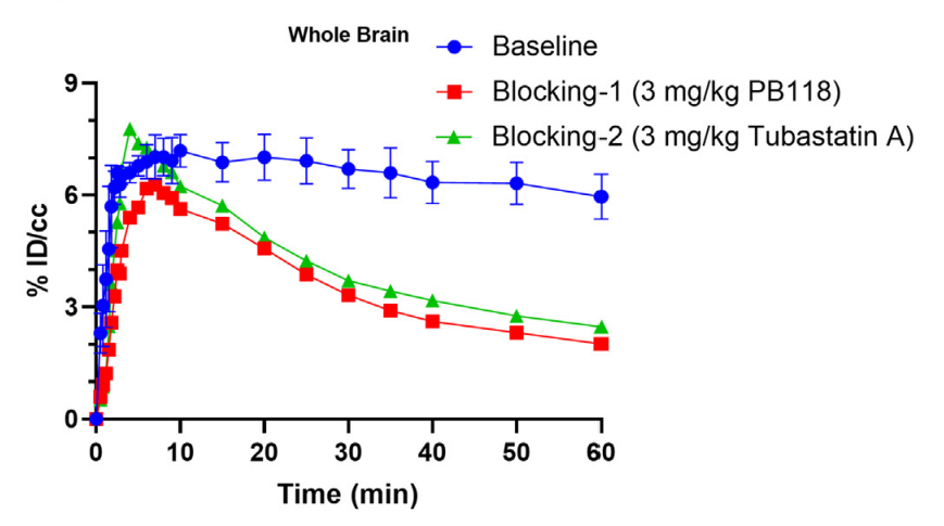
Tubastatin A purchased from MedChemExpress. Usage Cited in: Acta Pharm Sin B. 2022 Oct;12(10):3891-3904. [Abstract]
The baseline and blocking time-activity curves of PB118 in the mice whole brain. Compared with baseline mice, remarkably reduced radioactivity was observed in the brain of mice pretreated with Tubastatin A (3.0 mg/kg; 5 min before RI).
-
Adv Sci (Weinh)
The Probiotic Parabacteroides johnsonii Ameliorates Metabolic Disorders Through Promoting BCAAs to BSCFAs Conversion. [Abstract]2025 Aug 7:e02624. PMID: 40772426 -
J Biomed Sci
Targeting enolase 1 reverses bortezomib resistance in multiple myeloma through YWHAZ/Parkin axis. [Abstract]2025 Jan 20;32(1):9. PMID: 39828712 -
Cell Death Dis
HDAC6-dependent deacetylation of TAK1 enhances sIL-6R release to promote macrophage M2 polarization in colon cancer. [Abstract]2022 Oct 21;13(10):888. PMID: 36270986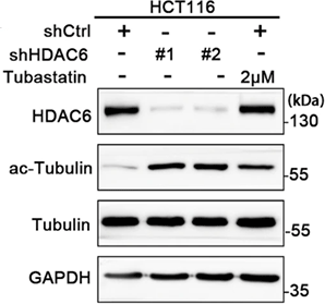
Tubastatin A purchased from MedChemExpress. Usage Cited in: Cell Death Dis. 2022 Oct 21;13(10):888. [Abstract]
Construction of HCT116 cell line with stable HDAC6 knockdown. Tubastatin A HCl is HDAC6 selective inhibitor.
-
Free Radic Biol Med
HDAC inhibition protects RPE cells from oxidative stress via enhanced mitochondrial fusion, cytoskeletal repair, and Nrf-2 activation. [Abstract]2025 Aug 13:240:59-70. PMID: 40816649 -
NPJ Precis Oncol
Connectivity mapping-based identification of pharmacological inhibitor targeting HDAC6 in aggressive pancreatic ductal adenocarcinoma. [Abstract]2024 Mar 7;8(1):66. PMID: 38454151 -
Neurotherapeutics
2023 Jul;20(4):1215-1228. PMID: 37268847
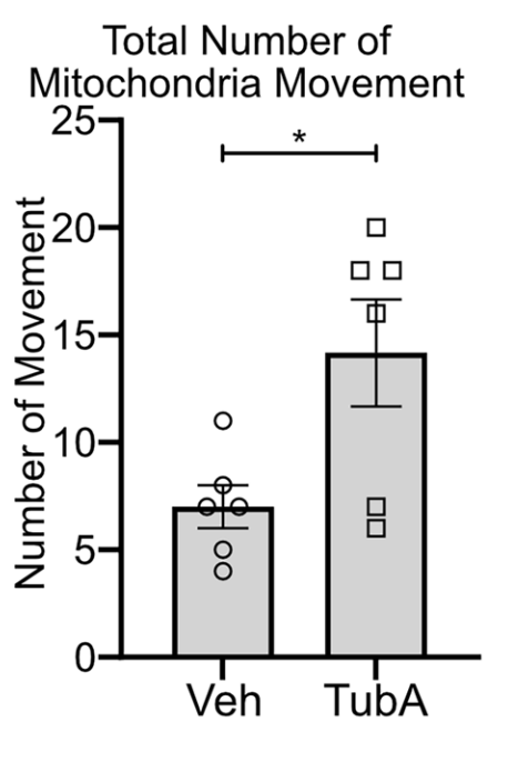
Tubastatin A purchased from MedChemExpress. Usage Cited in: Neurotherapeutics. 2023 Jul;20(4):1215-1228. [Abstract]
Tubastatin A (TubA) (1 μM; overnight) treatment restores mitochondrial transport in cultured Gan−/− DRG neurons. Graph represents the significant increase in the total movement of mitochondria in Tubastatin A (TubA)-treated Gan−/− DRG neurons.
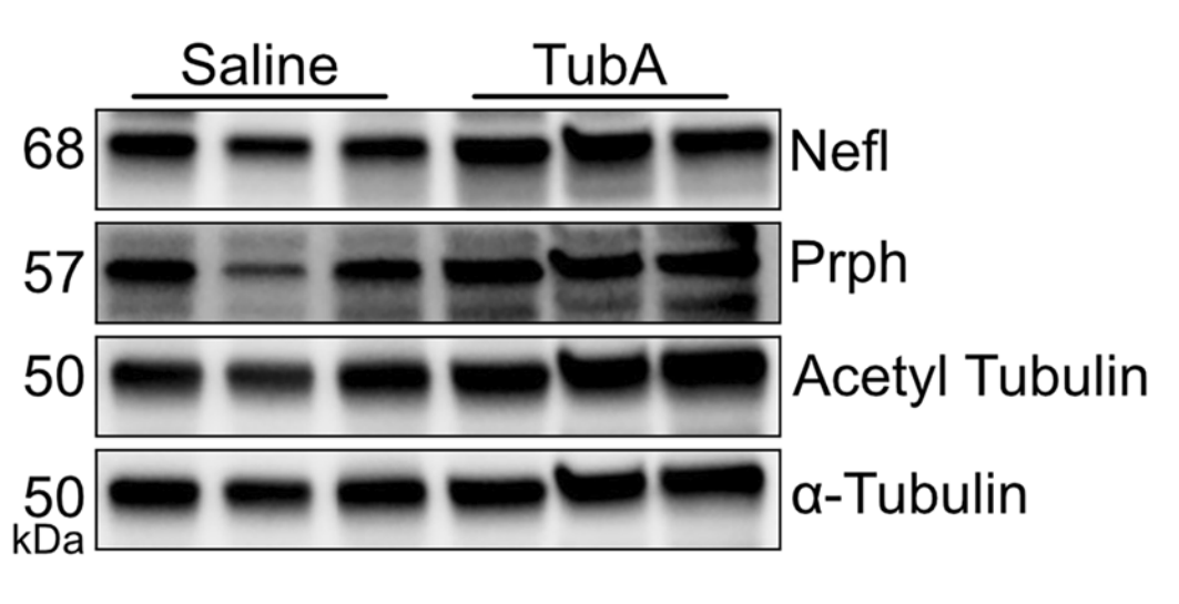
Tubastatin A purchased from MedChemExpress. Usage Cited in: Neurotherapeutics. 2023 Jul;20(4):1215-1228. [Abstract]
Tubastatin A (TubA) (1 μM; overnight) treatment increased levels of acetylated tubulin and Prph in the sciatic nerve of Gan−/−;TgPer mice. The figure represented the western blots for Nefl, Prph, acetyl tubulin, and α-tubulin, respectively, from the sciatic nerve of saline- and Tubastatin A (TubA)-treated mice.
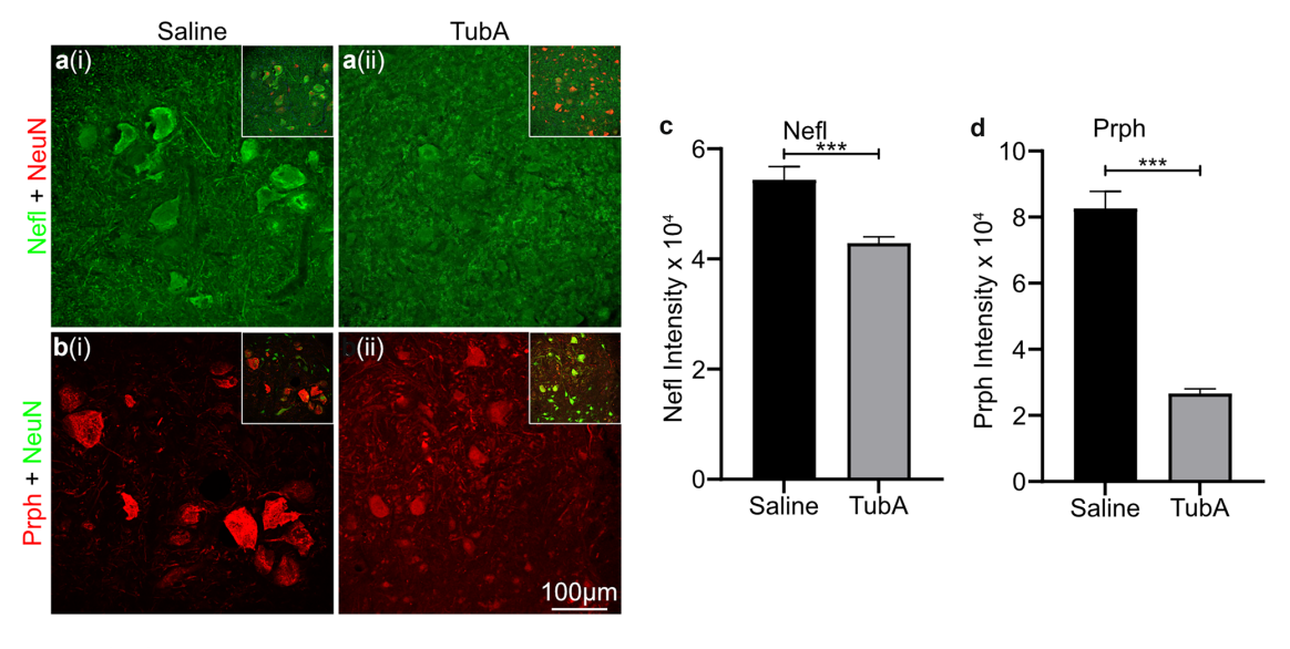
Tubastatin A purchased from MedChemExpress. Usage Cited in: Neurotherapeutics. 2023 Jul;20(4):1215-1228. [Abstract]
Immunostaining of Nefl and Prph was performed on the cell body area of spinal neurons (larger than 250 μm²) from saline- and Tubastatin A (TubA)-treated mice, followed by quantification of the signal intensity. There was a significant decrease in the signal intensity for Nefl and Prph proteins in the perikaryon of spinal neurons from Gan−/−;TgPer mice treated with Tubastatin A (TubA) when compared to saline-treated Gan−/−;TgPer mice.
-
Clin Transl Med
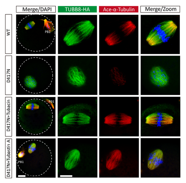
Pathogenic variants of TUBB8 cause oocyte spindle defects by disrupting with EB1/CAKP5 interactions and potential treatment targeting microtubule acetylation through HDAC6 inhibition. [Abstract]2025 Jan;15(1):e70193. PMID: 39834092
Tubastatin A purchased from MedChemExpress. Usage Cited in: Clin Transl Med. 2025 Jan;15(1):e70193. [Abstract]
The inhibition of HDAC6 restored the spindle assembly caused by TUBB8 missense variants. Representative confocal images showed spindle morphology in groups of WT, D417N (DMSO), D417N + tubacin (2 μM), and D417N + Tubastatin A (10 μM; DMSO).
-
J Med Chem
Discovery of a Novel Benzimidazole Derivative Targeting Histone Deacetylase to Induce Ferroptosis and Trigger Immunogenic Cell Death. [Abstract]2024 Sep 12;67(17):15098-15117. PMID: 39145486 -
J Med Chem
Development and Pharmacochemical Characterization Discover a Novel Brain-Permeable HDAC11-Selective Inhibitor with Therapeutic Potential by Regulating Neuroinflammation in Mice. [Abstract]2023 Dec 14;66(23):16075-16090. PMID: 37972387 -
Antioxidants (Basel)
Sp1 S-Sulfhydration Induced by Hydrogen Sulfide Inhibits Inflammation via HDAC6/MyD88/NF-κB Signaling Pathway in Adjuvant-Induced Arthritis. [Abstract]2022 Apr 7;11(4):732. PMID: 35453416 -
Antioxidants
Inhibition of HDAC6 Attenuates Diabetes-Induced Retinal Redox Imbalance and Microangiopathy. [Abstract]2020 Jul 9;9(7):599. PMID: 32660051 -
Atherosclerosis
Targeting HDAC6 attenuates nicotine-induced macrophage pyroptosis via NF-κB/NLRP3 pathway. [Abstract]2021 Jan:317:1-9. PMID: 33321327 -
Virulence
The inhibitory and anti-inflammatory effects of TMP269 on peste des petits ruminants virus replication. [Abstract]2025 Dec;16(1):2495838. PMID: 40275702 -
J Pathol
Histone deacetylase 6 inhibition attenuates pathological cardiac hypertrophy by promoting autophagy through MAP1LC3B ubiquitination. [Abstract]2025 Apr 11. PMID: 40212005 -
PLoS Pathog
PCV2 targets cGAS to inhibit type I interferon induction to promote other DNA virus infection. [Abstract]2021 Sep 20;17(9):e1009940. PMID: 34543359 -
Front Pharmacol
HDAC6 Inhibition Promotes Transcription Factor EB Activation and Is Protective in Experimental Kidney Disease. [Abstract]2018 Feb 1:9:34. PMID: 29449811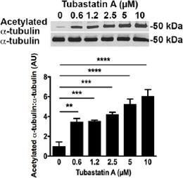
Tubastatin A purchased from MedChemExpress. Usage Cited in: Front Pharmacol. 2018 Feb 1:9:34. [Abstract]
HDAC6 inhibition with Tubastatin A acetylates TFEB, facilitates TFEB nuclear translocation and attenuates programmed cell death in NRK-52E cells. Immunoblotting for acetylated α-tubulin in NRK-52E cells treated with Tubastatin A.
-
Mol Cancer Res
Constitutive β-Catenin Overexpression Represses Lncrna MIR100HG Transcription via HDAC6-Mediated Histone Modification in Colorectal Cancer. [Abstract]2022 Jun 3;20(6):949-959. PMID: 35247921 -
Mol Cancer Res
NEDD9 regulates actin dynamics through cortactin deacetylation in an AURKA/HDAC6-dependent manner. [Abstract]2014 May;12(5):681-93. PMID: 24574519 -
BMC Biol
Microtubule polyglutamylation and acetylation drive microtubule dynamics critical for platelet formation. [Abstract]2018 Oct 18;16(1):116. PMID: 30336771 -
J Neurochem
Effects of Lysine Deacetylation Inhibition Alone or in Combination With Arimoclomol on TDP-43 Proteinopathy. [Abstract]2026 Jun;170(6):e70493. PMID: 42283221 -
J Dermatol Sci
2022 Jun;106(3):181-188. PMID: 35637111 -
Aging (Albany NY)
Inhibition of HDAC6 by Tubastatin A reduces chondrocyte oxidative stress in chondrocytes and ameliorates mouse osteoarthritis by activating autophagy. [Abstract]2021 Mar 19;13(7):9820-9837. PMID: 33744850 -
J Nutr
Dietary Glucose Increases Glucose Absorption and Lipid Deposition via SGLT1/2 Signaling and Acetylated ChREBP in the Intestine and Isolated Intestinal Epithelial Cells of Yellow Catfish. [Abstract]2020 Jul 1;150(7):1790-1798. PMID: 32470978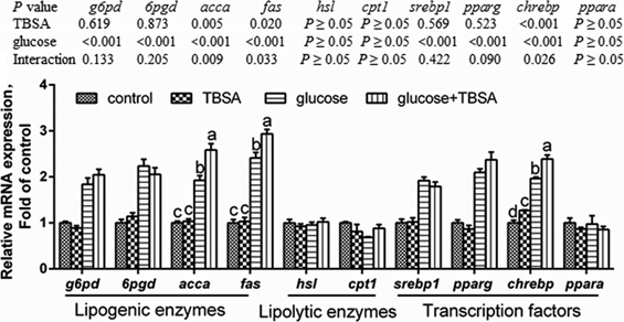
Tubastatin A purchased from MedChemExpress. Usage Cited in: J Nutr. 2020 Jul 1;150(7):1790-1798. [Abstract]
Effects of TBSA (Tubastatin A) pretreatment on glucose-induced changes in mRNA amounts of genes in IECs of yellow catfish.
-
Biochim Biophys Acta Mol Cell Res
Shear stress stimulates integrin β1 trafficking and increases directional migration of cancer cells via promoting deacetylation of microtubules. [Abstract]2020 May;1867(5):118676. PMID: 32044386 -
Am J Pathol
Inhibition of Histone Deacetylase 6 by Tubastatin A Attenuates the Progress of Osteoarthritis via Improving Mitochondrial Function. [Abstract]2020 Dec;190(12):2376-2386. PMID: 32926854 -
Viruses
2025 Jan 13;17(1):90. PMID: 39861880 -
Hum Mol Genet
E3 ligase Smurf1 protects against misfolded SOD1 in neuronal cells by promoting its K63 ubiquitylation and aggresome formation. [Abstract]2022 Jun 22;31(12):2035-2048. PMID: 35022748 -
J Mol Neurosci
Effects of the Glucocorticoid-Mediated Mitochondrial Translocation of Glucocorticoid Receptors on Oxidative Stress and Pyroptosis in BV-2 Microglia. [Abstract]2024 Mar 13;74(1):30. PMID: 38478195 -
J Muscle Res Cell Motil
Tubastatin A attenuates impaired autophagic degradation and promotes myogenic program in skeletal muscle following downhill running. [Abstract]2025 Nov 13. PMID: 41233637 -
-
-
bioRxiv
2025 Jul 12:2025.07.08.663754. PMID: 40672312 -
-
-
-
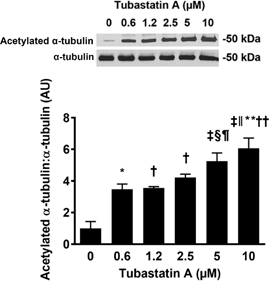
Tubastatin A purchased from MedChemExpress. Usage Cited in: University of Toronto. 2017 Nov.
Tubastatin A induces a dose-dependent increase in acetylated α-tubulin levels in NRK-52E cells.


Solvent & Solubility
DMSO : 12.5 mg/mL (37.27 mM; Need ultrasonic; Hygroscopic DMSO has a significant impact on the solubility of product, please use newly opened DMSO)
H2O : < 0.1 mg/mL (insoluble)
Please refer to the solubility information to select the appropriate solvent. Once prepared, please aliquot and store the solution to prevent product inactivation from repeated freeze-thaw cycles.
Storage method and period of stock solution: -80°C, 6 months; -20°C, 1 month. When stored at -80°C, please use it within 6 months. When stored at -20°C, please use it within 1 month.
Concentration (start) × Volume (start) = Concentration (final) × Volume (final)
Select the appropriate dissolution method based on your experimental animal and administration route.
- For the following dissolution methods, please ensure to first prepare a clear stock solution using an In Vitro approach and then sequentially add co-solvents:
- To ensure reliable experimental results, the clarified stock solution can be appropriately stored based on storage conditions. As for the working solution for In Vivo experiments, it is recommended to prepare freshly and use it on the same day.
- The percentages shown for the solvents indicate their volumetric ratio in the final prepared solution. If precipitation or phase separation occurs during preparation, heat and/or sonication can be used to aid dissolution.
Add each solvent one by one: 10% DMSO 40% PEG300 5% Tween-80 45% Saline
Solubility: ≥ 1.25 mg/mL (3.73 mM); Clear solution
This protocol yields a clear solution of ≥ 1.25 mg/mL (saturation unknown).
Taking 1 mL working solution as an example, add 100 μL DMSO stock solution (12.5 mg/mL) to 400 μL PEG300, and mix evenly; then add 50 μL Tween-80 and mix evenly; then add 450 μL Saline to adjust the volume to 1 mL.
Preparation of Saline: Dissolve 0.9 g sodium chloride in ddH₂O and dilute to 100 mL to obtain a clear Saline solution.
Add each solvent one by one: 10% DMSO 90% (20% SBE-β-CD in Saline)
Solubility: ≥ 1.25 mg/mL (3.73 mM); Clear solution
This protocol yields a clear solution of ≥ 1.25 mg/mL (saturation unknown).
Taking 1 mL working solution as an example, add 100 μL DMSO stock solution (12.5 mg/mL) to 900 μL 20% SBE-β-CD in Saline, and mix evenly.
Preparation of 20% SBE-β-CD in Saline (4°C, storage for one week): 2 g SBE-β-CD powder is dissolved in 10 mL Saline, completely dissolve until clear.
For the following dissolution methods, please prepare the working solution directly:
It is recommended to prepare fresh solutions and use them promptly within a short period of time.
The percentages shown for the solvents indicate their volumetric ratio in the final prepared solution. If precipitation or phase separation occurs during preparation, heat and/or sonication can be used to aid dissolution.
Add each solvent one by one: 50% PEG300 50% Saline
Solubility: 25 mg/mL (74.54 mM); Suspended solution; Need ultrasonic
Please enter the basic information of animal experiments:
-
-
-
-
Recommended: Prepare an additional quantity of animals to account for potential losses during experiments.
Please enter your animal formula composition:
-
%DMSO +
Recommended: Keep the proportion of DMSO in working solution below 2% if your animal is weak.
-
%+
-
+%Tween-80 + +
-
%Saline +
The co-solvents required include: DMSO, . All of co-solvents are available by MedChemExpress (MCE). , Tween 80. All of co-solvents are available by MedChemExpress (MCE).
Working solution concentration: 0.22 mg/mL
Method for preparing stock solution: mg drug dissolved in μL DMSO. Stock solution concentration: mg/mL.
1. Take μL DMSO stock solution;
2. Add μL .
μL , mix evenly;
3. Then add μL Tween 80, mix evenly;
4. Then add μL
Please ensure that the stock solution in the first step is dissolved to a clear state, and add co-solvents in sequence. You can use ultrasonic heating (ultrasonic cleaner, recommended frequency 20-40 kHz), vortexing, etc. to assist dissolution.

Protocol
Primary cortical neuron cultures are obtained from the cerebral cortex of fetal Sprague-Dawley rats (embryonic day 17) as described previously. All experiments are initiated 24 hours after plating. Under these conditions, the cells are not susceptible to glutamate-mediated excitotoxicity. For cytotoxicity studies, cells are rinsed with warm PBS and then placed in minimum essential medium containing 5.5 g/L glucose, 10% fetal calf serum, 2 mM L-glutamine, and 100 μM cystine. Oxidative stress is induced by the addition of the glutamate analogue homocysteate (HCA; 5 mM) to the media. HCA is diluted from 100-fold concentrated solutions that are adjusted to pH 7.5. In combination with HCA, neurons are treated with Tubastatin A at the indicated concentrations. Viability is assessed after 24 hours by MTT assay.
MedChemExpress (MCE) has not independently confirmed the accuracy of these methods. They are for reference only.
The effects of HDAC6 targeting in dextran sodium sulfate (DSS) and adoptive transfer models of colitis are evaluated, using 10 mice per group. Freshly prepared 4% (wt/vol) DSS (MP Biomedicals) is added daily for 5 days to the pH-balanced tap water of WT B6 mice. Mice are treated daily for 7 days with tubacin or niltubacin (0.5 mg/kg of body weight/day, i.p.), and colitis is assessed by daily monitoring of body weight, stool consistency, and fecal blood. Stool consistency is scored as 0 (hard), 2 (soft), or 4 (diarrhea), and fecal blood (Hemoccult) is scored as 0 (absent), 2 (occult), or 4 (gross). To assess prevention of colitis in a T cell-dependent model, CD4+ CD45RBhi T cells (1×10⁶) isolated from WT mice using magnetic beads (>95% cell purity, flow cytometry) are injected i.p. into B6/Rag1−/− mice plus CD4+ CD25+ Tregs (1.25×10⁵) isolated using magnetic beads from HDAC6−/− or WT mice (>90% Treg purity, flow cytometry) and mice are monitored biweekly for clinical evidence of colitis. To assess therapy of established T cell-dependent colitis, B6/Rag1−/− mice are injected i.p. with CD4+ CD45RBhi cells (1×10⁶). Once colitis has developed, mice also receive CD4+ CD25+ Tregs (5×10⁵ cells) isolated as described above from HDAC6−/− or WT mice or treatment with HDAC6i (tubastatin A) or HSP90i (17-AAG). Mice are monitored for continued weight loss and stool consistency. At the cessation of the study, paraffin sections of colons stained with Alcian Blue or hematoxylin and eosin are graded histologically or evaluated by immunoperoxidase staining for Foxp3+ Treg infiltration.
MedChemExpress (MCE) has not independently confirmed the accuracy of these methods. They are for reference only.


Purity & Documentation
-
Data Sheet (285 KB)
-
SDS (393 KB)
- English - EN (393 KB)
- Français - FR (393 KB)
- Deutsch - DE (393 KB)
- Norwegian - NO (393 KB)
- Español - ES (393 KB)
- Swedish - SV (393 KB)
- Italian - IT (393 KB)
- Portuguese - PT (393 KB)
-
Handling Instructions (2659 KB)
References
[1]. Kyle V. Butler et al. Rational Design and Simple Chemistry Yield a Superior, Neuroprotective HDAC6 Inhibitor, Tubastatin A J. Am. Chem. Soc., 2010, 132 (31), pp 10842-10846 [Content Brief]
[2]. Kozyreva VK, et al. NEDD9 regulates actin dynamics through cortactin deacetylation in an AURKA/HDAC6-dependent manner. Mol Cancer Res. 2014 May;12(5):681-93. [Content Brief]
[3]. de Zoeten EF, et al. Histone deacetylase 6 and heat shock protein 90 control the functions of Foxp3(+) T-regulatory cells. Mol Cell Biol. 2011 May;31(10):2066-78. [Content Brief]
[5]. Di Fulvio S, et al. Dysferlin interacts with histone deacetylase 6 and increases alpha-tubulin acetylation. PLoS One. 2011;6(12):e28563 [Content Brief]
[6]. Ketene AN, et al. Actin filaments play a primary role for structural integrity and viscoelastic response in cells. Integr Biol (Camb). 2012 May;4(5):540-9. [Content Brief]
[7]. Lechner S, Malgapo MIP, Grätz C, et al. Target deconvolution of HDAC pharmacopoeia reveals MBLAC2 as common off-target [published online ahead of print, 2022 Apr 28]. Nat Chem Biol. 2022;10.1038/s41589-022-01015-5. [Content Brief]
[8]. Géraldy M, Morgen M, Sehr P, et al. Selective Inhibition of Histone Deacetylase 10: Hydrogen Bonding to the Gatekeeper Residue is Implicated. J Med Chem. 2019;62(9):4426-4443. [Content Brief]
[9]. Severin Lechner, et al. Target deconvolution of HDAC pharmacopoeia reveals MBLAC2 as common off-target. Nat Chem Biol. 2022 Apr 28. [Content Brief]


Complete Stock Solution Preparation Table
Please refer to the solubility information to select the appropriate solvent. Once prepared, please aliquot and store the solution to prevent product inactivation from repeated freeze-thaw cycles.
Storage method and period of stock solution: -80°C, 6 months; -20°C, 1 month. When stored at -80°C, please use it within 6 months. When stored at -20°C, please use it within 1 month.
| Optional Solvent | Concentration Solvent Mass | 1 mg | 5 mg | 10 mg | 25 mg |
|---|---|---|---|---|---|
| DMSO | 1 mM | 2.9815 mL | 14.9076 mL | 29.8151 mL | 74.5379 mL |
| 5 mM | 0.5963 mL | 2.9815 mL | 5.9630 mL | 14.9076 mL | |
| 10 mM | 0.2982 mL | 1.4908 mL | 2.9815 mL | 7.4538 mL | |
| 15 mM | 0.1988 mL | 0.9938 mL | 1.9877 mL | 4.9692 mL | |
| 20 mM | 0.1491 mL | 0.7454 mL | 1.4908 mL | 3.7269 mL | |
| 25 mM | 0.1193 mL | 0.5963 mL | 1.1926 mL | 2.9815 mL | |
| 30 mM | 0.0994 mL | 0.4969 mL | 0.9938 mL | 2.4846 mL |


 Powered by Bioz