



hnRNP Q Antibody (YA1838)
(Synonyms: SYNCRIP; HNRPQ; NSAP1; Heterogeneous nuclear ribonucleoprotein Q; hnRNP Q; Glycine- and tyrosine-rich RNA-binding protein; GRY-RBP; NS1-associated protein 1; Synaptotagmin-binding; cytoplasmic RNA-interacting protein)
Based on 1 Customer Validation
hnRNP Q Antibody (YA1838) is a Rabbit-derived and non-conjugated IgG monoclonal antibody, targeting to hnRNP Q.
-
Host:
Rabbit
-
Isotype:
IgG
-
Application:
WB, IHC-P, ICC/IF
-
Reactivity :
Human, Mouse, Rat
-
Formulation:
Supplied in 50mM Tris-Glycine(pH 7.4), 0.15M NaCl, 40%Glycerol, 0.01% sodium azide and 0.05% BSA.
-
Conjugation:
Non-conjugated


To place orders, for customer services and technical support, please contact: MedChemExpress USA
Tel: 609-228-6898 E-mail: [email protected] [email protected]





Applications
| Application |
WB

WB: Western Blot
|
IHC-P
IHC-P: Immunohistochemistry-Paraffin
|
ICC/IF
ICC/IF: Immunocytochemistry/
Immunofluorescence |
|---|---|---|---|
| Dilution Ratio | 1:500-1:1000 | 1:50-1:100 | 1:50-1:200 |


Product Details
hnRNP Q Antibody (YA1838) is a Rabbit-derived and non-conjugated IgG monoclonal antibody, targeting to hnRNP Q.
-
Host Rabbit

-
Clonality Recombinant,Monoclonal

-
Species ReactivityHuman, Mouse, Rat
-
Observed Molecular WeightObserved band size: 65-82 kDa
Note: Due to possible protein modifications or aggregation, the molecular weight should be confirmed by actual measurement, and the predicted value is for reference only.

-
Calculated Molecular Weight Predicted band size: 70 kDa

Entrez Gene: 10492 Human ; 56403 Mouse ; 363113 Rat
SwissProt: O60506 Human ; Q7TMK9 Mouse ; Q7TP47 Rat
A synthesized peptide derived from human hnRNP Q aa570-623.
Endogenous
Affinity Chromatography
Non-conjugated
Unmodified
IgG


Product Properties
-
Appearance
Liquid
-
Formulation
Supplied in 50mM Tris-Glycine(pH 7.4), 0.15M NaCl, 40%Glycerol, 0.01% sodium azide and 0.05% BSA.
-
Concentration
Batch-dependent, Please check the COA for the concentration of each lot. Check Lot Concentration
-
Storage & Stability
Stored at -20°C for 1 year. Avoid repeated freeze / thaw cycles.
-
Shipping
Shipping with blue ice.


Verification Images
-
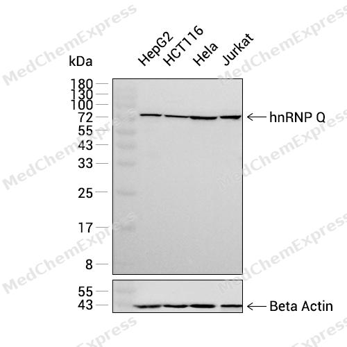 Western blot analysis was performed on extracts from HepG2 (lane 1, 15 μg), HCT116 (lane 2, 15 μg), Hela (lane 3, 15 μg), and Jurkat (lane 4, 15 μg) using hnRNP Q Rabbit mAb.Proteins were transferred to a PVDF membrane and blocked with 5% non-fat milk in TBST at 4°C overnight.The primary antibody (1:1000 dilution) and the loading control antibody (beta-Actin, HY-P83730, 1:20000 dilution) were incubated in 5% non-fat milk in TBST for 1 hour at 37°C.Goat Anti-Rabbit IgG-HRP Secondary Antibody (1:20000 dilution) was then applied for 40 minutes at 37°C.
 -
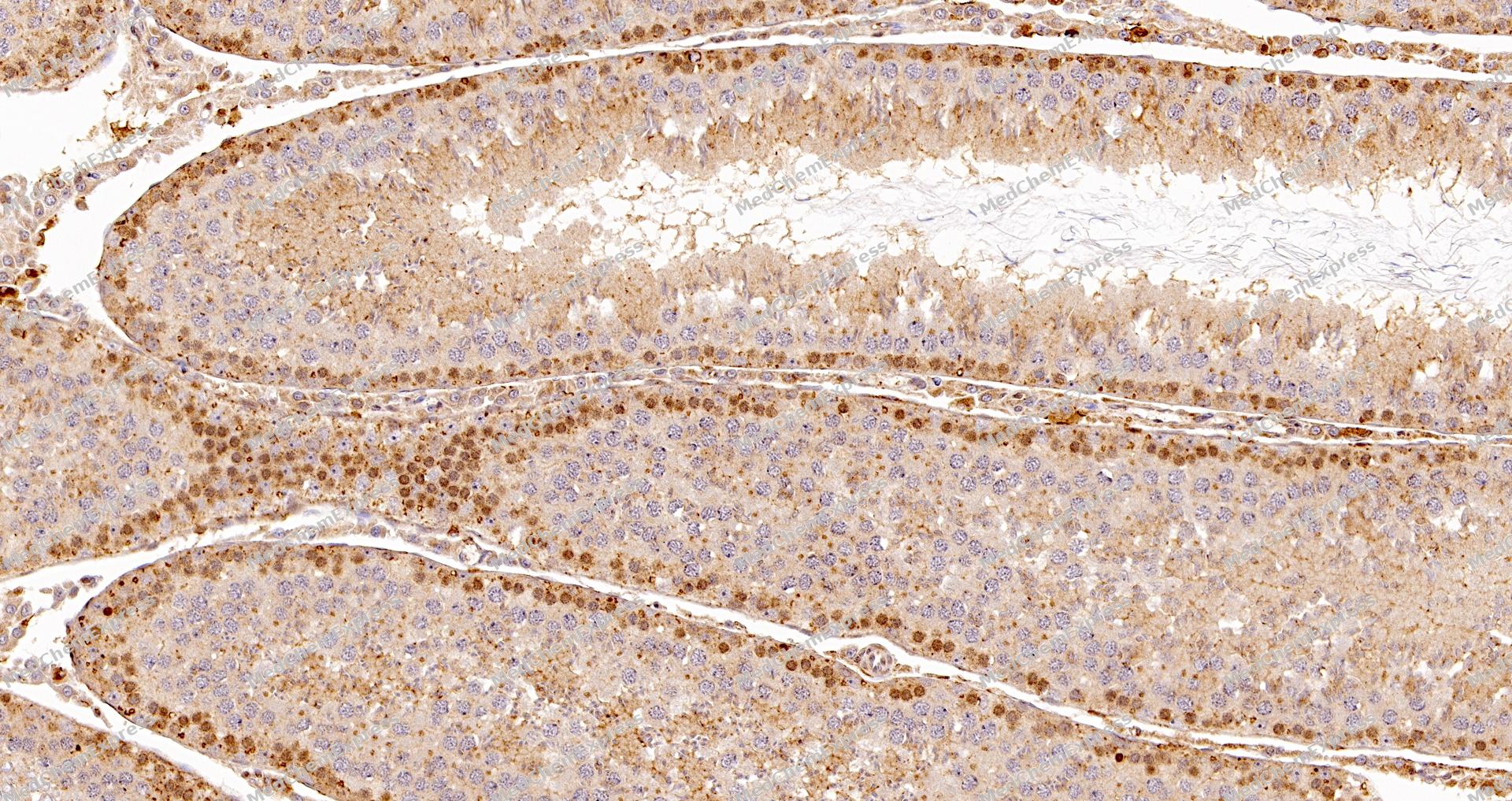 Immunohistochemical analysis of paraffin-embedded Rat testis tissue using hnRNP Q Antibody (YA1838). The section was pre-treated using heat mediated antigen retrieval with Tris-EDTA buffer (pH 9.0) for 8 minutes. The tissues were blocked in QuickBlock for 20 minutes at room temperature, washed with ddH2O and PBS, and then probed with the primary antibody (HY-P82093, 1/100) in 4℃ overnight. The detection was performed using an HRP conjugated compact polymer system. DAB was used as the chromogen. Tissues were counterstained with hematoxylin and mounted with DPX.
 -
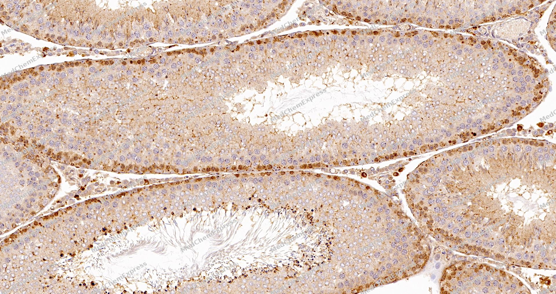 Immunohistochemical analysis of paraffin-embedded Rat testis tissue using hnRNP Q Antibody (YA1838). The section was pre-treated using heat mediated antigen retrieval with Tris-EDTA buffer (pH 9.0) for 8 minutes. The tissues were blocked in QuickBlock for 20 minutes at room temperature, washed with ddH2O and PBS, and then probed with the primary antibody (HY-P82093, 1/100) in 4℃ overnight. The detection was performed using an HRP conjugated compact polymer system. DAB was used as the chromogen. Tissues were counterstained with hematoxylin and mounted with DPX.
 -
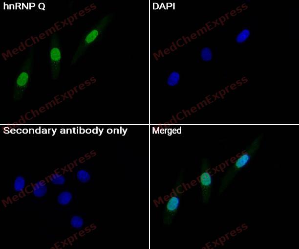 Immunocytochemistry analysis of HepG2 cells labeling hnRNP Q Antibody(HY-P82093) at 1/50 dilution. Cells were fixed in 4% paraformaldehyde for 15 minutes at room temperature, permeabilized with 0.1% Triton X-100 for 10 minutes at room temperature, then blocked with BSA for Immunol Staining for 10 min at room temperature. Cells were then incubated with hnRNP Q Antibody(HY-P82093) at 1/50 dilution in BSA for Immunol Staining at 4 ℃ Overnight. AF488-conjugated AffiniPure Goat Anti-Rabbit IgG H&L(HY-P8002, Green) was used as the secondary antibody at 1/1,000 dilution. PBS instead of the primary antibody was used as the secondary antibody only control. The Nuclear counterstain was DAPI (Blue).
 -
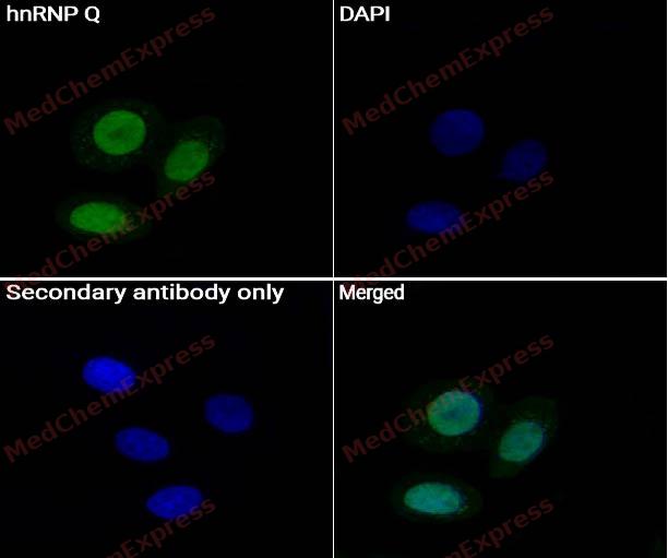 Immunocytochemistry analysis of SiHa cells labeling hnRNP Q Antibody(HY-P82093) at 1/50 dilution. Cells were fixed in 4% paraformaldehyde for 15 minutes at room temperature, permeabilized with 0.1% Triton X-100 for 10 minutes at room temperature, then blocked with BSA for Immunol Staining for 10 min at room temperature. Cells were then incubated withhnRNP Q Antibody(HY-P82093) at 1/50 dilution in BSA for Immunol Staining at 4 ℃ Overnight. AF488-conjugated AffiniPure Goat Anti-Rabbit IgG H&L(HY-P8002, Green) was used as the secondary antibody at 1/1,000 dilution. PBS instead of the primary antibody was used as the secondary antibody only control. The Nuclear counterstain was DAPI (Blue).


Background
-
Function
hnRNP Q is a Heterogenous nuclear ribonucleoprotein (hnRNP) implicated in mRNA processing mechanisms. Component of the CRD-mediated complex that promotes MYC mRNA stability. Isoform 1, isoform 2 and isoform 3 are associated in vitro with pre-mRNA, splicing intermediates and mature mRNA protein complexes. Isoform 1 binds to apoB mRNA AU-rich sequences. Isoform 1 is part of the APOB mRNA editosome complex and may modulate the postranscriptional C to U RNA-editing of the APOB mRNA through either by binding to A1CF (APOBEC1 complementation factor), to APOBEC1 or to RNA itself. May be involved in translationally coupled mRNA turnover. Implicated with other RNA-binding proteins in the cytoplasmic deadenylation/translational and decay interplay of the FOS mRNA mediated by the major coding-region determinant of instability (mCRD) domain. Interacts in vitro preferentially with poly(A) and poly(U) RNA sequences. Isoform 3 may be involved in cytoplasmic vesicle-based mRNA transport through interaction with synaptotagmins. Component of the GAIT (gamma interferon-activated inhibitor of translation) complex which mediates interferon-gamma-induced transcript-selective translation inhibition in inflammation processes. Upon interferon-gamma activation assembles into the GAIT complex which binds to stem loop-containing GAIT elements in the 3'-UTR of diverse inflammatory mRNAs (such as ceruplasmin) and suppresses their translation; seems not to be essential for GAIT complex function
-
Subcellular Localization
Cytoplasm; Microsome; Endoplasmic reticulum; Nucleus; Nucleus, nucleoplasm; Nucleus, nucleoplasm; Nucleus, nucleoplasm
-
Expression
Tissue_specificity:Ubiquitously expressed. Detected in heart, brain, pancreas, placenta, spleen, lung, liver, skeletal muscle, kidney, thymus, prostate, uterus, small intestine, colon, peripheral blood and testis -
Isoforms & Post-Translational Modification
O60506 has 5 isomers: O60506-1: 69603 Da (predicted); O60506-2: 65682 Da (predicted); O60506-3: 62656 Da (predicted); O60506-4: 58736 Da (predicted); O60506-5: 46328 Da (predicted).
Phosphorylated on tyrosine. The membrane-bound form found in microsomes is phosphorylated in vitro by insulin receptor tyrosine kinase (INSR). Phosphorylation is inhibited upon binding to RNA, whereas the cytoplasmic form is poorly phosphorylated (By similarity) -
Subunit
Isoform 1 is a component of the APOB mRNA editosome complex and interacts with APOBEC1 and A1CF (APOBEC1 complementation factor). Part of a complex associated with the FOS mCRD domain and consisting of PABPC1, PAIP1, CSDE1/UNR, HNRPD and SYNCRIP. Isoform 3 interacts with HNRPR. Interacts with POLR2A hyperphosphorylated C-terminal domain. Isoform 1, isoform 2 and isoform 3 interact with SMN. Isoform 3 interacts through its C-terminal domain with SYT7, SYT8 and SYT9 (By similarity). The non-phosphorylated and phosphorylated forms are colocalized with PAIP1 in polysomes (By similarity). Interacts with HABP4 (PubMed:19523114). Identified in a histone pre-mRNA complex, at least composed of ERI1, LSM11, SLBP, SNRPB, SYNCRIP and YBX1 (By similarity). Identified in the spliceosome C complex. Component of the coding region determinant (CRD)-mediated complex, composed of DHX9, HNRNPU, IGF2BP1, SYNCRIP and YBX1. Identified in a mRNP complex, at least composed of DHX9, DDX3X, ELAVL1, HNRNPU, IGF2BP1, ILF3, PABPC1, PCBP2, PTBP2, STAU1, STAU2, SYNCRIP and YBX1. Identified in a mRNP granule complex, at least composed of ACTB, ACTN4, DHX9, ERG, HNRNPA1, HNRNPA2B1, HNRNPAB, HNRNPD, HNRNPL, HNRNPR, HNRNPU, HSPA1, HSPA8, IGF2BP1, ILF2, ILF3, NCBP1, NCL, PABPC1, PABPC4, PABPN1, RPLP0, RPS3, RPS3A, RPS4X, RPS8, RPS9, SYNCRIP, YBX1 and untranslated mRNAs. Interacts with GTPBP1. Component of the GAIT complex; in humans the complex assembly seems to be a two-step process in which EPRS1 first associates with SYNCRIP to form a pre-GAIT complex which is deficient in GAIT element binding
-
SwissProt ID
-
Synonyms
SYNCRIP; HNRPQ; NSAP1; Heterogeneous nuclear ribonucleoprotein Q; hnRNP Q; Glycine- and tyrosine-rich RNA-binding protein; GRY-RBP; NS1-associated protein 1; Synaptotagmin-binding; cytoplasmic RNA-interacting protein
-
Research Field
Microbiology


Documentation
-
Data Sheet (258 KB)
-
SDS (252 KB)
- English - EN (252 KB)
- Français - FR (252 KB)
- Deutsch - DE (252 KB)
- Norwegian - NO (252 KB)
- Español - ES (252 KB)
- Swedish - SV (252 KB)
- Italian - IT (252 KB)
- Korean - KR (252 KB)
- Portuguese - PT (252 KB)
-
User Guide for Antibodies (1077 KB)

 Powered by Bioz
