



Moesin Antibody (YA284)

Based on 1 Customer Validation
Moesin Antibody (YA284) is a Rabbit-derived and non-conjugated IgG monoclonal antibody, targeting to Moesin.
-
Host:
Rabbit
-
Isotype:
IgG
-
Application:
WB, IHC-P, ICC/IF
-
Reactivity :
Human, Mouse
-
Formulation:
Supplied in 1*TBS (pH7.4), 0.05% BSA and 40% Glycerol. Preservative: 0.05% Sodium Azide.
-
Conjugation:
Non-conjugated


To place orders, for customer services and technical support, please contact: MedChemExpress USA
Tel: 609-228-6898 E-mail: [email protected] [email protected]





Applications
| Application |
WB

WB: Western Blot
|
ICC/IF
ICC/IF: Immunocytochemistry/
Immunofluorescence |
IHC-P
IHC-P: Immunohistochemistry-Paraffin
|
|---|---|---|---|
| Dilution Ratio | 1:1000-1:2000 | 1:50-1:200 | 1:50-1:200 |


Product Details
Moesin Antibody (YA284) is a Rabbit-derived and non-conjugated IgG monoclonal antibody, targeting to Moesin.
-
Host Rabbit

-
Clonality Recombinant,Monoclonal

-
Species ReactivityHuman, Mouse
-
Observed Molecular WeightObserved band size: 75 kDa
Note: Due to possible protein modifications or aggregation, the molecular weight should be confirmed by actual measurement, and the predicted value is for reference only.

-
Calculated Molecular Weight Predicted band size: 68 kDa

Entrez Gene: 4478 Human ; 17698 Mouse ; 81521 Rat
SwissProt: P26038 Human ; P26041 Mouse ; O35763 Rat
OMIM: 300988 Human
Synthetic peptide corresponding to Human Moesin.AA range:455-500.
Endogenous
Protein A affinity purified.
Non-conjugated
Unmodified
IgG


Product Properties
-
Appearance
Liquid
-
Formulation
Supplied in 1*TBS (pH7.4), 0.05% BSA and 40% Glycerol. Preservative: 0.05% Sodium Azide.
-
Concentration
Batch-dependent, Please check the COA for the concentration of each lot. Check Lot Concentration
-
Storage & Stability
Stored at -20°C for 1 year. Avoid repeated freeze / thaw cycles.
-
Shipping
Shipping with blue ice.


Verification Images
-
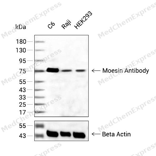 Western blot analysis of extracts from C6(lane2(20μg) , Raji(lane 3(20μg) and HEK293(lane 4(20ug) using Moesin Antibody (HY-P80228) Rabbit mAb. Proteins were transferred to a PVDF membrane and blocked with 5% non-fat milk in TBST for 2 hour at room temperature. The primary antibody (1/1000) and Loading control antibody (Beta Actin, HY-P80438, 1/10000) was used in 5% non-fat milk in TBST at 4°C overnight. Goat Anti-Mouse/Rabbit IgG-HRP Secondary Antibody (1/10000) was used for 1 hour at room temperature.
 -
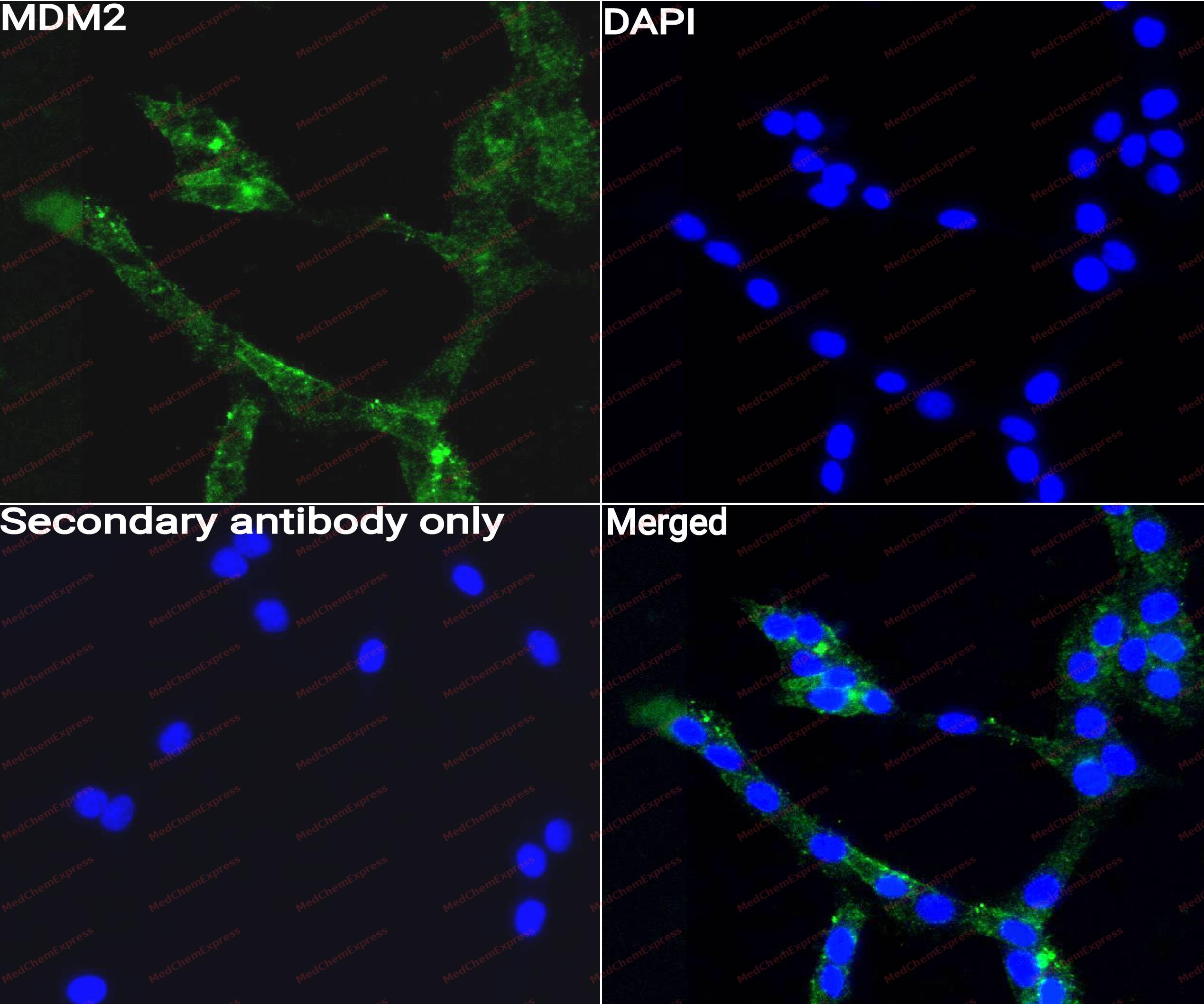 Immunocytochemistry analysis of Hela cells labeling Moesin with Moesin Antibody (HY-P80228) at 1/50 dilution. Cells were fixed in 4% paraformaldehyde for 15 minutes at room temperature, permeabilized with 0.1% Triton X-100 for 10 minutes at room temperature, then blocked with QuickBlock™ Blocking Buffer for Immunol Staining for 10 min at room temperature. Cells were then incubated with Moesin Antibody (HY-P80228) at 1/50 dilution in QuickBlock™ Blocking Buffer for Immunol Staining at 4 ℃. Alexa Fluor® 488-conjugated AffiniPure Goat Anti-Rabbit IgG H&L(HY-P8002, Green) was used as the secondary antibody at 1/1,000 dilution. PBS instead of the primary antibody was used as the secondary antibody only control. The Nuclear counterstain was DAPI (Blue).
 -
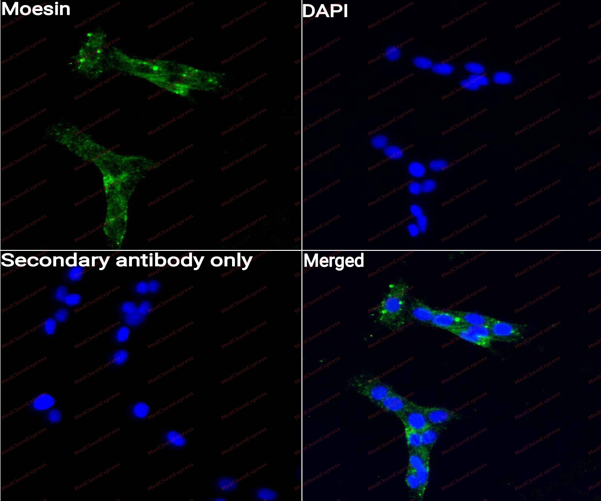 Immunocytochemistry analysis of C6 cells labeling Moesin with Moesin Antibody (HY-P80228) at 1/50 dilution. Cells were fixed in 4% paraformaldehyde for 15 minutes at room temperature, permeabilized with 0.1% Triton X-100 for 10 minutes at room temperature, then blocked with QuickBlock™ Blocking Buffer for Immunol Staining for 10 min at room temperature. Cells were then incubated with Moesin Antibody (HY-P80228)at 1/50 dilution in QuickBlock™ Blocking Buffer for Immunol Staining at 4 ℃. Alexa Fluor® 488-conjugated AffiniPure Goat Anti-Rabbit IgG H&L(HY-P8002,Green) was used as the secondary antibody at 1/1,000 dilution. PBS instead of the primary antibody was used as the secondary antibody only control. The Nuclear counterstain was DAPI (Blue).
 -
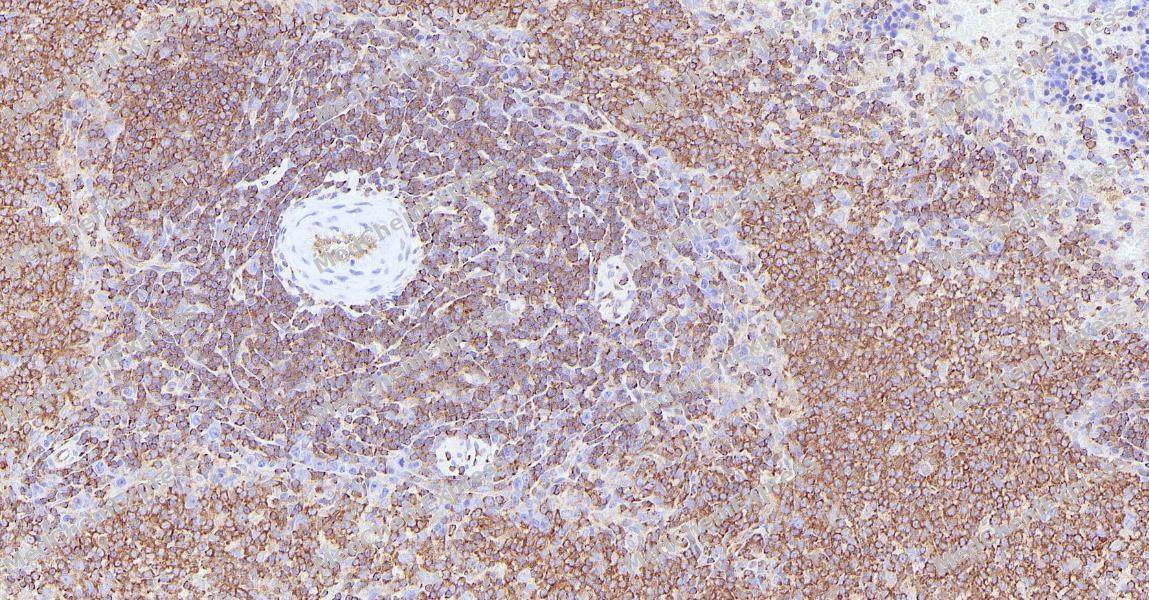 Immunohistochemical analysis of paraffin-embedded rat spleen tissue using Moesin Antibody. The section was pre-treated using heat mediated antigen retrieval with Tris-EDTA buffer (pH 9.0) for 8 minutes. The tissues were blocked in QuickBlock for 20 minutes at room temperature, washed with ddH2O and PBS, and then probed with the primary antibody (HY-P80228, 1:400) in 4℃ overnight. The detection was performed using an HRP conjugated compact polymer system. DAB was used as the chromogen. Tissues were counterstained with hematoxylin and mounted with DPX.
 -
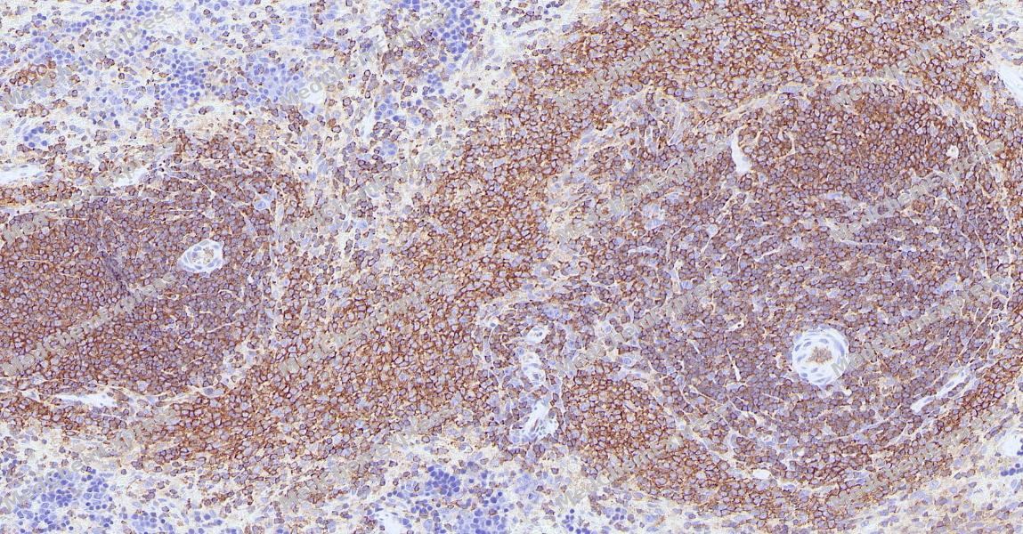 Immunohistochemical analysis of paraffin-embedded rat spleen tissue using Moesin Antibody. The section was pre-treated using heat mediated antigen retrieval with Tris-EDTA buffer (pH 9.0) for 8 minutes. The tissues were blocked in QuickBlock for 20 minutes at room temperature, washed with ddH2O and PBS, and then probed with the primary antibody (HY-P80228, 1:400) in 4℃ overnight. The detection was performed using an HRP conjugated compact polymer system. DAB was used as the chromogen. Tissues were counterstained with hematoxylin and mounted with DPX.


Background
-
Function
Moesin is an Ezrin-radixin-moesin (ERM) family protein that connects the actin cytoskeleton to the plasma membrane and thereby regulates the structure and function of specific domains of the cell cortex. Tethers actin filaments by oscillating between a resting and an activated state providing transient interactions between moesin and the actin cytoskeleton. Once phosphorylated on its C-terminal threonine, moesin is activated leading to interaction with F-actin and cytoskeletal rearrangement. These rearrangements regulate many cellular processes, including cell shape determination, membrane transport, and signal transduction. The role of moesin is particularly important in immunity acting on both T and B-cells homeostasis and self-tolerance, regulating lymphocyte egress from lymphoid organs. Modulates phagolysosomal biogenesis in macrophages. Also participates in immunologic synapse formation[1][2][3][4][5][6].
-
Subcellular Localization
Cell membrane; Peripheral membrane protein; Cytoplasmic side; Cytoplasm, cytoskeleton; Apical cell membrane; Peripheral membrane protein; Cytoplasmic side; Cell projection, microvillus membrane; Peripheral membrane protein; Cytoplasmic side; Cell projection, microvillus
-
Expression
Tissue_specificity:In all the tissues and cultured cells studied -
Subunit
In resting T-cells, part of a PAG1-NHERF1-MSN complex which is disrupted upon TCR activation. Interacts with NHERF1 (PubMed:15020681, PubMed:9314537). Interacts with PPP1R16B (PubMed:18586956). Interacts with SELPLG and SYK; these interactions mediate the activation of SYK by SELPLG (PubMed:12387735). Interacts with PDPN (via cytoplasmic domain); this interaction activates RHOA and promotes epithelial-mesenchymal transition (PubMed:17046996). Interacts with SPN/CD43 cytoplasmic tail (PubMed:11728332, PubMed:9616160). Interacts with CD44 (PubMed:9298994). Interacts with ICAM2 (By similarity). Interacts with ICAM3 (via C-terminus) (PubMed:9298994). Interacts with PDZD8 (PubMed:21549406). Interacts with F-actin (PubMed:10212266). Interacts with CD46 (PubMed:7884872). Interacts with PTPN6 (By similarity)
-
SwissProt ID
-
Research Field
Signal Transduction


Documentation
-
Data Sheet (259 KB)
-
SDS (251 KB)
- English - EN (251 KB)
- Français - FR (251 KB)
- Deutsch - DE (251 KB)
- Norwegian - NO (251 KB)
- Español - ES (251 KB)
- Swedish - SV (251 KB)
- Italian - IT (251 KB)
- Korean - KR (251 KB)
- Portuguese - PT (251 KB)
-
User Guide for Antibodies (1077 KB)

 Powered by Bioz
