



Phospho-FAK (Tyr397) Antibody (YA198)
(Synonyms: FAK, FAK1, PTK2, Focal adhesion kinase 1, FADK 1, Focal adhesion kinase-related nonkinase, Protein phosphatase 1 regulatory subunit 71, Protein-tyrosine kinase 2, p125FAK, pp125FAK, FRNK, PPP1R71)Based on 1 publication(s) in Google Scholar
Phospho-FAK (Tyr397) Antibody (YA198) is a Rabbit-derived and non-conjugated IgG monoclonal antibody, targeting to Phospho-FAK (Tyr397).
-
Host:
Rabbit
-
Isotype:
IgG
-
Application:
WB, ICC/IF
-
Reactivity :
Human, Mouse
-
Formulation:
Supplied in PBS (pH7.4), 0.1% BSA, 40% Glycerol, 0.2% Proclean 950.
-
Conjugation:
Non-conjugated


To place orders, for customer services and technical support, please contact: MedChemExpress USA
Tel: 609-228-6898 E-mail: [email protected] [email protected]



Publications Citing Use of MedChemExpress (MCE) Phospho-FAK (Tyr397) Antibody (YA198)
More

Applications
| Application |
WB

WB: Western Blot
|
ICC/IF
ICC/IF: Immunocytochemistry/
Immunofluorescence |
|---|---|---|
| Dilution Ratio | 1:500-1:1000 | 1:50-1:100 |


Product Details
Phospho-FAK (Tyr397) Antibody (YA198) is a Rabbit-derived and non-conjugated IgG monoclonal antibody, targeting to Phospho-FAK (Tyr397).
-
Host Rabbit

-
Clonality Recombinant,Monoclonal

-
Species ReactivityHuman, Mouse
-
Observed Molecular WeightObserved band size: 119 kDa
Note: Due to possible protein modifications or aggregation, the molecular weight should be confirmed by actual measurement, and the predicted value is for reference only.

-
Calculated Molecular Weight Predicted band size: 119 kDa

Entrez Gene: 5747 Human ; 14083 Mouse ; 25614 Rat
SwissProt: Q05397 Human ; P34152 Mouse ; O35346 Rat
OMIM: 600758 Human
Synthetic phosphopeptide corresponding to residues surrounding Tyr397 of Human FAK.The exact sequence is proprietary to MCE.
Endogenous
Protein A affinity purified.
Non-conjugated
Phosphorylated
IgG


Product Properties
-
Appearance
Solution
-
Formulation
Supplied in PBS (pH7.4), 0.1% BSA, 40% Glycerol, 0.2% Proclean 950.
-
Concentration
Batch-dependent, Please check the COA for the concentration of each lot. Check Lot Concentration
-
Storage & Stability
Stored at -20°C for 1 year. Avoid repeated freeze / thaw cycles.
-
Shipping
Shipping with blue ice.


Publications (1)
-
Journal Impact Factor
-
Most Recent
-
J Immunother Cancer
Gasdermin D antagonizes immunosuppression in prostate cancer by inducing LAMC2 degradation to block M2 macrophage polarization. [Abstract]2026 May 27;14(5):e014533. PMID: 42203263


Verification Images
-
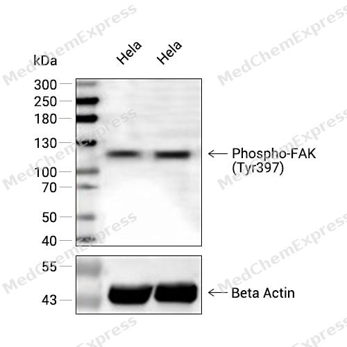 Western blot analysis of extracts from Hela (lane 2(20μg) and Hela (lane 3(40μg) using Phospho-FAK (Tyr397) (HY-P80460) Rabbit mAb. Proteins were transferred to a PVDF membrane and blocked with 5% non-fat milk in TBST for 2 hour at room temperature. The primary antibody (1/1000) and Loading control antibody (Beta Actin, HY-P80438, 1/10000) was used in 5% non-fat milk in TBST at 4°C overnight. Goat Anti-Mouse/Rabbit IgG-HRP Secondary Antibody (1/10000) was used for 1 hour at room temperature.
 -
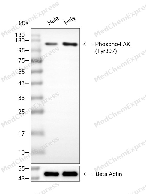 Western blot analysis of extracts from Hela (lane 2(20μg), Hela (lane 3(40μg), using Phospho-FAK (Tyr397) Antibody. Proteins were transferred to a PVDF membrane and blocked with 5% BSA in TBST for 2 hour at room temperature. The primary antibody and Loading control antibody (Beta Actin, HY-P80438, 1/3000) was used in 5% BSA in TBST at 4°C overnight. Goat Anti-Mouse/Rabbit IgG-HRP Secondary Antibody (HY-P8004/HY-P8001, 1/10,000) was used for 1 hour at room temperature.
 -
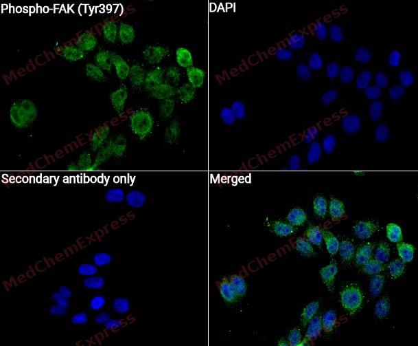 Immunocytochemistry analysis of Hela cells labeling Phospho-FAK (Tyr397) with Phospho-FAK (Tyr397) Antibody (HY-P80460)at 1/50 dilution. Cells were fixed in 4% paraformaldehyde for 15 minutes at room temperature, permeabilized with 0.1% Triton X-100 for 10 minutes at room temperature, then blocked with QuickBlock™ Blocking Buffer for Immunol Staining for 10 min at room temperature. Cells were then incubated with Phospho-FAK (Tyr397) Antibody (HY-P80460) at 1/50 dilution in QuickBlock™ Blocking Buffer for Immunol Staining at 4 ℃. Alexa Fluor® 488-conjugated AffiniPure Goat Anti-Rabbit IgG H&L(HY-P8002, Green) was used as the secondary antibody at 1/1,000 dilution. PBS instead of the primary antibody was used as the secondary antibody only control. The Nuclear counterstain was DAPI (Blue).
 -
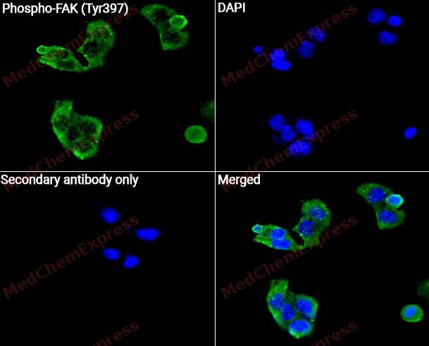 Immunocytochemistry analysis of NIH/3T3 cells labeling Phospho-FAK (Tyr397) with Phospho-FAK (Tyr397) Antibody (HY-P80460) at 1/50 dilution. Cells were fixed in 4% paraformaldehyde for 15 minutes at room temperature, permeabilized with 0.1% Triton X-100 for 10 minutes at room temperature, then blocked with QuickBlock™ Blocking Buffer for Immunol Staining for 10 min at room temperature. Cells were then incubated with Phospho-FAK (Tyr397) Antibody (HY-P80460) at 1/50 dilution in QuickBlock™ Blocking Buffer for Immunol Staining at 4 ℃. Alexa Fluor® 488-conjugated AffiniPure Goat Anti-Rabbit IgG H&L(HY-P8002, Green) was used as the secondary antibody at 1/1,000 dilution. PBS instead of the primary antibody was used as the secondary antibody only control. The Nuclear counterstain was DAPI (Blue).


Background
-
Function
FAK (Focal Adhesion Kinase or PTK2) is a non-receptor and non-membrane associated protein tyrosine kinase that is activated at the sites of cell-matrix adhesions and integrin clustering by auto-phosphorylation (at Tyr397), Src, and other tyrosine kinases. FAK mediates integrin-based cell signaling by transferring signals regulating cell migration, adhesion, and survival from the extracellular matrix to the cytoplasm. FAK is overexpressed in many tumors, including those derived from the head and neck, colon, breast, prostate, liver, and thyroid. Furthermore, FAK overexpression is highly correlated with an invasive phenotype in these tumors. Inhibition of FAK signaling by overexpression of dominant-negative fragments of FAK reduces invasion of glioblastomas and ovarian cancer cells. FAK therefore represents an important target for the development of anti-neoplastic and anti-metastatic drugs.
-
Subcellular Localization
Cell junction, focal adhesion; Cell membrane; Peripheral membrane protein; Cytoplasmic side; Cytoplasm, perinuclear region; Cytoplasm, cell cortex; Cytoplasm, cytoskeleton; Cytoplasm, cytoskeleton, microtubule organizing center, centrosome; Nucleus; Cytoplasm, cytoskeleton, cilium basal body; Cytoplasm
-
Expression
Tissue_specificity:Detectable in both B cells and T lymphocytes. Isomers 1 and 6 are detectable in lung fibroblasts (protein level) . Widely present. Expressed in epithelial cells (protein level) (PubMed:31630787) -
Subunit
Interacts (via first Pro-rich region) with CAS family members (via SH3 domain), including BCAR1, BCAR3, and CASS4. Interacts with NEDD9 (via SH3 domain) (PubMed:9360983). Interacts with GIT1. Interacts with SORBS1. Interacts with ARHGEF28. Interacts with SHB. Part of a complex composed of THSD1, PTK2/FAK1, TLN1 and VCL (PubMed:29069646). Interacts with PXN and TLN1. Interacts with STAT1. Interacts with DCC. Interacts with WASL. Interacts with ARHGEF7. Interacts with GRB2 and GRB7 (By similarity). Component of a complex that contains at least FER, CTTN and PTK2/FAK1. Interacts with BMX. Interacts with TGFB1I1. Interacts with STEAP4. Interacts with ZFYVE21. Interacts with ESR1. Interacts with PIK3R1 or PIK3R2. Interacts with SRC, FGR, FLT4 and RET. Interacts with EPHA2 in resting cells; activation of EPHA2 recruits PTPN11, leading to dephosphorylation of PTK2/FAK1 and dissociation of the complex. Interacts with EPHA1 (kinase activity-dependent). Interacts with CD4; this interaction requires the presence of HIV-1 gp120. Interacts with PIAS1. Interacts with ARHGAP26 and SHC1. Interacts with RB1CC1; this inhibits PTK2/FAK1 activity and activation of downstream signaling pathways. Interacts with P53/TP53 and MDM2. Interacts with LPXN (via LD motif 3). Interacts with MISP. Interacts with CIB1 isoform 2. Interacts with CD36. Interacts with EMP2; regulates PTK2 activation and localization (PubMed:19494199). Interacts with DSCAM (By similarity). Interacts with AMBRA1 (By similarity). Interacts (when tyrosine-phosphorylated) with tensin TNS1; the interaction is increased by phosphorylation of TNS1 (PubMed:20798394)
-
SwissProt ID
-
Synonyms
FAK, FAK1, PTK2, Focal adhesion kinase 1, FADK 1, Focal adhesion kinase-related nonkinase, Protein phosphatase 1 regulatory subunit 71, Protein-tyrosine kinase 2, p125FAK, pp125FAK, FRNK, PPP1R71
-
Research Field
Cardiovascular
Documentation
-
Data Sheet (235 KB)
-
SDS (252 KB)
- English - EN (252 KB)
- Français - FR (252 KB)
- Deutsch - DE (252 KB)
- Norwegian - NO (252 KB)
- Español - ES (252 KB)
- Swedish - SV (252 KB)
- Italian - IT (252 KB)
- Korean - KR (252 KB)
- Portuguese - PT (252 KB)
-
User Guide for Antibodies (1077 KB)

 Powered by Bioz
