



PDGFRA Protein, Rat (HEK293, His)

Based on 1 Customer Validation
PDGFRA Protein is a tyrosine protein kinase receptor for PDGFA, PDGFB, and PDGFC that coordinates embryonic development, cell proliferation, survival, and chemotaxis. Their functions vary, promoting or inhibiting cell proliferation and migration depending on the situation. PDGFRA Protein, Rat (HEK293, His) is the recombinant rat-derived PDGFRA Protein, expressed by HEK293 , with C-His labeled tag.
- Species: Rat
- Source: HEK293
-
Storage:Stored at -20°C for 2 years from date of receipt. After reconstitution, it is stable at 4°C for 1 week or -20°C for longer (with carrier protein). It is recommended to freeze aliquots at -20°C or -80°C for extended storage.


To place orders, for customer services and technical support, please contact: MedChemExpress USA
Tel: 609-228-6898 E-mail: [email protected] [email protected]


-
 Biological Activity
-
Technical Parameters
-
Product Properties
- Documentation
- Help & FAQs




Biological Activity
Description
PDGFRA Protein is a tyrosine protein kinase receptor for PDGFA, PDGFB, and PDGFC that coordinates embryonic development, cell proliferation, survival, and chemotaxis. Their functions vary, promoting or inhibiting cell proliferation and migration depending on the situation. PDGFRA Protein, Rat (HEK293, His) is the recombinant rat-derived PDGFRA Protein, expressed by HEK293 , with C-His labeled tag.
Background
PDGFRA Protein, a tyrosine-protein kinase acting as a cell-surface receptor for PDGFA, PDGFB, and PDGFC, plays a pivotal role in the orchestration of embryonic development, cell proliferation, survival, and chemotaxis. Its multifaceted functions include both promotion and inhibition of cell proliferation and migration depending on the cellular context. PDGF R alpha is integral to the differentiation of bone marrow-derived mesenchymal stem cells, essential for normal skeletal development, and crucial for cephalic closure during embryonic development. Furthermore, it is indispensable for the development of the gastrointestinal tract mucosa, playing a role in recruiting mesenchymal cells and facilitating the normal development of intestinal villi. In the context of wound healing, PDGF R alpha contributes to cell migration and chemotaxis. Its involvement in platelet activation, secretion of agonists from platelet granules, and thrombin-induced platelet aggregation highlights its significance in hemostasis. Binding to its cognate ligands activates several signaling cascades, with the response modulated by ligand nature and heterodimer formation between PDGFRA and PDGFRB. PDGF R alpha phosphorylates PIK3R1, PLCG1, and PTPN11, leading to the activation of diverse signaling pathways, including AKT1, HRAS, and MAP kinases. Additionally, it promotes the activation of STAT family members, such as STAT1, STAT3, STAT5A, and/or STAT5B. Receptor signaling is tightly regulated by protein phosphatases and rapid internalization of the activated receptor. The intricate functions of PDGF R alpha underscore its crucial role in diverse physiological processes.
Verified Bioactivity
1.This product does not contain protein kinase domain.
2.Measured by its ability to inhibit the biological activity of PDGF-AA using NIH-3T3 mouse fibroblast cells. The ED50 for this effect is 0.6932 μg/mL in the presence of 100 ng/mL recombinant human PDGF-AA, corresponding to a specific activity is 1.443×10³ U/mg.
MCE Validation Data
-
Purity - SDS-PAGE
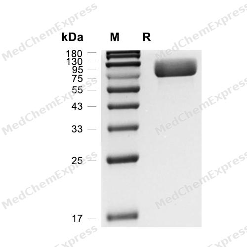
Purity - SDS-PAGE
-
Bioactivity - Cell-Based Assay
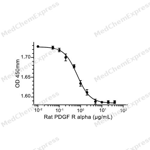
Bioactivity - Cell-Based Assay


Technical Parameters
-
Species Rat
-
Source HEK293
-
Tag C-His
-
Accession
P20786 (L24-E523)
-
Molecular Construction
-
N-term
-
PDGF R alpha (L24-E523)
Accession # P20786 -
His
-
C-term
-
-
Protein Length
Extracellular Domain
-
Synonyms
PDGFRA; CD140a Antigen; Platelet Derived Growth Factor Receptor Alpha; PDGF-R-Alpha; PDGFR2; PDGFR-2; Platelet-Derived Growth Factor Receptor Alpha; GAS9; CD140a; Platelet-Derived Growth Factor Receptor-Like Protein; Platelet-Derived Growth Factor Recepto
-
AA Sequence
LLLPSILPNENEKIVPLSSSFSLRCFGESEVSWQHPMSEEEDPNVEIRTEENNSSLFVTVLEVVNASAAHTGWYTCYYNHTQTEESEIEGRHIYIYVPDPDMAFVPLGMTDSLVIVEEDDSAIIPCLTTDPDTEVTLHNNGRLVPASYDSRQGFNGTFSVGPYICEATVRGRTFKTSEFNVYALKATSELNLEMDTRQTVYKAGETIVVTCAVFNNEVVDLQWTYPGEVRNKGITMLEEIKLPSIKLVYTLTVPKATVKDSGDYECAARQATKEVKEMKTVTISVHEKGFVQIRPTFGHLETVNLHQVREFVVEVQAYPTPRISWLKDNLTLIENLTEITTDVQRSQETRYQSKLKLIRAKEEDSGHYTIIVQNDDDMKSYTFELSTLVPASILELVDDHHGSGGGQTVRCTAEGTPLPNIEWMICKDIKKCNNDTSWTVLASNVSNIITEFHQRGRSTVEGRVSFAKVEETIAVRCLAKNDLGIGNRELKLVAPSLRSE
-
Molecular Weight
Approximately 70-110 kDa, based on SDS-PAGE under reducing conditions.
-
Glycosylation
Yes
-
Purity
≥ 95%, as determined by reducing SDS-PAGE.


Product Properties
Lyophilized powder.
Lyophilized from a 0.22 μm filtered solution of PBS, pH 7.4.
<1 EU/μg, determined by LAL method.
It is not recommended to reconstitute to a concentration less than 100 μg/mL in ddH2O. For long term storage it is recommended to add a carrier protein (0.1% BSA, 5% HSA, 10% FBS or 5% Trehalose).
Stored at -20°C for 2 years from date of receipt. After reconstitution, it is stable at 4°C for 1 week or -20°C for longer (with carrier protein). It is recommended to freeze aliquots at -20°C or -80°C for extended storage.
Room temperature in continental US; may vary elsewhere.


Documentation
-
Data Sheet (239 KB)
-
SDS (251 KB)
- English - EN (251 KB)
- Français - FR (251 KB)
- Deutsch - DE (251 KB)
- Norwegian - NO (251 KB)
- Español - ES (251 KB)
- Swedish - SV (251 KB)
- Italian - IT (251 KB)
- Korean - KR (251 KB)
- Portuguese - PT (251 KB)
-
Handling Instructions (2659 KB)

Calculators
Concentration (start) × Volume (start) = Concentration (final) × Volume (final)


 Powered by Bioz

