1. Protein Tyrosine Kinase/RTK Apoptosis
  2. c-Met/HGFR Apoptosis
  3. Capmatinib

Capmatinib  (Synonyms: INC280; INCB28060)

Cat. No.: HY-13404 Purity: 99.92%
COA Handling Instructions

Capmatinib (INC280; INCB28060) is a potent, orally active, selective, and ATP competitive c-Met kinase inhibitor (IC50=0.13 nM). Capmatinib can inhibit phosphorylation of c-MET as well as c-MET pathway downstream effectors such as ERK1/2, AKT, FAK, GAB1, and STAT3/5. Capmatinib potently inhibits c-MET-dependent tumor cell proliferation and migration and effectively induces apoptosis. Antitumor activity. Capmatinib is largely metabolized by CYP3A4 and aldehyde oxidase.

For research use only. We do not sell to patients.

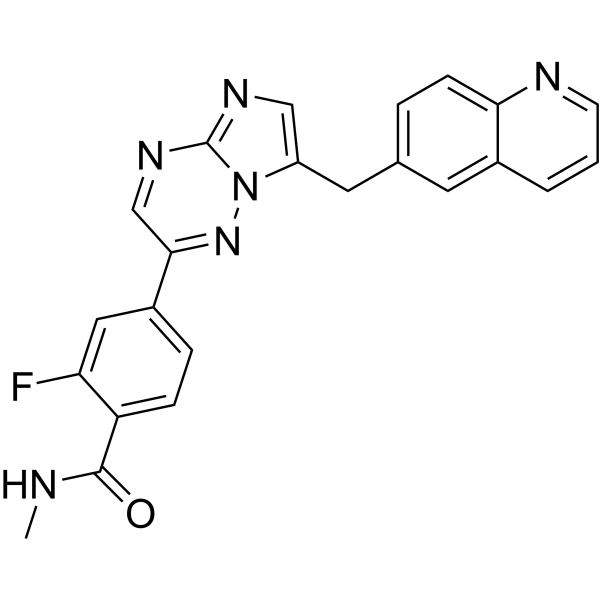
Capmatinib Chemical Structure

Capmatinib Chemical Structure

CAS No. : 1029712-80-8

Size Price Stock Quantity
Free Sample (0.1 - 0.5 mg)   Apply Now  
Solid + Solvent (Highly Recommended)
10 mM * 1 mL in DMSO
ready for reconstitution
USD 61 In-stock
Solution
10 mM * 1 mL in DMSO USD 61 In-stock
Solid
5 mg USD 55 In-stock
10 mg USD 77 In-stock
50 mg USD 121 In-stock
100 mg USD 165 In-stock
200 mg USD 209 In-stock
500 mg   Get quote  
1 g   Get quote  

* Please select Quantity before adding items.

This product is a controlled substance and not for sale in your territory.

Customer Review

Based on 11 publication(s) in Google Scholar

Other Forms of Capmatinib:

Top Publications Citing Use of Products

    Capmatinib purchased from MedChemExpress. Usage Cited in: Department Cancer Biology. Wayne State University. 2014 Jan.

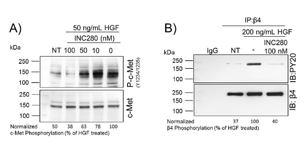
    The c-Met Inhibitor INC280 Reveals HGF Activation of c-Met Leads to β4 Activation. (A) Dose-dependent assay to determine the concentration of INC280 required to prevent HGF-induced c-Met phosphorylation. Cells are pre-treated for two hours with INC280 at the indicated concentrations and then stimulated with 50 ng/mL HGF for 30 minutes. Phosphorylated (upper panel) and total c-Met (lower panel) are analyzed by Western blot. Densitometry represents the ratio of phosphorylated to total c-Met as a p
    • Biological Activity

    • Purity & Documentation

    • References

    • Customer Review

    Description

    Capmatinib (INC280; INCB28060) is a potent, orally active, selective, and ATP competitive c-Met kinase inhibitor (IC50=0.13 nM). Capmatinib can inhibit phosphorylation of c-MET as well as c-MET pathway downstream effectors such as ERK1/2, AKT, FAK, GAB1, and STAT3/5. Capmatinib potently inhibits c-MET-dependent tumor cell proliferation and migration and effectively induces apoptosis. Antitumor activity. Capmatinib is largely metabolized by CYP3A4 and aldehyde oxidase[1][2][3].

    IC50 & Target

    IC50: 0.13 nM (c-MET)[1]

    In Vitro

    Capmatinib (INCB28060) inhibits c-MET phosphorylation with an IC50 value of approximately 1 nM and a concentration of approximately 4 nM inhibits c-MET more than 90%, which is? reversible and the effect is significantly reduced in several hours after the compound is removed and completely disappeared by 48 hours[1].
    ? Capmatinib (INCB28060) (0-10000 nM; 72 h) inhibits the proliferation of SNU-5, S114, H441 and U-87MG[1].
    ? Capmatinib (INCB28060) (0.06-62.25 nM; 2h) effectively inhibits phosphorylation of c-MET as well as c-MET pathway downstream effectors such as ERK1/2, AKT, FAK, GAB1, and STAT3/5[1].
    ? Capmatinib (INCB28060) (0.24-63 nM; over night) prevents HGF-stimulated H441 cell migration[1].
    ? Capmatinib (INCB28060) (0.5-50 nM; 20 min) suppresses phosphorylation of both EGFR and HER-3 rapidly[1].
    ? Capmatinib (INCB28060) (0-333 nM; 24 h) induces apoptosis in SNU-5 cells[1].

    MedChemExpress (MCE) has not independently confirmed the accuracy of these methods. They are for reference only.

    Cell Viability Assay[1]

    Cell Line: SNU-5, S114, H441 and U-87MG
    Concentration: 0-10000 nM
    Incubation Time: 72 h
    Result: Inhibited the cell viability of SNU-5 and S114, as well as the colony formation of H441 and U-87MG, with IC50 values of 1.2 nM, 12.4 nM, ~0.5 nM and 2 nM, respectively.

    Cell Migration Assay [1]

    Cell Line: H441 (stimulated with 50 ng/mL recombinant human HGF for 24h)
    Concentration: 0.24, 1, 4, 16 and 63 nM
    Incubation Time: Over night
    Result: Prevented HGF-stimulated H441 cell migration, with IC50 of approximately 2 nM, and less cell migration at 16 nM.

    Western Blot Analysis[1]

    Cell Line: SNU-5
    Concentration: 0.06, 0.24, 0.98, 3.91, 15.63 and 62.25 nM
    Incubation Time: 2 h
    Result: Effectively inhibited phosphorylation of c-MET as well as c-MET pathway downstream effectors such as ERK1/2, AKT, FAK, GAB1, and STAT3/5.

    Western Blot Analysis[1]

    Cell Line: H1993 cells
    Concentration: 0.5, 5 and 50 nM
    Incubation Time: 20 min
    Result: Suppressed phosphorylation of both EGFR and HER-3 rapidly and as effectively as the compound inhibited c-MET phosphorylation in H1993 cells.

    Apoptosis Analysis[1]

    Cell Line: SNU-5 cells
    Concentration: 0.017, 0.15, 1.37, 12.33, 111 and 333 nM
    Incubation Time: 24 h
    Result: Effectively induced DNA fragmentation.
    In Vivo

    Capmatinib (INCB28060) (1-30 mg/kg; PO, twice daily, for 2 weeks) exhibits dose-dependent inhibition of tumor growth, and shows well tolerance at all doses during the treatment periods, with no evidence of overt toxicity or weight loss in U-87MG tumor mice model[1].
    ? Capmatinib (INCB28060) (0.03-10 mg/kg; PO, single dosage) causes inhibition of c-MET phosphorylation in S114 tumor mice model[1].

    MedChemExpress (MCE) has not independently confirmed the accuracy of these methods. They are for reference only.

    Animal Model: Female Balb/c nu/nu mice (inoculated subcutaneously with 5×10⁶ U-87MG glioblastoma cells)[1]
    Dosage: 1, 3, 10 and 30 mg/kg
    Administration: PO, twice daily, for 2 weeks
    Result: Exhibited dose-dependent inhibition of tumor growth with 35% and 76% at 1 and 3 mg/kg once daily; resulted in partial regressions in 6 of 10 U-87MG tumor-bearing mice at 10 mg/kg once daily; and showed well tolerance at all doses during the treatment periods, with no evidence of overt toxicity or weight loss.
    Animal Model: Female Balb/c nu/nu mice (inoculated subcutaneously with 4×10⁶ S114 tumor cells)[1]
    Dosage: 0.03, 0.1, 0.3, 1, 3 and 10 mg/kg
    Administration: PO, single dosage
    Result: Caused approximately 50% and 90% inhibition of c-MET phosphorylation at 0.03 and 0.3 mg/kg after administration of 30 min, and inhibition of phospho-c-MET exceeded 90% after 7 hours.
    Clinical Trial
    Molecular Weight

    412.42

    Formula

    C23H17FN6O

    CAS No.
    Appearance

    Solid

    Color

    Light yellow to yellow

    SMILES

    O=C(NC)C1=CC=C(C2=NN3C(N=C2)=NC=C3CC4=CC=C5N=CC=CC5=C4)C=C1F

    Shipping

    Room temperature in continental US; may vary elsewhere.

    Storage
    Powder -20°C 3 years
    4°C 2 years
    In solvent -80°C 2 years
    -20°C 1 year
    Solvent & Solubility
    In Vitro: 

    DMSO : 5 mg/mL (12.12 mM; ultrasonic and warming and heat to 60°C; Hygroscopic DMSO has a significant impact on the solubility of product, please use newly opened DMSO)

    H2O : < 0.1 mg/mL (insoluble)

    Preparing
    Stock Solutions
    Concentration Solvent Mass 1 mg 5 mg 10 mg
    1 mM 2.4247 mL 12.1236 mL 24.2471 mL
    5 mM 0.4849 mL 2.4247 mL 4.8494 mL
    View the Complete Stock Solution Preparation Table

    * Please refer to the solubility information to select the appropriate solvent. Once prepared, please aliquot and store the solution to prevent product inactivation from repeated freeze-thaw cycles.
    Storage method and period of stock solution: -80°C, 2 years; -20°C, 1 year. When stored at -80°C, please use it within 2 years. When stored at -20°C, please use it within 1 year.

    • Molarity Calculator

    • Dilution Calculator

    Mass (g) = Concentration (mol/L) × Volume (L) × Molecular Weight (g/mol)

    Mass
    =
    Concentration
    ×
    Volume
    ×
    Molecular Weight *

    Concentration (start) × Volume (start) = Concentration (final) × Volume (final)

    This equation is commonly abbreviated as: C1V1 = C2V2

    Concentration (start)

    C1

    ×
    Volume (start)

    V1

    =
    Concentration (final)

    C2

    ×
    Volume (final)

    V2

    In Vivo:

    For the following dissolution methods, please prepare the working solution directly. It is recommended to prepare fresh solutions and use them promptly within a short period of time.
    The percentages shown for the solvents indicate their volumetric ratio in the final prepared solution. If precipitation or phase separation occurs during preparation, heat and/or sonication can be used to aid dissolution.

    • Protocol 1

      Add each solvent one by one:  0.5% CMC-Na/saline water

      Solubility: 10 mg/mL (24.25 mM); Suspended solution; Need ultrasonic

    In Vivo Dissolution Calculator
    Please enter the basic information of animal experiments:

    Dosage

    mg/kg

    Animal weight
    (per animal)

    g

    Dosing volume
    (per animal)

    μL

    Number of animals

    Recommended: Prepare an additional quantity of animals to account for potential losses during experiments.
    Calculation results:
    Working solution concentration: mg/mL
    Purity & Documentation

    Purity: 99.92%

    References

    Complete Stock Solution Preparation Table

    * Please refer to the solubility information to select the appropriate solvent. Once prepared, please aliquot and store the solution to prevent product inactivation from repeated freeze-thaw cycles.
    Storage method and period of stock solution: -80°C, 2 years; -20°C, 1 year. When stored at -80°C, please use it within 2 years. When stored at -20°C, please use it within 1 year.

    Optional Solvent Concentration Solvent Mass 1 mg 5 mg 10 mg 25 mg
    DMSO 1 mM 2.4247 mL 12.1236 mL 24.2471 mL 60.6178 mL
    5 mM 0.4849 mL 2.4247 mL 4.8494 mL 12.1236 mL
    10 mM 0.2425 mL 1.2124 mL 2.4247 mL 6.0618 mL
    • No file chosen (Maximum size is: 1024 Kb)
    • If you have published this work, please enter the PubMed ID.
    • Your name will appear on the site.
    Help & FAQs
    • Do most proteins show cross-species activity?

      Species cross-reactivity must be investigated individually for each product. Many human cytokines will produce a nice response in mouse cell lines, and many mouse proteins will show activity on human cells. Other proteins may have a lower specific activity when used in the opposite species.

    Your Recently Viewed Products:

    Inquiry Online

    Your information is safe with us. * Required Fields.

    Product Name


    Salutation

    Applicant Name *


    Email Address *

    Phone Number *


    Organization Name *

    Department *


    Requested quantity *

    Country or Region *


    Remarks

    Bulk Inquiry

    Inquiry Information

    Product Name:
    Capmatinib
    Cat. No.:
    HY-13404
    Quantity:
    MCE Japan Authorized Agent: