



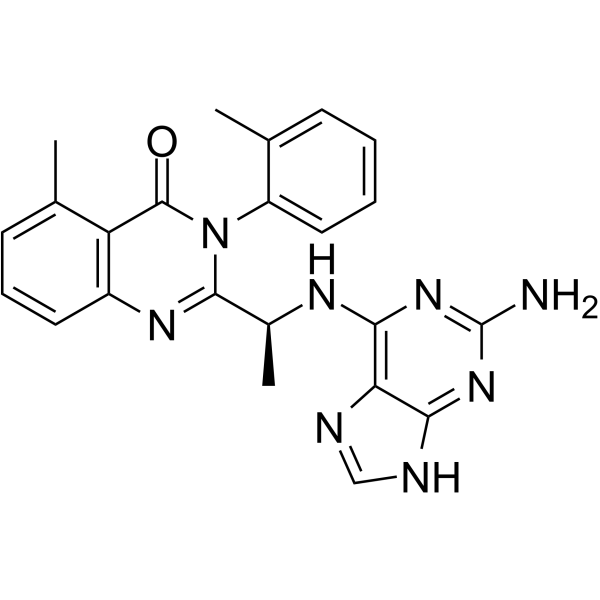
CAL-130
CAL-130 is a PI3Kδ and PI3Kγ inhibitor with IC50s of 1.3 and 6.1 nM, respectively.
For research use only. We do not sell to patients.
- CAS No.: 1431697-74-3
- Formula: C23H22N8O
- Molecular Weight:426.47
-
Storage:
Please store the product under the recommended conditions in the Certificate of Analysis.

To place orders, for customer services and technical support, please contact: MedChemExpress USA
Tel: 609-228-6898 E-mail: [email protected] [email protected]


-
 Biological Activity
-
Chemical Information
- Purity & Documentation
- References
-
 Help & FAQs


-
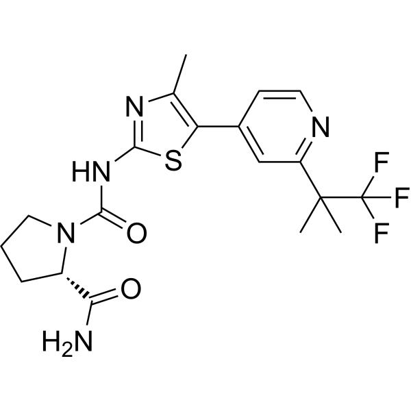
HY-15244
AlpelisibPI3Kα Inhibitor -
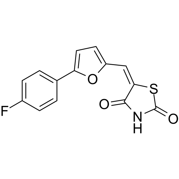
HY-13530
CAY10505 -
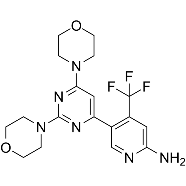
HY-70063
BuparlisibPI3K Inhibitor -
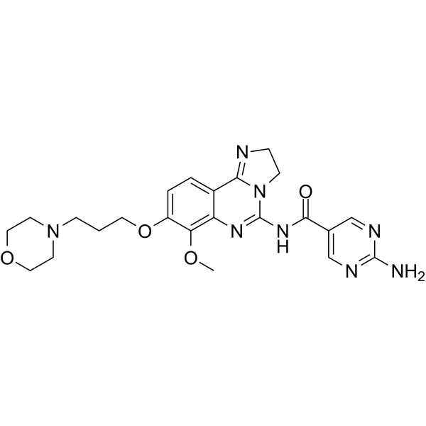
HY-15346
CopanlisibPI3K Inhibitor -
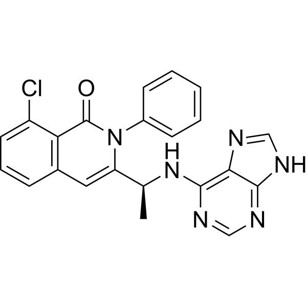
HY-17044
DuvelisibPI3Kδ Inhibitor -
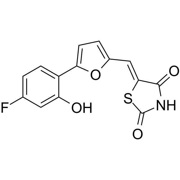
HY-13532
AS-252424PI3K Inhibitor -
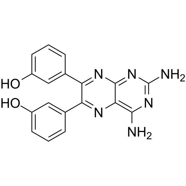
HY-10111
TG100-115PI3Kγ/PI3Kδ Inhibitor -
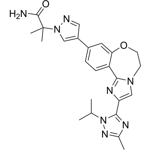
HY-13898
TaselisibPIK3CA Inhibitor -
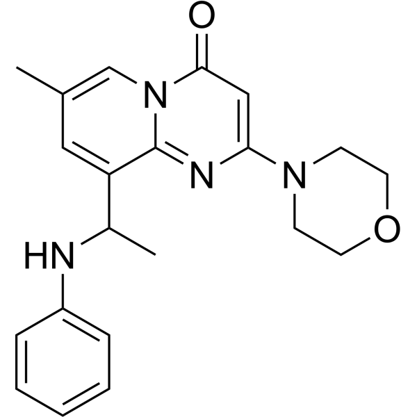
HY-10114
TGX-221Keap1-Nrf2 Interaction Inhibitor -
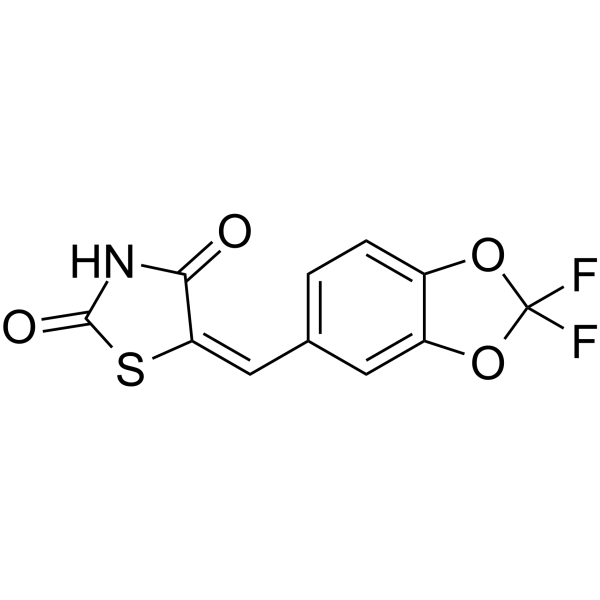
HY-13531
AS-604850PI3Kγ Inhibitor -
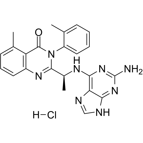
HY-16122B
CAL-130 HydrochloridePI3Kδ/γ Inhibitor -
HY-12330
AZD8186PI3K Inhibitor


Biological Activity
Description
IC50 & Target
[1]|
p110δ 1.3 nM (IC50) |
p110γ 6.1 nM (IC50) |
p110β 56 nM (IC50) |
p110α 115 nM (IC50) |
In Vitro
CAL-130 preferentially inhibits the function of both p110γ and p110δ catalytic domains. IC50 values of CAL-130 are 1.3 and 6.1 nM for p110δ and p110γ, respectively, as compared to 115 and 56 nM for p110α and p110β. CAL-130 does not inhibit additional intracellular signaling pathways (i.e., p38 MAPK or insulin receptor tyrosine kinase) that are critical for general cell function and survival[1].
MedChemExpress (MCE) has not independently confirmed the accuracy of these methods. They are for reference only. .
In Vivo
MedChemExpress (MCE) has not independently confirmed the accuracy of these methods. They are for reference only.


Chemical Information
-
CAS No. 1431697-74-3
-
Molecular Weight 426.47
-
Formula C23H22N8O
-
SMILES
O=C1N(C2=CC=CC=C2C)C([C@H](C)NC3=C4N=CNC4=NC(N)=N3)=NC5=C1C(C)=CC=C5
-
Shipping
Room temperature in continental US; may vary elsewhere.
-
Storage
Please store the product under the recommended conditions in the Certificate of Analysis.


Purity & Documentation
References

Calculators
Concentration (start) × Volume (start) = Concentration (final) × Volume (final)


CAL-130 Related Classifications
HY-16122A Related Classifications
 Powered by Bioz