



EGF Protein, Human
Based on 107 publication(s) in Google Scholar
EGF proteins act as potent stimulators of growth in a variety of epidermal and epithelial tissues in both in vivo and in vitro settings, while promoting the proliferation of specific fibroblasts in cell culture. This multifaceted protein also acts as a magnesium stimulating hormone, driving magnesium reabsorption in the renal distal tubule through engagement of EGFR and activation of the magnesium channel TRPM6. EGF Protein, Human is the recombinant human-derived EGF protein, expressed by E. coli , with tag free.
- Species: Human
- Source: E. coli
-
Storage:Stored at -20°C for 2 years from date of receipt. After reconstitution, it is stable at 4°C for 1 week or -20°C for longer (with carrier protein). It is recommended to freeze aliquots at -20°C or -80°C for extended storage.


To place orders, for customer services and technical support, please contact: MedChemExpress USA
Tel: 609-228-6898 E-mail: [email protected] [email protected]


-
 Biological Activity
-
Technical Parameters
-
Product Properties
- Documentation
- References
- Help & FAQs


-
HY-N0583
HydrocortisoneGlucocorticoid Receptor Agonist -

HY-P7004
FGF basic/bFGF Protein, Human (146a.a) -

HY-10071
Y-27632ROCK Inhibitor -
HY-10432
A 83-01ALK4/5/7 Inhibitor -

HY-P0035
Insulin (human)Recombinant Human Insulin -

HY-10182
LaduviglusibGSK-3 Inhibitor -

HY-10431
SB-431542TGF-β Receptor Kinase Inhibitor -

HY-15371
ForskolinAdenylate Cyclase Activator -

HY-B0215
AcetylcysteineMucolytic Agent -

HY-10583
Y-27632 dihydrochlorideROCK Inhibitor -
HY-B0150
NicotinamideSIRT Inhibitor -
HY-P7331
FGF basic/bFGF Protein, Human (154a.a)


Biological Activity
Description
EGF proteins act as potent stimulators of growth in a variety of epidermal and epithelial tissues in both in vivo and in vitro settings, while promoting the proliferation of specific fibroblasts in cell culture. This multifaceted protein also acts as a magnesium stimulating hormone, driving magnesium reabsorption in the renal distal tubule through engagement of EGFR and activation of the magnesium channel TRPM6. EGF Protein, Human is the recombinant human-derived EGF protein, expressed by E. coli , with tag free.
Background
EGF Protein emerges as a potent stimulator of the growth of diverse epidermal and epithelial tissues in both in vivo and in vitro environments, along with fostering the proliferation of specific fibroblasts in cell culture. This multifaceted protein also acts as a magnesiotropic hormone, driving magnesium reabsorption in the renal distal convoluted tubule through the engagement of EGFR and activation of the magnesium channel TRPM6. Notably, EGF Protein possesses the capability to induce neurite outgrowth in motoneurons of the pond snail Lymnaea stagnalis in vitro. Its molecular interactions include binding to EGFR, thereby promoting EGFR dimerization, and interacting with RHBDF2, suggesting involvement in diverse cellular processes. Furthermore, EGF Protein engages with RHBDF1, potentially influencing the retention of EGF in the endoplasmic reticulum and regulating its degradation through endoplasmic reticulum-associated degradation (ERAD).
In Vivo
EGF Protein (Human) (10 μg/g, cream, a single dose for 7 days) reduces inflammatory infiltrate and results in wound re-epithelialization in rats with full thicknessskin wounds[1].
EGF Protein (Human) (50 μg/g, cream, every 24 h for 7 days) significantly stimulates re-epithelialization at high levels, preferably in combination with a protease inhibitor in pigs with thickness wounds on the back[1].
EGF Protein (Human) (30 μg/kg, s.c., daily for 3 weeks) attenuates the oesophageal damage normally caused by the sclerotherapy (surgical dilation of the oesophagus) procedure in Gottingen minipigs[1].
Verified Bioactivity
Measured in a cell proliferation assay using BALB/c 3T3 cells. The ED50 for this effect is <200 pg/mL.
MCE Validation Data
-
Purity - SDS-PAGE
Purity - SDS-PAGE
-
Bioactivity - Cell-Based Assay
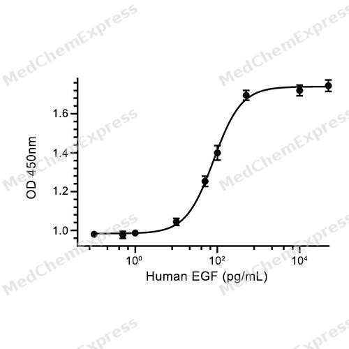
Bioactivity - Cell-Based Assay


Publications (107)
-
Journal Impact Factor
-
Most Recent
-
Cell
2024 Nov 14;187(23):6566-6583.e22. PMID: 39332412 -
Mol Cancer
A positive feedback loop between PFKP and c-Myc drives head and neck squamous cell carcinoma progression. [Abstract]2024 Jul 9;23(1):141. PMID: 38982480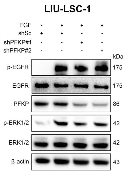
EGF Protein, Human purchased from MedChemExpress. Usage Cited in: Mol Cancer. 2024 Jul 9;23(1):141. [Abstract]
LIU-LSC-1 cells transfected with PFKP shRNA were stimulated with or without EGF (50 ng/mL) for 15 min. PFKP is involved in EGFR-mediated activation of ERK1/2.
-
Mol Cancer
Organoids derived from patients provide a new opportunity for research and individualized treatment of malignant peritoneal mesothelioma. [Abstract]2024 Jan 10;23(1):12. PMID: 38200517 -
Cancer Commun (Lond)
RNA m1A methyltransferase TRMT61A promotes colorectal tumorigenesis by enhancing ONECUT2 mRNA stability and is a potential therapeutic target. [Abstract]2025 Oct 16. PMID: 41103012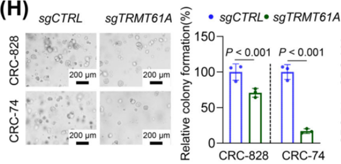
EGF Protein, Human purchased from MedChemExpress. Usage Cited in: Cancer Commun (Lond). 2025 Oct 16. [Abstract]
Primary CRC organoids (CRC‐74 and CRC‐828) were cultured in DMEM/F12 supplemented with N2, B27, 10 mmol/L HEPES, 1.25 mmol/L N‐acetyl cysteine, 10 mmol/L SB202190‐monohydrochloride, 500 ng/mL R‐spondin‐1, 100 ng/mL Noggin, 100 ng/mL Wnt3a (HY‐P70453A, MedChemExpress), 50 ng/mL EGF (HY‐P7109, MedChemExpress), and 100 units/mL penicillin/streptomycin. The growth of patient‐derived CRC organoids was suppressed upon silencing of TRMT61A.
-
Cancer Commun (Lond)
N6-methyladenosine reader hnRNPA2B1 recognizes and stabilizes NEAT1 to confer chemoresistance in gastric cancer. [Abstract]2024 Apr;44(4):469-490. PMID: 38512764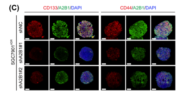
EGF Protein, Human purchased from MedChemExpress. Usage Cited in: Cancer Commun (Lond). 2024 Apr;44(4):469-490. [Abstract]
Cells were seeded in ultra‐low attachment plates and cultured in serum‐free RPMI‐1640 supplemented with 2% B27 supplemen, 20 ng/mL EGF (MCE), and 10 ng/mL bFGF (MCE) in a humidified 5% CO2 at 37°C. Additionally, 25 µM ICG‐001 or 100 ng/mL Wnt3A (MCE) was added in the culture medium. After one‐week incubation, tumor sphere numbers were counted under a phase‐contrast microscope. The role of hnRNPA2B1 in maintenance of gastric cancer stemness was evaluated.
-
Nat Genet
Single-nucleus multi-omic profiling of human placental syncytiotrophoblasts identifies cellular trajectories during pregnancy. [Abstract]2024 Feb;56(2):294-305. PMID: 38267607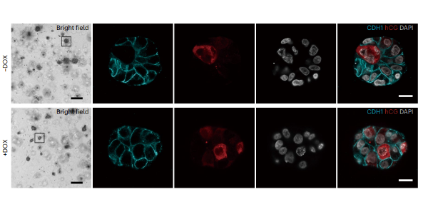
EGF Protein, Human purchased from MedChemExpress. Usage Cited in: Nat Genet. 2024 Feb;56(2):294-305. [Abstract]
Trophoblast organoids constructed from hTSCs-BL. Trophoblast markers: CDH1 and hCG. Briefly, hTSCs were cultured in hTSCs medium (DMEM/F12 supplemented with 0.1 mM 2-mercaptoethanol, 0.2% FBS, 0.5% Penicillin–Streptomycin, 0.3% BSA, 1% ITS-X supplement, 1.5 μg/ml L-ascorbicacid, 50 ng/mL Epidermal Growth Factor (EGF)(MedChemExpress), 2 μM CHIR99021 (MedChemExpress), 0.5 μM A83-01 (MedChemExpress), 1 μM SB431542 (MedChemExpress), 0.8 mM Valproic acid and 5 μM Y27632 (MedChemExpress).
-
Bioact Mater
Melatonin-incorporated brain extracellular matrix hydrogel enhances NSCs mitochondrial metabolism to promote neuroregeneration via the AMPK-PGC-1α-NRF1/TFAM axis after spinal cord injury. [Abstract]2026 Apr 9:63:373-389. PMID: 42006003 -
Cancer Res
Targeting Sialylation Enhances the Therapeutic Efficacy of the Nectin-4-Targeted Antibody-Drug Conjugate Enfortumab Vedotin in Bladder Cancer. [Abstract]2025 Oct 9. PMID: 41066596 -
Drug Resist Updat
Acetate utilization promotes hormone therapy resistance in prostate cancer through neuroendocrine differentiation. [Abstract]2024 Nov:77:101158. PMID: 39395327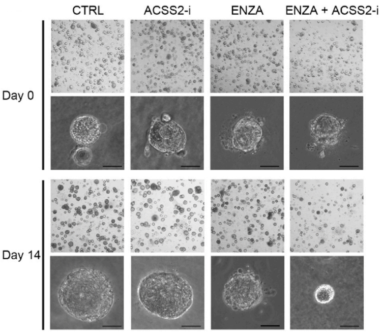
EGF Protein, Human purchased from MedChemExpress. Usage Cited in: Drug Resist Updat. 2024 Nov:77:101158. [Abstract]
PCa-derived organoids were established and cultured in Matrigel. The culture medium consisted of Advanced DMEM/F-12 supplemented with B27, N-acetylcysteine, EGF (5 ng/ml, MCE, HY-P7109), Noggin (100 ng/ml), R-spondin 1 (500 ng/ml), A83-01 (500 nM), FGF10 (10 ng/ml), FGF2 (5 ng/ml), Prostaglandin E2 (1 μM), Nicotinamide (10 mM), SB202190 (10 μM), DHT (1 nM), and Y-27632 (10 μM). The combination of an ACSS2 inhibitor and ENZA resulted in the death of over 90 % of organoids compared with organoids treated with ENZA.
-
Nat Commun
Sensight enables quantitative multivariate engineering of high-performance chemical imaging tools. [Abstract]2026 Jan 27;17(1):2061. PMID: 41593056 -
Nat Commun
Single-cell and spatial transcriptome analyses reveal tumor heterogeneity and immune remodeling involved in pituitary neuroendocrine tumor progression. [Abstract]2025 May 30;16(1):5007. PMID: 40442104 -
Nat Commun
MYG1 drives glycolysis and colorectal cancer development through nuclear-mitochondrial collaboration. [Abstract]2024 Jun 11;15(1):4969. PMID: 38862489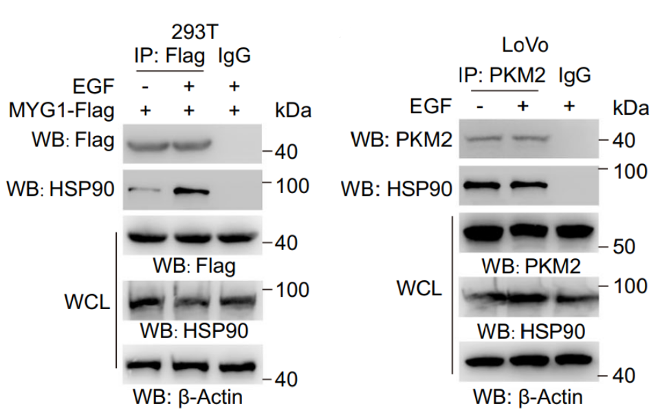
EGF Protein, Human purchased from MedChemExpress. Usage Cited in: Nat Commun. 2024 Jun 11;15(1):4969. [Abstract]
EGF (100 ng/mL; 10 h) increases the interaction between MYG1 and HSP90 in 293 T cells instead of LoVo cells with KRAS mutation.
-
Theranostics
2024 May 27;14(9):3423-3438. PMID: 38948056 -
J Exp Clin Cancer Res
NOTCH3 regulates myofibroblastic CAF differentiation via the P62-ROS signaling axis to promote bladder cancer progression. [Abstract]2026 Jun 6. PMID: 42251414 -
J Exp Clin Cancer Res
Targeting DNM1L/DRP1-FIS1 axis inhibits high-grade glioma progression by impeding mitochondrial respiratory cristae remodeling. [Abstract]2024 Sep 30;43(1):273. PMID: 39350223 -
J Exp Clin Cancer Res
Kremen2 drives the progression of non-small cell lung cancer by preventing SOCS3-mediated degradation of EGFR. [Abstract]2023 Jun 3;42(1):140. PMID: 37270563 -
Adv Sci (Weinh)
DTX3L Inhibits the EMT, Metastasis, and Stem-Like Features of Gastric Cancer Through Promoting GSK-3β Dependent SNAI1 Decay. [Abstract]2026 Jul;13(37):e24036. PMID: 41972409 -
Adv Sci (Weinh)
Branched-Chain α Keto-Acid Dehydrogenase Kinase-Mediated AKT Phosphorylation Promotes RCC Tumorigenesis and Drug Resistance. [Abstract]2025 Aug 11:e11081. PMID: 40789057 -
MedComm (2020)
Feedback loop LINC00511-YTHDF2-SOX2 regulatory network drives cholangiocarcinoma progression and stemness. [Abstract]2024 Oct 22;5(11):e743. PMID: 39445001 -
Adv Sci (Weinh)
mRNA-Lipid Nanoparticle-Mediated Restoration of PTPN14 Exhibits Antitumor Effects by Overcoming Anoikis Resistance in Triple-Negative Breast Cancer. [Abstract]2024 Aug;11(32):e2309988. PMID: 39189475 -
Adv Sci (Weinh)
Oxidative Stress Promotes Liver Cancer Metastasis via RNF25-Mediated E-Cadherin Protein Degradation. [Abstract]2024 Apr;11(13):e2306929. PMID: 38286671 -
Sci Adv
Wearing bacteriophages individually with an adhesive drug-loadable nanohelmet for treating ocular infections. [Abstract]2025 Jul 11;11(28):eadx4183. PMID: 40644532 -
Med
Real-time diagnosis of cholangiocarcinoma by combining digestive endoscopy and optic fiber biosensors based on bile clusterin. [Abstract]2025 Sep 2:100843. PMID: 40930098 -
Alzheimers Dement
Human iPSC-derived GABAergic interneuron transplantation restores circuit balance and cognitive function in an Alzheimer's disease model. [Abstract]2026 Apr;22(4):e71378. PMID: 42043780 -
Cell Death Dis
2026 Jun 23. PMID: 42336842 -
Cell Death Dis
N6-methyladenosine-modified GPX2 impacts cancer cell stemness and TKI resistance through regulating of redox metabolism. [Abstract]2025 Jun 18;16(1):458. PMID: 40533443 -
Cell Death Dis
MAL2 reprograms lipid metabolism in intrahepatic cholangiocarcinoma via EGFR/SREBP-1 pathway based on single-cell RNA sequencing. [Abstract]2024 Jun 12;15(6):411. PMID: 38866777 -
Cell Death Dis
LncRNA LHFPL3-AS1 contributes to tumorigenesis of melanoma stem cells via the miR-181a-5p/BCL2 pathway. [Abstract]2020 Nov 4;11(11):950. PMID: 33149126 -
Cancer Lett
2024 Aug 10:597:217068. PMID: 38901665 -
Cancer Lett
PTPN23[Thr] variant reduces susceptibility and tumorigenesis in esophageal squamous cell carcinoma through dephosphorylation of EGFR. [Abstract]2024 Jun 28:592:216936. PMID: 38704135 -
Phytomedicine
Targeting UBE2T by β-elemene inhibits prostate cancer stem cells and bone metastasis by blocking the TRIM28/pGSK3β/β-catenin signaling. [Abstract]2026 Jul:156:158242. PMID: 42070337 -
Phytomedicine
Baicalein specifically suppresses microsatellite instability colorectal cancer by targeting adenosylhomocysteinase to inhibit histone H3 lysine 4 trimethylation-mediated cancer stemness. [Abstract]2025 Sep 22:148:157291. PMID: 41038143 -
EBioMedicine
VPS35 promotes cell proliferation via EGFR recycling and enhances EGFR inhibitors response in gastric cancer. [Abstract]2023 Mar:89:104451. PMID: 36738481 -
Adv Healthc Mater
Antitumor Activity of a Bispecific Chimera Targeting EGFR and Met in Gefitinib-Resistant Non-Small Cell Lung Cancer. [Abstract]2025 Jan;14(2):e2402884. PMID: 39586988 -
-
J Transl Med
Pleiotrophin modulates cell proliferation, prostate smooth muscle contraction and fibrosis in hyperplastic prostate. [Abstract]2025 Oct 17;23(1):1128. PMID: 41107820 -
-
-
-
Oncogene
Targeting YRDC blocks codon-biased FABP7 translation and lipid droplet formation to overcome chemoresistance in glioblastoma. [Abstract]2026 Jun;45(22):2155-2170. PMID: 42014887 -
Oncogene
MTPN drives noncanonical ERK hyperactivation in colorectal cancer and provides a promising therapeutic approach for precision medicine in CRC. [Abstract]2026 Jun;45(22):2137-2154. PMID: 42009970 -
Oncogene
Par6/SOX2 interact to modulate stemness maintenance in glioma by regulating the EGFR/PI3K/AKT signaling cascade. [Abstract]2025 Oct 11. PMID: 41076461 -
EMBO J
2025 Mar;44(5):1464-1487. PMID: 39838173 -
Clin Transl Med
Exosomal miR-3126-5p derived from cancer-associated fibroblasts facilitates glycolysis to accelerate NSCLC progression by targeting KLF13 to activate the SH2B1/IRS1 axis. [Abstract]2025 Dec;15(12):e70554. PMID: 41410190 -
Br J Cancer
Inhibition of the AURKA/YAP1 axis is a promising therapeutic option for overcoming cetuximab resistance in colorectal cancer stem cells. [Abstract]2024 May;130(8):1402-1413. PMID: 38467828 -
Cell Rep
SLC6A8-mediated creatine uptake suppresses ERK2-FSP1 signaling and induces ferroptosis in colorectal cancer. [Abstract]2025 Sep 1;44(9):116139. PMID: 40892544 -
Cell Rep
Pro-prion, as a membrane adaptor protein for E3 ligase c-Cbl, facilitates the ubiquitination of IGF-1R, promoting melanoma metastasis. [Abstract]2022 Dec 20;41(12):111834. PMID: 36543142 -
Cell Rep
TRIM32/USP11 Balances ARID1A Stability and the Oncogenic/Tumor-Suppressive Status of Squamous Cell Carcinoma. [Abstract]2020 Jan 7;30(1):98-111.e5. PMID: 31914402 -
Cell Prolif
ISRIB facilitates the co-culture of human trophoblast stem cells and embryonic stem cells. [Abstract]2024 Jun;57(6):e13599. PMID: 38217296 -
Sci Signal
PARP1-mediated PARylation of TEAD4 stabilizes the YAP1-TEAD4 complex and promotes growth and immune evasion in breast cancer cells. [Abstract]2025 Oct 21;18(909):eadx2532. PMID: 41118450 -
Front Immunol
A Novel Ferroptosis-Related Long Non-Coding RNA Prognostic Signature Correlates With Genomic Heterogeneity, Immunosuppressive Phenotype, and Drug Sensitivity in Hepatocellular Carcinoma. [Abstract]2022 Jul 8;13:929089. PMID: 35874689 -
Cancer Cell Int
Annexin A1 promotes the progression of bladder cancer via regulating EGFR signaling pathway. [Abstract]2022 Jan 6;22(1):7. PMID: 34991599 -
JCI Insight
The AURKA inhibitor alters the immune microenvironment and enhances targeting B7-H3 immunotherapy in glioblastoma. [Abstract]2025 Feb 10;10(5):e173700. PMID: 39928563 -
Cell Biosci
2025 Feb 5;15(1):14. PMID: 39910656 -
Inflammopharmacology
Pseudolaric acid B exerts an antifungal effect and targets SIRT1 to ameliorate inflammation by regulating Nrf2/NF-κB pathways in fungal keratitis. [Abstract]2024 Apr;32(2):1133-1146. PMID: 38150134 -
J Cell Biol
2024 Nov 4;223(11):e202308099. PMID: 39196068 -
Commun Biol
CTBP1 links metabolic syndrome to polycystic ovary syndrome through interruption of aromatase and SREBP1. [Abstract]2024 Sep 18;7(1):1174. PMID: 39294274 -
Commun Biol
A small-molecule compound D6 overcomes EGFR-T790M-mediated resistance in non-small cell lung cancer. [Abstract]2021 Dec 13;4(1):1391. PMID: 34903832 -
Cell Biol Toxicol
Celastrol modulates IRS1 expression to alleviate ovarian aging and to enhance follicular development. [Abstract]2025 Sep 2;41(1):129. PMID: 40897829 -
J Zhejiang Univ Sci B
Chronic exposure to hexavalent chromium induces esophageal tumorigenesis via activating the Notch signaling pathway. [Abstract]2024 Oct 21;26(1):76-91. PMID: 39815612 -
Int Immunopharmacol
Isorhapontigenin alleviates inflammatory responses in periodontitis through the EGFR/PI3K/AKT signaling pathway. [Abstract]2026 Sep 1:184:116933. PMID: 42225001 -
Bioorg Chem
A novel Quinazoline derivative exerts anti-tumor effects in non-small cell lung cancer through Wnt/β-catenin pathway inhibition. [Abstract]2025 Jun 15:160:108481. PMID: 40253763 -
Biochim Biophys Acta Mol Basis Dis
Targeted inhibition of the H3K27ac/OGT/O-GlcNAcylation axis to overcome gemcitabine-induced chemotherapy resistance in bladder cancer. [Abstract]2026 Aug;1872(6):168281. PMID: 42069092 -
Biochim Biophys Acta Mol Basis Dis
LRRC8A as a central mediator promotes colon cancer metastasis by regulating PIP5K1B/PIP2 pathway. [Abstract]2024 Feb 11;1870(4):167066. PMID: 38350542 -
Mol Med Rep
miR‑302d‑3p regulates the viability, migration and apoptosis of breast cancer cells through regulating the TMBIM6‑mediated ERK signaling pathway. [Abstract]2021 Dec;24(6):853. PMID: 34651659 -
-
Sci Rep
Comparative analysis of organoid, air-liquid interface, and direct infection models for studying pathogen-host interactions in endometrial tissue. [Abstract]2025 Mar 12;15(1):8531. PMID: 40075187 -
Sci Rep
Cutaneous wound healing functions of novel milk-derived antimicrobial peptides, hLFT-68 and hLFT-309 from human lactotransferrin, and bLGB-111 from bovine β-lactoglobulin. [Abstract]2025 Mar 22;15(1):9965. PMID: 40121253 -
BMC Biotechnol
Optimized zymogram protocol from 3D spheroid cultures to study MMP-2 and -9 activities in tumor cells. [Abstract]2025 Apr 11;25(1):28. PMID: 40217226 -
J Mol Biol
2025 Sep 1;437(17):169261. PMID: 40466906 -
Mol Nutr Food Res
2024 May;68(10):e2300737. PMID: 38700077 -
Mol Oncol
L1CAM promotes vasculogenic mimicry formation by miR-143-3p-induced expression of hexokinase 2 in glioma. [Abstract]2023 Apr;17(4):664-685. PMID: 36708044 -
FASEB J
Propofol Inhibits the Stem Characteristics and Tumor Growth of NSCLC by Altering the Expression and Localization of HDAC1. [Abstract]2025 Dec 31;39(24):e71337. PMID: 41427950 -
Antiviral Res
Identification of structurally re-engineered rocaglates as inhibitors against hepatitis E virus replication. [Abstract]2022 Aug:204:105359. PMID: 35728703 -
J Virol
2025 Jul 22;99(7):e0074125. PMID: 40530850 -
BMC Cancer
Systematic optimization and evaluation of culture conditions for the construction of circulating tumor cell clusters using breast cancer cell lines. [Abstract]2024 Apr 23;24(1):507. PMID: 38654231 -
Front Biosci (Landmark Ed)
Increased expression of PD-L1 in endometrial cancer stem-like cells is regulated by hypoxia. [Abstract]2022 Jan 17;27(1):23. PMID: 35090328 -
Naunyn Schmiedebergs Arch Pharmacol
Integrated experimental and network pharmacology analyses reveal inhibitory effects of albiflorin on renal cell carcinoma cells. [Abstract]2026 May 27. PMID: 42201344 -
J Pharm Pharmacol
Aucubin inhibits the activity of lung cancer stem-like cells by targeting degradation of β-catenin. [Abstract]2025 Sep 10:rgaf081. PMID: 40928458 -
J Bioenerg Biomembr
E2F7 transcriptionally upregulates SPC24 to mediate aerobic Glycolysis and facilitate stemness of breast cancer. [Abstract]2025 Jul 15. PMID: 40663204 -
Cancer Med
MCPIP1 Controls Hybrid EMT and Tumor Stemness via the IL6/JAK2/STAT3 Axis in Pancreatic Cancer. [Abstract]2025 Sep;14(17):e71179. PMID: 40874680 -
Transl Lung Cancer Res
Development of a 3D-3 co-culture microbead consisting of cancer-associated fibroblasts and human umbilical vein endothelial cells for the anti-tumor drug assessment of lung cancer. [Abstract]2025 Jun 30;14(6):2159-2179. PMID: 40673102 -
Front Oncol
C12orf59 Promotes Esophageal Squamous Cell Carcinoma Progression via YAP-Mediated Epithelial-Mesenchymal Transition. [Abstract]2022 Jul 4:12:927249. PMID: 35860553 -
Animal Model Exp Med
Disruption of ovarian function and induction of apoptosis in female mice by Brefeldin A: Mechanistic insights into reproductive toxicity. [Abstract]2025 Nov 11. PMID: 41216763 -
PeerJ
Three distinct classes of myenteric ganglia in mice and humans: insights from quantitative analyses. [Abstract]2025 Apr 24:13:e19329. PMID: 40292108 -
World J Surg Oncol
IFIT3 accelerates the progression of head and neck squamous cell carcinoma by targeting PD-L1 to activate PI3K/AKT signaling pathway. [Abstract]2024 Jan 25;22(1):34. PMID: 38273364 -
Toxicol In Vitro
Transcription Factor yin-Yang 1 augments nucleoporin 93 oncogene activity and modulates bladder Cancer malignancy. [Abstract]2024 Jun 8:105875. PMID: 38857852 -
J Mol Histol
SOX4 promotes triple-negative breast cancer progression by promoting SRPX2 transcription to regulate AKR1C1. [Abstract]2025 Jul 15;56(4):226. PMID: 40663167 -
Oncol Lett
Investigating the roles of microRNAs associated with cancer stem cells, drug resistance, metastasis and recurrence in hepatocellular carcinoma: A systematic review, network analysis and experimental verification. [Abstract]2025 Oct 23;31(1):3. PMID: 41221502 -
J Gastrointest Oncol
Promotion of hepatocellular carcinoma stemness and progression by abnormal spindle-like microcephaly-associated protein via the Wnt/β-catenin pathway. [Abstract]2024 Aug 31;15(4):1613-1626. PMID: 39279956 -
Oncol Lett
Identification of immunogenic cell death‑related prognostic signatures in pancreatic cancer. [Abstract]2023 Sep 20;26(5):473. PMID: 37809045 -
-
-
-
-
-
Methods Mol Biol
2024:2767:43-52. PMID: 36515896 -
-
Methods Mol Biol
Workflow for Performing Genetic Manipulation in Human Trophoblast Stem Cells Using CRISPR/Cas9 Technology. [Abstract]2024:2767:53-62. PMID: 36515893 -
-
Heliyon
Role of HDAC3 in the epithelial-mesenchymal transition of retinal pigment epithelium cells: Implications for proliferative vitreoretinopathy. [Abstract]2024 Oct 11;10(21):e39333. PMID: 39524785 -
-
-
Biomed Pharmacother
XS-2, a novel potent dual PI3K/mTOR inhibitor, exhibits high in vitro and in vivo anti-breast cancer activity and low toxicity with the potential to inhibit the invasion and migration of triple-negative breast cancer. [Abstract]2022 Nov:155:113537. PMID: 36113258 -
Aging (Albany NY)
The comprehensive expression and functional analysis of m6A modification "readers" in hepatocellular carcinoma. [Abstract]2022 Aug 12;14(15):6269-6298. PMID: 35963644 -
-


Technical Parameters
-
Species Human
-
Source E. coli
-
Tag Tag Free
-
Accession
P01133-1 (N971-R1023)
-
Molecular Construction
-
N-term
-
EGF (N971-R1023)
Accession # P01133 -
C-term
-
-
Protein Length
Full Length of EGF
-
Synonyms
EGF; Alternative Protein EGF; Epidermal Growth Factor; Beta-Urogastrone; Pro-Epidermal Growth Factor; HOMG4; Epidermal Growth Factor (Beta-Urogastrone); URG
-
AA Sequence
NSDSECPLSHDGYCLHDGVCMYIEALDKYACNCVVGYIGERCQYRDLKWWELR
-
Predicted Molecular Mass
6.2 kDa
-
Molecular Weight
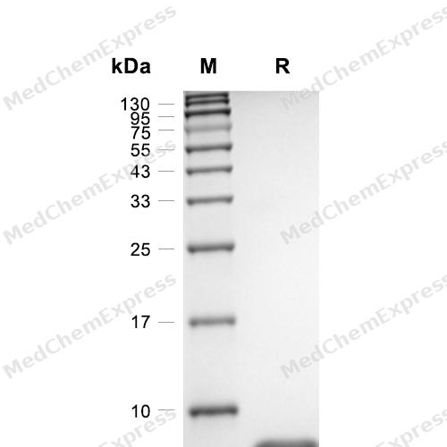
Approximately 6-11 kDa, based on SDS-PAGE under reducing conditions, due to the glycosylation.
-
Purity
≥ 95%, as determined by reducing SDS-PAGE.


Product Properties
Lyophilized powder
1.Lyophilized from a 0.22 μm filtered solution of 20 mM PB, 150 mM NaCl, pH 7.4.
2.Lyophilized from a 0.22 μm filtered solution of 10 mM PB, 200 mM NaCl, pH 7.0.
3.Lyophilized from a 0.22 μm filtered solution of 50 mM Tris-HCl, 200 mM NaCl, pH 8.0.
4.Lyophilized from a 0.22 μm filtered solution of PBS, pH 7.4, 5% trehalose, 5% mannitol, 0.01% Tween 80.
5.Lyophilized from a 0.22 μm filtered solution of PBS, pH 7.4, 8% trehalose.
6.Lyophilized from a 0.22 μm filtered solution of 20 mM Tris-HCl, 200 mM NaCl, pH 8.0.
Please refer to the lot-specific COA for specific buffer information.
Note: For SPR assay, please replace the buffer. Primary amine components (e.g., Tris, imidazole) can affect protein-coupled chips.
<1 EU/μg, determined by LAL method.
It is not recommended to reconstitute to a concentration less than 100 μg/mL in ddH2O. For long term storage it is recommended to add a carrier protein (0.1% BSA, 5% HSA, 10% FBS or 5% Trehalose).
Stored at -20°C for 2 years from date of receipt. After reconstitution, it is stable at 4°C for 1 week or -20°C for longer (with carrier protein). It is recommended to freeze aliquots at -20°C or -80°C for extended storage.
Room temperature in continental US; may vary elsewhere.


Documentation
-
Data Sheet (262 KB)
-
SDS (252 KB)
- English - EN (252 KB)
- Français - FR (252 KB)
- Deutsch - DE (252 KB)
- Norwegian - NO (252 KB)
- Español - ES (252 KB)
- Swedish - SV (252 KB)
- Italian - IT (252 KB)
- Korean - KR (252 KB)
- Portuguese - PT (252 KB)
-
Handling Instructions (2659 KB)


References
[1]. Wong WR, et al. Applications, and efficient large-scale production, of recombinant human epidermal growth factor. Biotechnol Genet Eng Rev. 2001;18:51-71. [Content Brief]
[2]. Lao G, et al. Controlled release of epidermal growth factor from hydrogels accelerates wound healing in diabetic rats. J Am Podiatr Med Assoc. 2012 Mar-Apr;102(2):89-98. [Content Brief]
[3]. Pouranvari S, et al., Cloning, Expression, and Cost Effective Purification of Authentic Human Epidermal Growth Factor With High Activity. Iran Red Crescent Med J. 2016 Mar 20;18(3):e24966. [Content Brief]

Calculators
Concentration (start) × Volume (start) = Concentration (final) × Volume (final)


 Powered by Bioz

