



BETd-260
Based on 5 publication(s) in Google Scholar
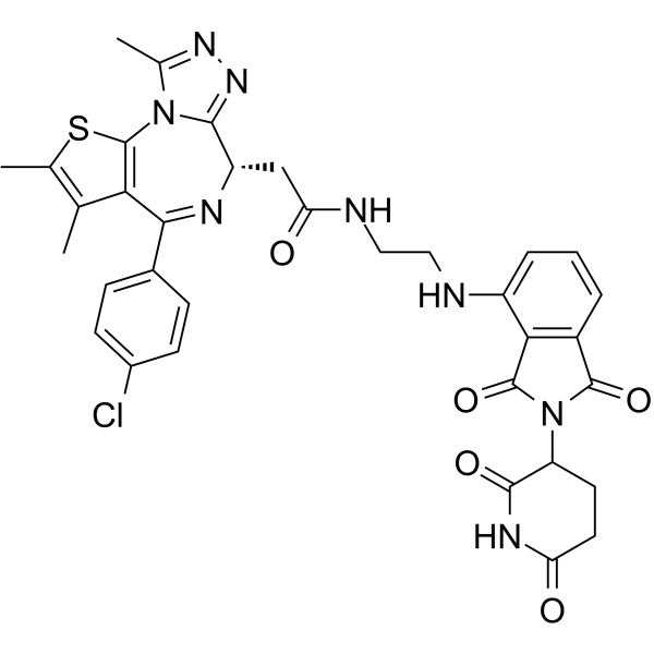
BETd-260 (ZBC 260) is a BET PROTAC degrader. BETd-260 recruits BRD2, BRD3, and BRD4 to the CUL4-RBX1-DDB1-CRBN E3 ubiquitin ligase complex, driving cereblon-, proteasome-, and NEDD8-activating enzyme-dependent ubiquitination and degradation, with a DC50 of approximately 30-100 pM in RS4;11 cells. BETd-260 induces cancer cell apoptosis via endogenous signaling pathways, regulates the expression of the Bcl-2 family, inhibits the oncogene c-Myc, and reduces cell viability. BETd-260 suppresses tumor growth in mouse xenograft models with good biosafety. BETd-260 can be used in research related to acute leukemia, hepatocellular carcinoma, osteosarcoma, and triple-negative breast cancer.
(Pink: BET ligand (HY-112429); Blue: Cereblon ligand (HY-43722); Black: linker).
For research use only. We do not sell to patients.
- Purity : 99.68%
- CAS No.: 2093388-62-4
- Formula: C43H46N10O6
- Molecular Weight:798.89
-
Storage:
-80°C, protect from light, stored under nitrogen

To place orders, for customer services and technical support, please contact: MedChemExpress USA
Tel: 609-228-6898 E-mail: [email protected] [email protected]


-
 Biological Activity
-
Chemical Information
-
Solvent & Solubility
- Protocols
- Purity & Documentation
- References
-
 Help & FAQs


-
HY-112609
QCA570pan-BET-BRD PROTAC Degrader -
HY-112588
dBET6PROTAC BET Degrader -
HY-114228
PROTAC BET degrader-2BRD2/BRD3/BRD4 PROTAC Degrader -
HY-100972
ARV-771BET PROTAC Degrader -
HY-16954
ARV-825PROTAC BRD2/BRD3/BRD4 Degrader -
HY-107425
MZ 1PROTAC BRD4 Degrader -
HY-101838
dBET1PROTAC BRD4 Degrader -
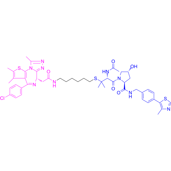
HY-111433
BRD4 degrader AT1BRD4 PROTAC Degrader -
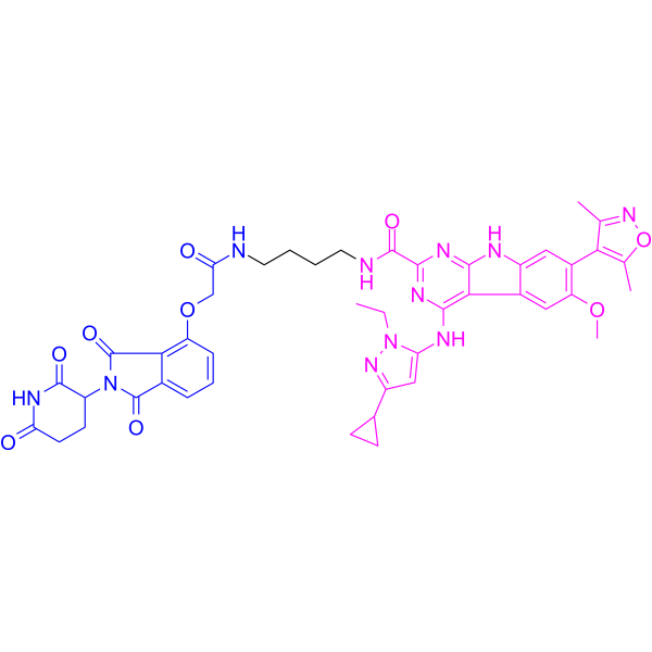
HY-103633
PROTAC BET Degrader-1BRD2/BRD3/BRD4 PROTAC Degrader -
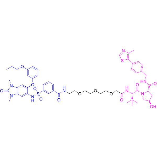
HY-111519
dTRIM24PROTAC TRIM24 Degrader -
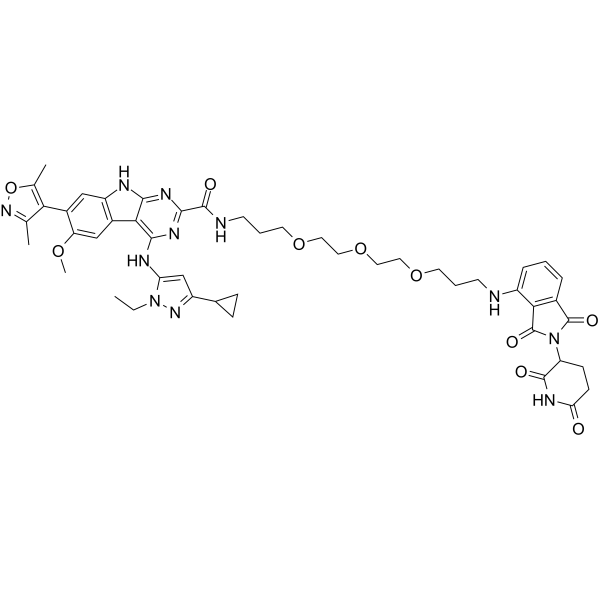
HY-115568
BETd-246BRD2/3/4 PROTAC Degrader -
HY-123844
dBET57BRD4 PROTAC Degrader
Publications Citing Use of MedChemExpress (MCE) BETd-260
MoreAll PROTACs Isoforms
More

Biological Activity
Description
|
BRD4 <30 pM (DC50) |
BRD2 30-100 pM (DC50) |
BRD3 30-100 pM (DC50) |
Cellular Effect
|
Cell Line

|
Type | Value | Description | References |
|---|---|---|---|---|
| RS4-11 | IC50 |
0.051 nM
|
Cell growth inhibition against human RS4;11 acute leukemia cells assessed via lactate dehydrogenase-based WST-8 assay after 4 days of incubation.
Cell growth inhibition against human RS4;11 acute leukemia cells assessed via lactate dehydrogenase-based WST-8 assay after 4 days of incubation.
|
28339196 |
| MOLM-13 | IC50 |
2.2 nM
|
Cell growth inhibition against human MOLM-13 acute leukemia cells assessed via lactate dehydrogenase-based WST-8 assay after 4 days of incubation.
Cell growth inhibition against human MOLM-13 acute leukemia cells assessed via lactate dehydrogenase-based WST-8 assay after 4 days of incubation.
|
28339196 |
| HepG2 | EC50 |
10.3 nM
|
Inhibition of cell viability against human HepG2 cells assessed by CCK-8 assay after 72 hrs of treatment.
Inhibition of cell viability against human HepG2 cells assessed by CCK-8 assay after 72 hrs of treatment.
|
31993368 |
| Bel-7402 | EC50 |
5.4 nM
|
Inhibition of cell viability against human BEL-7402 cells assessed by CCK-8 assay after 72 hrs of treatment.
Inhibition of cell viability against human BEL-7402 cells assessed by CCK-8 assay after 72 hrs of treatment.
|
31993368 |
| SK-HEP1 | EC50 |
24.3 nM
|
Inhibition of cell viability against human SK-HEP-1 cells assessed by CCK-8 assay after 72 hrs of treatment.
Inhibition of cell viability against human SK-HEP-1 cells assessed by CCK-8 assay after 72 hrs of treatment.
|
31993368 |
| SMMC-7721 | EC50 |
23.3 nM
|
Inhibition of cell viability against human SMMC-7721 cells assessed by CCK-8 assay after 72 hrs of treatment.
Inhibition of cell viability against human SMMC-7721 cells assessed by CCK-8 assay after 72 hrs of treatment.
|
31993368 |
| Huh-7 | EC50 |
63.3 nM
|
Inhibition of cell viability against human HuH-7 cells assessed by CCK-8 assay after 72 hrs of treatment.
Inhibition of cell viability against human HuH-7 cells assessed by CCK-8 assay after 72 hrs of treatment.
|
31993368 |
| MHCC97H | EC50 |
64.9 nM
|
Inhibition of cell viability against human MHCC97H cells assessed by CCK-8 assay after 72 hrs of treatment.
Inhibition of cell viability against human MHCC97H cells assessed by CCK-8 assay after 72 hrs of treatment.
|
31993368 |
| SAOS-2 | EC50 |
1.1 nM
|
Inhibition of cell viability against human Saos-2 osteosarcoma cells incubated for 72 h by CCK-8 assay.
Inhibition of cell viability against human Saos-2 osteosarcoma cells incubated for 72 h by CCK-8 assay.
|
31653826 |
| MG-63 | EC50 |
7.2 nM
|
Inhibition of cell viability against human MG-63 osteosarcoma cells incubated for 72 h by CCK-8 assay.
Inhibition of cell viability against human MG-63 osteosarcoma cells incubated for 72 h by CCK-8 assay.
|
31653826 |
| SJSA-1 | EC50 |
16.1 nM
|
Inhibition of cell viability against human SJSA-1 osteosarcoma cells incubated for 72 h by CCK-8 assay.
Inhibition of cell viability against human SJSA-1 osteosarcoma cells incubated for 72 h by CCK-8 assay.
|
31653826 |
In Vitro
BETd-260 (for 4 days) inhibits the proliferation of human acute leukemia cell line MOLM-13, with an IC50 of 2.2 nM[1].
BETd-260 (0.1-1 nM; 3 h) potently degrades BRD2, BRD3 and BRD4 proteins in the human acute leukemia cell line RS4;11[1].
BETd-260 (0.03-10 nM; 24 h) degrades BRD2, BRD3 and BRD4 proteins in RS4;11 human acute leukemia cells and downregulates c-Myc[1].
BETd-260 (1 nM; 3 h, preceded by 2 h pretreatment) induces the degradation of BRD2, BRD3 and BRD4 in human acute leukemia cell line RS4;11, and this process depends on binding to BET proteins, cereblon, the proteasome and NEDD8-activating enzyme[1].
BETd-260 (10-100 nM; 1-48 h) rapidly and potently degrades BRD2, BRD3 and BRD4 proteins in human hepatocellular carcinoma HepG2 cells[2].
BETd-260 (100 nM; 24 h) exhibits broad BET protein degradation activity in human hepatocellular carcinoma (HCC) cell lines, completely eliminating BRD2, BRD3 and BRD4 in BEL-7402, SK-HEP-1 and SMMC-7721 cells, and reducing the levels of these proteins to low levels in HuH-7 and MHCC97H cells[2].
BETd-260 (72 h) potently inhibits the cell viability of MNNG/HOS, Saos-2, MG-63 and SJSA-1 osteosarcoma cell lines, with EC50 values ranging from 1.1 nM to 16.1 nM[3].
BETd-260 (3-30 nM; 1-24 h) potently degrades BRD2, BRD3 and BRD4 in MNNG/HOS, Saos-2, MG-63 and SJSA-1 osteosarcoma cell lines[3].
BETd-260 (10 nM; 2 h, preceded by 1 h pre-treatment with inhibitors) induces the degradation of BRD2, BRD3 and BRD4 in MNNG/HOS osteosarcoma cells via a pathway mediated by the Cullin-dependent E3 ligase and ubiquitin-proteasome system, and this degradation process depends on specific binding to BET proteins[3].
BETd-260 potently inhibits the growth of human triple-negative breast cancer (TNBC) cell lines[4].
BETd-260 efficiently degrades BRD2, BRD3 and BRD4 proteins in human triple-negative breast cancer (TNBC) cell lines[4].
BETd-260 (10-100 nM; 48 h) induces extensive apoptosis in human HepG2, BEL-7402, SK-HEP-1, SMMC-7721, HuH-7 and MHCC97H hepatocellular carcinoma cells[2].
BETd-260 (10-100 nM; 24 h) activates the apoptotic signaling pathway in human HepG2, BEL-7402, SK-HEP-1, SMMC-7721, HuH-7 and MHCC97H hepatocellular carcinoma cells, and induces PARP cleavage and caspase-3 activation[2].
BETd-260 (10-100 nM; 24 h) bidirectionally regulates the expression of apoptosis-related proteins, inhibits c-Myc expression, upregulates Bad expression, and downregulates the expressions of Mcl-1, Bcl-2, XIAP and c-Myc in human hepatocellular carcinoma cells[2].
BETd-260 (100 nM; 48 h following 24 h siRNA transfection) induces cell death in human HepG2 and BEL-7402 hepatocellular carcinoma cells, and this process depends on the endogenous caspase-9-mediated apoptotic pathway rather than the exogenous caspase-8 pathway[2].
BETd-260 (100 nM; 48 h after 24 h siRNA transfection) induces cell death in human HepG2 and BEL-7402 hepatocellular carcinoma cells in a Bad-dependent manner[2].
BETd-260 (10-100 nM; 24 h) disrupts mitochondrial membrane integrity in human HepG2 and BEL-7402 hepatocellular carcinoma cells, and induces the release of cytochrome c from mitochondria into the cytosol[2].
BETd-260 (10-100 nM; 4-24 h) upregulates the expression of Bad mRNA in human HepG2 and BEL-7402 hepatocellular carcinoma cells in a dose-dependent manner[2].
BETd-260 (3-30 nM; 24 h) induces dose-dependent apoptosis in MNNG/HOS and Saos-2 osteosarcoma cells[3].
BETd-260 (3-30 nM; 24 h) activates the mitochondrial apoptotic pathway in MNNG/HOS and Saos-2 osteosarcoma cells, which is evidenced by the cleavage of caspase-3, caspase-9 and PARP-1[3].
BETd-260 (10 nM; 24-48 h, with 1 h pre-treatment with inhibitors) induces death and apoptosis in MNNG/HOS and Saos-2 osteosarcoma cells, a process that mainly relies on the endogenous pathway mediated by the caspase-9 and caspase-3 cascade[3].
BETd-260 (3-30 nM; 1-24 h) regulates the expression of Bcl-2 family members in MNNG/HOS and Saos-2 osteosarcoma cells, and inhibits c-Myc (without affecting p53), specifically characterized by depletion of Mcl-1 and Bcl-xl, upregulation of Noxa levels, and reduction of c-Myc levels[3].
MedChemExpress (MCE) has not independently confirmed the accuracy of these methods. They are for reference only.
-
Cell Line:RS4;11 human acute leukemia cells
-
Concentration:0.1, 0.3 and 1 nM
-
Incubation Time:3 h
-
Result:Decreased levels of BRD2 and BRD4 at concentrations as low as 0.3 nM.
Decreased BRD3 levels at 0.1 nM.
-
Cell Line:RS4;11 human acute leukemia cells
-
Concentration:0.03, 0.1, 0.3, 1, 3 and 10 nM
-
Incubation Time:24 h
-
Result:Decreased BRD4 levels at 0.03 nM.
Decreased BRD2 and BRD3 levels at 0.1 nM.
Decreased c-Myc levels at 0.1 nM.
-
Cell Line:RS4;11 human acute leukemia cells
-
Concentration:1 nM (this compound); 10-30 μM (Lenalidomide (HY-A0003)); 20 μM (MG-132 (HY-13259)); 1 μM (MLN4924 (HY-70062))
-
Incubation Time:3 h (BETd-260); 2 h (pretreatment)
-
Result:Completely blocked this compound-induced degradation of BRD2, BRD3, and BRD4 following pretreatment with 8, lenalidomide, MG-132, or MLN4924.
-
Cell Line:RS4;11 human acute leukemia cells; MOLM-13 human acute leukemia cells
-
Concentration:0.3, 1, 3, 10 and 30 nM
-
Incubation Time:24 h
-
Result:Induced strong cell cycle arrest at 0.3 nM.
Induced robust apoptosis at 3-10 nM.
-
Cell Line:human HepG2 hepatocellular carcinoma (HCC) cells
-
Concentration:10, 30 and 100 nM (24 h time point); 100 nM (time course)
-
Incubation Time:24 h (10-100 nM); 1 h, 3 h, 12 h, 24 h, 48 h (100 nM time course)
-
Result:Almost completely eliminated BRD2, BRD3, and BRD4 protein levels at 10-100 nM after 24 h.
Reduced BET protein levels within 1 h and achieved complete elimination by 12 h at 100 nM.
-
Cell Line:human BEL-7402, SK-HEP-1, SMMC-7721, HuH-7, and MHCC97H HCC cells
-
Concentration:100 nM
-
Incubation Time:24 h
-
Result:Completely degraded BRD2, BRD3, and BRD4 proteins in BEL-7402, SK-HEP-1, and SMMC-7721 cell lines.
Reduced BET protein levels to very low levels in HuH-7 and MHCC97H cell lines.
-
Cell Line:human HepG2, BEL-7402, SK-HEP-1, SMMC-7721, HuH-7, and MHCC97H HCC cells
-
Concentration:10, 30 and 100 nM
-
Incubation Time:48 h
-
Result:Triggered apoptosis in HepG2 and BEL-7402 cells at 10 nM.
Induced 86% apoptosis in HepG2 cells and 77% apoptosis in BEL-7402 cells at 100 nM.
Induced 45-85% cell death in SK-HEP-1, SMMC-7721, HuH-7, and MHCC97H cells.
-
Cell Line:human HepG2, BEL-7402, SK-HEP-1, SMMC-7721, HuH-7, and MHCC97H HCC cells
-
Concentration:10, 30 and 100 nM
-
Incubation Time:24 h
-
Result:Caused extensive cleavage of PARP in all 6 HCC cell lines.
Induced robust activation of caspase-3 in all 6 HCC cell lines.\nIncreased Bad protein levels in HepG2 and BEL-7402 cells.
Reduced Mcl-1, Bcl-2, and XIAP protein levels in HepG2 and BEL-7402 cells.
Distinctly reduced c-Myc protein levels in 5 of the 6 HCC cell lines.
-
Cell Line:human HepG2 and BEL-7402 HCC cells
-
Concentration:100 nM
-
Incubation Time:48 h (after 24 h siRNA transfection)
-
Result:Significantly attenuated triggered cell death when caspase-9 was knocked down.
Showed no effect on triggered cell death when caspase-8 was knocked down.\nSignificantly attenuated induced cell death in both HepG2 and BEL-7402 cell lines when Bad was knocked down.
-
Cell Line:human HepG2 and BEL-7402 HCC cells
-
Concentration:10, 30 and 100 nM
-
Incubation Time:24 h
-
Result:Increased cytochrome c levels in the cytosolic fraction.
Decreased cytochrome c levels in the mitochondria-enriched fraction.
-
Cell Line:human HepG2 and BEL-7402 HCC cells
-
Concentration:10, 30 and 100 nM (4 h time point); 100 nM (4 h, 12 h, 24 h time course)
-
Incubation Time:4 h (10-100 nM); 4 h, 12 h, 24 h (100 nM time course)
-
Result:Increased Bad mRNA levels 3-fold at 4 h and 8-fold at 12 h in HepG2 cells at 100 nM.
Increased Bad mRNA levels 3-fold at 4 h, 7-fold at 12 h, and 2-fold at 24 h in BEL-7402 cells at 100 nM.
Dose-dependently upregulated Bad mRNA levels in both cell lines at 10-100 nM after 4 h.
-
Cell Line:MNNG/HOS, Saos-2, MG-63, SJSA-1
-
Concentration:3 nM, 10 nM, 30 nM
-
Incubation Time:24 h (3 nM, 10 nM, 30 nM); 1 h, 4 h, 14 h, 24 h (30 nM)
-
Result:Potently degraded BRD2, BRD3, and BRD4 in all four osteosarcoma cell lines.
Completely depleted BRD3 and BRD4, and largely suppressed BRD2 levels in MNNG/HOS and Saos-2 cells at 3 nM for 24 h.
Achieved maximum degradation of BET proteins within 1 h in MNNG/HOS cells at 30 nM, with the effect lasting up to 24 h.
-
Cell Line:MNNG/HOS
-
Concentration:10 nM
-
Incubation Time:2 h (preceded by 1 h pre-treatment with inhibitors)
-
Result:Largely had its BRD2, BRD3, and BRD4 degradation activity abrogated when cells were pre-treated with proteasome inhibitor MG-132, NEDD8-activating enzyme inhibitor MLN4924, or excess BET inhibitor HJB-97.
-
Cell Line:MNNG/HOS, Saos-2
-
Concentration:3 nM, 10 nM, 30 nM
-
Incubation Time:24 h
-
Result:Induced apoptosis in 43%, 62%, and 84% of MNNG/HOS cells at 3 nM, 10 nM, and 30 nM respectively.
Induced apoptosis in 25%, 57%, and 75% of Saos-2 cells at 3 nM, 10 nM, and 30 nM respectively.
-
Cell Line:MNNG/HOS, Saos-2
-
Concentration:3 nM, 10 nM, 30 nM
-
Incubation Time:24 h
-
Result:Induced activation of caspase-3 and caspase-9, and cleavage of PARP-1 in both MNNG/HOS and Saos-2 cells.
Increased effects of caspase-3, caspase-9 activation, and PARP-1 cleavage with higher concentrations.
In Vivo
BETd-260 (5 mg/kg; intravenous injection; single administration / three times per week; for 3 consecutive weeks) exerts potent anti-hepatocellular carcinoma activity in vivo. It inhibits tumor growth in HepG2 and BEL-7402 xenograft models by degrading BET proteins, regulating apoptosis regulators, and inducing tumor cell apoptosis[2].
BETd-260 (5 mg/kg; intravenous injection; three times per week; for 4 consecutive weeks) inhibits tumor growth in an osteosarcoma PDX xenograft model in NOD SCID mice, with no observed toxicity[3].
MedChemExpress (MCE) has not independently confirmed the accuracy of these methods. They are for reference only.
-
Animal Model:severe combined immunodeficient (SCID) mice[1]
-
Dosage:5 mg/kg (tumor regression); 5 mg/kg (pharmacodynamic analysis)
-
Administration:i.v.; every other day, three times a week; 3 weeks (tumor regression); i.v.; single dose (pharmacodynamic analysis)
-
Result:Achieved >90% tumor regression in RS4;11 xenografts, with no signs of toxicity or animal weight loss.
Induced near-complete degradation of BRD2, BRD3, and BRD4 proteins in tumor tissue starting at 1 hour, with effects persisting for >24 hours.
Caused strong down-regulation of c-Myc protein for at least 6 hours.
Detected robust cleavage of PARP and caspase-3 starting at 3 hours, peaking at 6 hours.
Reached tumor concentrations of 166.3 ng/g at 1 hour, 98.5 ng/g at 3 hours, and 35.8 ng/g at 6 hours after a single dose.
-
Animal Model:Balb/c (6-week-old; subcutaneous xenograft model injected with HepG2 or BEL-7402 cells)[2]
-
Dosage:5 mg/kg (pharmacodynamic analysis); 5 mg/kg (efficacy analysis)
-
Administration:i.v.; single dose (pharmacodynamic analysis); 3 times per week; 3 weeks (efficacy analysis)
-
Result:Suppressed BRD2, BRD3, and BRD4 expression in HepG2 and BEL-7402 xenograft tumor tissues.
Reduced Mcl-1 expression, increased Bad expression, augmented cleaved PARP and activated caspase-3 levels in HepG2 and BEL-7402 xenograft tumor tissues.
Reduced Ki-67 expression by 57% (HepG2) and 71% (BEL-7402).
Achieved a tumor growth inhibition (TGI) of 49% in HepG2 xenografts and 78% in BEL-7402 xenografts.
Caused only slight effects on mouse body weight.
-
Animal Model:BALB/c (6-week-old; subcutaneous xenograft induced by injecting 5 million MNNG/HOS cells suspended in Matrigel)[3]
-
Dosage:5 mg/kg
-
Administration:i.v.; single dose (pharmacodynamic study); i.v.; three times per week; 3 weeks (efficacy study)
-
Result:Completely depleted BRD2, BRD3, and BRD4 proteins in tumor tissue starting at 1 h post-treatment, with the effect lasting more than 24 h.
Triggered PARP-1 cleavage and reduced Ki67-positive cells, indicating massive apoptosis and inhibited tumor cell proliferation.
Achieved ~94% tumor growth inhibition (TGI).
Reduced tumor volume from 164 mm3 to 155 mm3 after 3 doses, with this partial reduction persisting for 69 days after treatment cessation.
Resulted in an average tumor volume of 246.9 mm3 at study termination (day 121), compared to 1540.1 mm3 in control mice, with 5 out of 7 treated tumors remaining smaller than their initial volume.
Caused no significant weight loss or other signs of toxicity.
-
Animal Model:NOD SCID (6-week-old; subcutaneous patient-derived xenograft induced by implanting 300-1000 mg PDX tumor masses)[3]
-
Dosage:5 mg/kg (pharmacodynamic study); 5 mg/kg (efficacy study)
-
Administration:i.v.; single dose (pharmacodynamic study); i.v.; three times per week; 4 weeks (efficacy study)
-
Result:Markedly reduced BRD2, BRD3, and BRD4 protein levels, increased cleaved PARP-1-positive cells, and reduced Ki67-positive cells in PDX tumor tissue.
Achieved 57% tumor growth inhibition (TGI).
Resulted in an average tumor volume of 740.9 mm3 at study termination (day 39), compared to 1623.4 mm3 in control mice.
Caused no significant weight loss or apparent toxicity.
-
Animal Model:SCID mice bearing subcutaneous xenograft tumors were established by injecting 5 million MDA-MB-231 cells or 5 million MDA-MB-468 cells, respectively[4]
-
Dosage:5 mg/kg
-
Administration:i.v.; 3 times per week; 3 weeks
-
Result:Exerted stronger antitumor activity than BETd-246 at 10 mg/kg with the same dosing schedule.
Reduced BET protein levels in tumors at 1-3 hours post-exposure.
Upregulated p21WAF1 in tumors at 1-3 hours post-exposure.
Downregulated MCL1 in tumors at 1-3 hours post-exposure.
Caused no significant weight loss or overt toxicity.\nExerted stronger antitumor activity than BETd-246 at 10 mg/kg with the same dosing schedule.
Reduced BET protein levels in tumors at 1-3 hours post-exposure.
Upregulated p21WAF1 in tumors at 1-3 hours post-exposure.
Downregulated MCL1 in tumors at 1-3 hours post-exposure.
Caused no significant weight loss or overt toxicity.
- RNA extraction experimental
- Mammalian live/dead viability and cytotoxicity staining
- Annexin V plus membrane-impermeant dye apoptosis staining
- Patient-Derived Orthotopic Xenograft (PDOX)
- CCK-8/WST-8 Cell Proliferation Assay
- Liver Cancer Modeling
- Breast Cancer Modeling
- Cell Viability Determination by MTT Colorimetric Assay
- Detection of Apoptosis
- TUNEL staining for apoptotic DNA fragmentation
- Cell Cytotoxicity Assay
- Patient-Derived Xenograft (PDX)
- Apoptosis Solutions
- Splenic/Portal-Vein Liver Metastasis Xenograft
- MTT Cell Proliferation Assay
- Subcutaneous Cell-Line-Derived Xenograft
- Orthotopic Cell-Line Xenograft
- Intraperitoneal/Peritoneal Dissemination Xenograft


Chemical Information
-
CAS No. 2093388-62-4
-
Appearance Solid
-
Molecular Weight 798.89
-
Formula C43H46N10O6
-
Color Off-white to yellow
-
SMILES
CCN1N=C(C2CC2)C=C1NC3=NC(C(NCCCCCC4=CC=CC5=C4CN(C6C(NC(CC6)=O)=O)C5=O)=O)=NC7=C3C8=CC(OC)=C(C9=C(C)ON=C9C)C=C8N7
-
Synonyms
ZBC 260
-
Shipping
Shipping with dry ice.
-
Storage
-80°C, protect from light, stored under nitrogen


Publications (5)
-
Journal Impact Factor
-
Most Recent
-
PLoS Biol
The cell cycle regulator PLK1 promotes murine melanoma progression by regulating the transcription factor BACH1. [Abstract]2025 Nov 24;23(11):e3003490. PMID: 41284701 -
Structure
PROTAC-mediated activation, rather than degradation, of a nuclear receptor reveals complex ligand-receptor interaction network. [Abstract]2024 Dec 5;32(12):2352-2363.e8. PMID: 39389062 -
ACS Pharmacol Transl Sci
Developing MYC Degraders Bearing the Von Hippel-Lindau Ligand to Target the "Undruggable" MYC. [Abstract]2024 Nov 15;7(12):3955-3968. PMID: 39698270 -
-


Solvent & Solubility
In Vitro:
DMSO : 25 mg/mL (31.29 mM; Need ultrasonic; Hygroscopic DMSO has a significant impact on the solubility of product, please use newly opened DMSO)
Please refer to the solubility information to select the appropriate solvent. Once prepared, please aliquot and store the solution to prevent product inactivation from repeated freeze-thaw cycles.
Storage method and period of stock solution: -80°C, 6 months (protect from light, stored under nitrogen). When stored at -80°C, please use it within 6 months.
Please refer to the solubility information to select the appropriate solvent. Once prepared, please aliquot and store the solution to prevent product inactivation from repeated freeze-thaw cycles.
Storage method and period of stock solution: -80°C, 6 months (protect from light, stored under nitrogen). When stored at -80°C, please use it within 6 months.
Concentration (start) × Volume (start) = Concentration (final) × Volume (final)
In Vivo:
Select the appropriate dissolution method based on your experimental animal and administration route.
- For the following dissolution methods, please ensure to first prepare a clear stock solution using an In Vitro approach and then sequentially add co-solvents:
- To ensure reliable experimental results, the clarified stock solution can be appropriately stored based on storage conditions. As for the working solution for In Vivo experiments, it is recommended to prepare freshly and use it on the same day.
- The percentages shown for the solvents indicate their volumetric ratio in the final prepared solution. If precipitation or phase separation occurs during preparation, heat and/or sonication can be used to aid dissolution.
Add each solvent one by one: 10% DMSO 40% PEG300 5% Tween-80 45% Saline
Solubility: ≥ 0.83 mg/mL (1.04 mM); Clear solution
This protocol yields a clear solution of ≥ 0.83 mg/mL (saturation unknown).
Taking 1 mL working solution as an example, add 100 μL DMSO stock solution (8.3 mg/mL) to 400 μL PEG300, and mix evenly; then add 50 μL Tween-80 and mix evenly; then add 450 μL Saline to adjust the volume to 1 mL.
Preparation of Saline: Dissolve 0.9 g sodium chloride in ddH₂O and dilute to 100 mL to obtain a clear Saline solution.
In Vivo Dissolution Calculator
Please enter the basic information of animal experiments:
-
-
-
-
Recommended: Prepare an additional quantity of animals to account for potential losses during experiments.
Please enter your animal formula composition:
-
%DMSO +
Recommended: Keep the proportion of DMSO in working solution below 2% if your animal is weak.
-
%+
-
+%Tween-80 + +
-
%Saline +
The co-solvents required include: DMSO, . All of co-solvents are available by MedChemExpress (MCE). , Tween 80. All of co-solvents are available by MedChemExpress (MCE).
Working solution concentration: 0.22 mg/mL
Method for preparing stock solution: mg drug dissolved in μL DMSO. Stock solution concentration: mg/mL.
1. Take μL DMSO stock solution;
2. Add μL .
μL , mix evenly;
3. Then add μL Tween 80, mix evenly;
4. Then add μL
Please ensure that the stock solution in the first step is dissolved to a clear state, and add co-solvents in sequence. You can use ultrasonic heating (ultrasonic cleaner, recommended frequency 20-40 kHz), vortexing, etc. to assist dissolution.


Purity & Documentation
-
Data Sheet (310 KB)
-
SDS (394 KB)
- English - EN (394 KB)
- Français - FR (394 KB)
- Deutsch - DE (394 KB)
- Norwegian - NO (394 KB)
- Español - ES (394 KB)
- Swedish - SV (394 KB)
- Italian - IT (394 KB)
- Korean - KR (394 KB)
- Portuguese - PT (394 KB)
-
Handling Instructions (2659 KB)
References
[1]. Zhou B, et al. Discovery of a Small-Molecule Degrader of Bromodomain and Extra-Terminal (BET) Proteins with Picomolar Cellular Potencies and Capable of Achieving Tumor Regression. Journal of medicinal chemistry. 2018 Jan 25;61(2):462-481. [Content Brief]
[2]. Zhang H, et al. Targeting BET Proteins With a PROTAC Molecule Elicits Potent Anticancer Activity in HCC Cells. Frontiers in oncology. 2019;9:1471. [Content Brief]
[4]. Shi C, et al. Targeted Degradation of BET Proteins in Triple-Negative Breast Cancer. Cancer Res. 2017 May 1;77(9):2476-2487. [Content Brief]


Complete Stock Solution Preparation Table
Please refer to the solubility information to select the appropriate solvent. Once prepared, please aliquot and store the solution to prevent product inactivation from repeated freeze-thaw cycles.
Storage method and period of stock solution: -80°C, 6 months (protect from light, stored under nitrogen). When stored at -80°C, please use it within 6 months.
| Optional Solvent | Concentration Solvent Mass | 1 mg | 5 mg | 10 mg | 25 mg |
|---|---|---|---|---|---|
| DMSO | 1 mM | 1.2517 mL | 6.2587 mL | 12.5174 mL | 31.2934 mL |
| 5 mM | 0.2503 mL | 1.2517 mL | 2.5035 mL | 6.2587 mL | |
| 10 mM | 0.1252 mL | 0.6259 mL | 1.2517 mL | 3.1293 mL | |
| 15 mM | 0.0834 mL | 0.4172 mL | 0.8345 mL | 2.0862 mL | |
| 20 mM | 0.0626 mL | 0.3129 mL | 0.6259 mL | 1.5647 mL | |
| 25 mM | 0.0501 mL | 0.2503 mL | 0.5007 mL | 1.2517 mL | |
| 30 mM | 0.0417 mL | 0.2086 mL | 0.4172 mL | 1.0431 mL |


BETd-260 Related Classifications
HY-101519 Related Classifications
 Powered by Bioz