



Rebastinib
Based on 13 publication(s) in Google Scholar
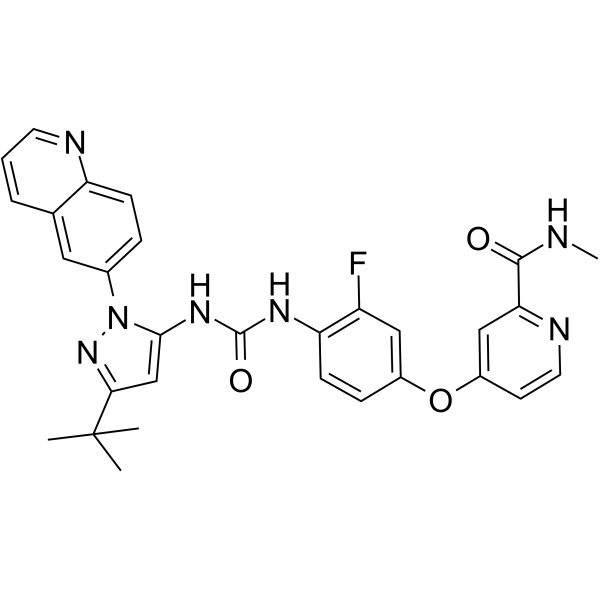
Rebastinib (DCC-2036) is an orally active, non-ATP-competitive Bcr-Abl inhibitor, with IC50 values of 0.8 nM and 4 nM against Abl1WT and Abl1T315I, respectively. Rebastinib potently inhibits CYP2C8 (IC50 = 0.0533 μM) and CYP2C9 (IC50 = 2.54 μM). Rebastinib allosterically inhibits Tie2 with an IC50 of 0.63 nM. Rebastinib inhibits the transcriptional activity of FoxO1. Rebastinib inhibits SRC family kinases, KDR and FLT3 kinases. Rebastinib induces Apoptosis. Rebastinib exerts anti-tumor activity in breast cancer. Rebastinib exerts anti-angiogenic effects. Rebastinib increases myotube diameter and improves reduced contractility. Rebastinib can be used in research related to triple-negative breast cancer, luminal breast cancer, pancreatic neuroendocrine tumors, muscle atrophy, cancer cachexia, chronic myeloid leukemia, and B-lymphocytic leukemia.
商品は「研究用試薬」です。人や動物の医療用・臨床診断用・食品用の製品ではありません。
研究用途以外に使用した場合、当社は一切の責任を負いかねます。
- 純度 : 99.74%
- CAS 番号: 1020172-07-9
- 分子式: C30H28FN7O3
- 分子量:553.59
-
保管条件:Powder -20°C, 3 years , 4°C, 2 years ; In solvent -80°C, 2 years , -20°C, 1 year

To place orders, for customer services and technical support, please contact: MedChemExpress USA
Tel: 609-228-6898 E-mail: [email protected] [email protected]


-
 生物活性
生物活性
-
化学情報
-
溶剤 & 溶解度
- プロトコル
- 純度とドキュメンテーション
- 参考文献
-
 Help & FAQs


-
HY-10159
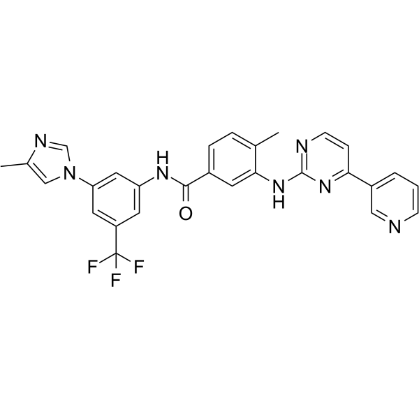
NilotinibBcr-Abl Inhibitor -
HY-15666
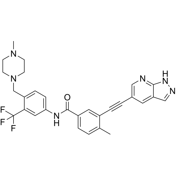
OlverembatinibBcr-Abl Inhibitor -
HY-12047
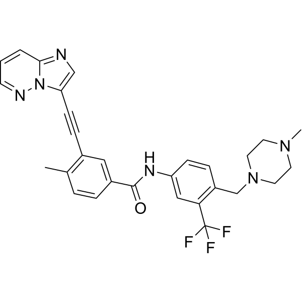
PonatinibMulti-targeted Kinase Inhibitor -
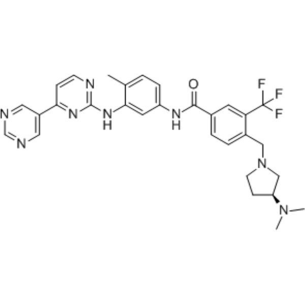
HY-50868
BafetinibLyn/Bcr-Abl Inhibitor -
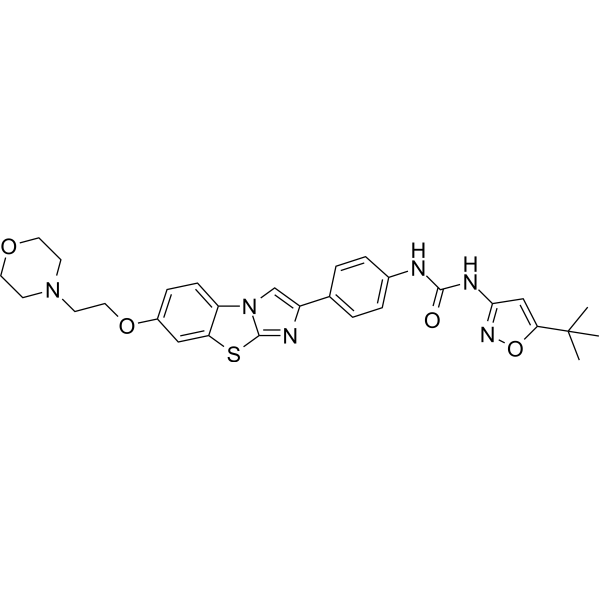
HY-13001
QuizartinibFLT3 Inhibitor -
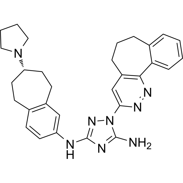
HY-15150
BemcentinibAXL Inhibitor -
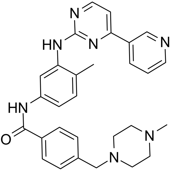
HY-15463
ImatinibTyrosine Kinases Inhibitor -
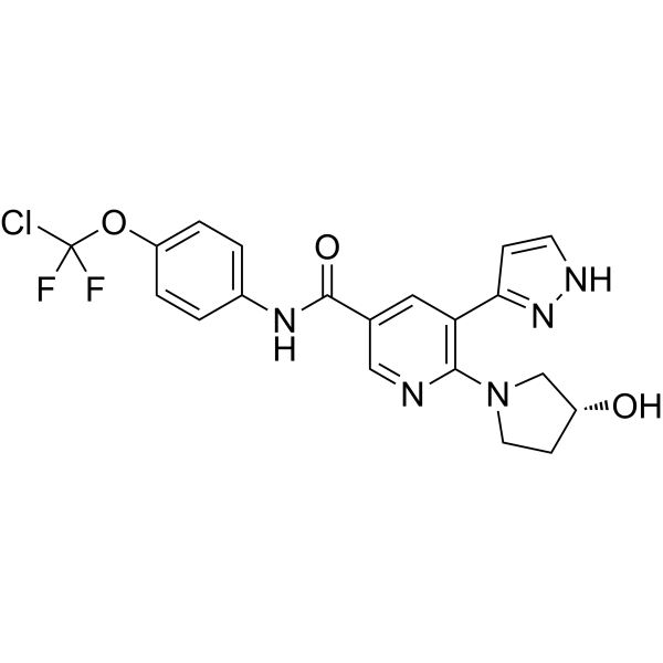
HY-104010
AsciminibBCR-ABL1 Inhibitor -
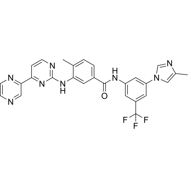
HY-15728
RadotinibBcr-Abl1 Inhibitor -
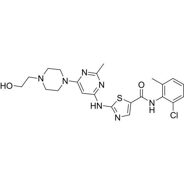
HY-10181
DasatinibSrc/Bcr-Abl Inhibitor -
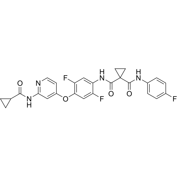
HY-B0791
AltiratinibMET/TIE2/VEGFR2/FLT3/Trk Inhibitor -
HY-10320
Doramapimodp38 MAPK Inhibitor
MedChemExpress(MCE)の使用を引用している文献 Rebastinib
More- Nat Commun. 2021 Jan 25;12(1):504. [Abstract]
- Sci Transl Med. 2018 Jul 18;10(450):eaaq1093. [Abstract]
- J Clin Invest. 2026 Jan 6;136(4):e193758. [Abstract]
- J Exp Clin Cancer Res. 2022 Apr 21;41(1):149. [Abstract]
- Brain. 2025 May 13;148(5):1723-1739. [Abstract]
- J Exp Med. 2026 May 4;223(5):e20251374. [Abstract]
- J Med Chem. 2015 Jan 8;58(1):466-79. [Abstract]
- J Cell Sci. 2026 May 28:jcs.264977. [Abstract]
- Anticancer Drugs. 2024 Jan 1;35(1):46-54. [Abstract]
- Res Sq. 2025 Sep 1.
- bioRxiv. 2025 August 23.
- University of Washington. 2025.
- Patent. US20170349880A1.
 Customer Validation & Images

-
Histological Imaging/Staining
-
Histological Imaging/Staining
-
Histological Imaging/Staining
-
Histological Imaging/Staining
-
Cell Imaging/Staining


生物活性
製品説明
IC50 & Target
|
CYP2C8 0.0533 μM (IC50) |
CYP2C9 2.54 μM (IC50) |
Tie2 0.63 nM (IC50) |
Abl1WT 0.8 nM (IC50) |
Abl1T315I 4 nM (IC50) |
FOXO1 |
Cellular Effect
|
Cell Line

|
Type | Value | Description | References |
|---|---|---|---|---|
| BaF3 | IC50 |
120 nM
Compound: DCC-2036
|
Antiproliferative activity against mouse BaF3 cells harbouring BCR-ABL1 F359C mutant assessed as inhibition of cell proliferation measured after 72 hrs by MTT assay
Antiproliferative activity against mouse BaF3 cells harbouring BCR-ABL1 F359C mutant assessed as inhibition of cell proliferation measured after 72 hrs by MTT assay
|
[PMID: 21481795] |
| BaF3 | IC50 |
127 nM
Compound: DCC-2036
|
Antiproliferative activity against mouse BaF3 cells harbouring BCR-ABL1 E255K mutant assessed as inhibition of cell proliferation measured after 72 hrs by MTT assay
Antiproliferative activity against mouse BaF3 cells harbouring BCR-ABL1 E255K mutant assessed as inhibition of cell proliferation measured after 72 hrs by MTT assay
|
[PMID: 21481795] |
| BaF3 | IC50 |
13 nM
Compound: DCC-2036
|
Antiproliferative activity against mouse BAF3 cells harboring wild type BCR-ABL measured after 72 hrs by MTT assay
Antiproliferative activity against mouse BAF3 cells harboring wild type BCR-ABL measured after 72 hrs by MTT assay
|
[PMID: 35551036] |
| BaF3 | IC50 |
13 nM
Compound: DCC-2036
|
Antiproliferative activity against mouse BaF3 cells harbouring BCR-ABL1 T315I mutant assessed as inhibition of cell proliferation measured after 72 hrs by MTT assay
Antiproliferative activity against mouse BaF3 cells harbouring BCR-ABL1 T315I mutant assessed as inhibition of cell proliferation measured after 72 hrs by MTT assay
|
[PMID: 21481795] |
| BaF3 | IC50 |
14 nM
Compound: DCC-2036
|
Antiproliferative activity against mouse BaF3 cells harbouring BCR-ABL1 M351T mutant assessed as inhibition of cell proliferation measured after 72 hrs by MTT assay
Antiproliferative activity against mouse BaF3 cells harbouring BCR-ABL1 M351T mutant assessed as inhibition of cell proliferation measured after 72 hrs by MTT assay
|
[PMID: 21481795] |
| BaF3 | IC50 |
150 nM
Compound: DCC-2036
|
Antiproliferative activity against mouse BaF3 cells harbouring BCR-ABL1 E255V mutant assessed as inhibition of cell proliferation measured after 72 hrs by MTT assay
Antiproliferative activity against mouse BaF3 cells harbouring BCR-ABL1 E255V mutant assessed as inhibition of cell proliferation measured after 72 hrs by MTT assay
|
[PMID: 21481795] |
| BaF3 | IC50 |
19 nM
Compound: DCC-2036
|
Antiproliferative activity against mouse BAF3 cells harboring BCR-ABL T315I mutant measured after 72 hrs by MTT assay
Antiproliferative activity against mouse BAF3 cells harboring BCR-ABL T315I mutant measured after 72 hrs by MTT assay
|
[PMID: 35551036] |
| BaF3 | IC50 |
19 nM
Compound: DCC-2036
|
Antiproliferative activity against mouse BaF3 cells harbouring BCR-ABL1 T315A mutant assessed as inhibition of cell proliferation measured after 72 hrs by MTT assay
Antiproliferative activity against mouse BaF3 cells harbouring BCR-ABL1 T315A mutant assessed as inhibition of cell proliferation measured after 72 hrs by MTT assay
|
[PMID: 21481795] |
| BaF3 | IC50 |
24 nM
Compound: DCC-2036
|
Antiproliferative activity against mouse BaF3 cells harbouring BCR-ABL1 Q252H mutant assessed as inhibition of cell proliferation measured after 72 hrs by MTT assay
Antiproliferative activity against mouse BaF3 cells harbouring BCR-ABL1 Q252H mutant assessed as inhibition of cell proliferation measured after 72 hrs by MTT assay
|
[PMID: 21481795] |
| BaF3 | IC50 |
3500 nM
Compound: DCC-2036
|
Antiproliferative activity against parent mouse BaF3 cells assessed as inhibition of cell proliferation measured after 72 hrs in presence of IL-3 by MTT assay
Antiproliferative activity against parent mouse BaF3 cells assessed as inhibition of cell proliferation measured after 72 hrs in presence of IL-3 by MTT assay
|
[PMID: 21481795] |
| BaF3 | IC50 |
36 nM
Compound: DCC-2036
|
Antiproliferative activity against mouse BaF3 cells harbouring BCR-ABL1 F317L mutant assessed as inhibition of cell proliferation measured after 72 hrs by MTT assay
Antiproliferative activity against mouse BaF3 cells harbouring BCR-ABL1 F317L mutant assessed as inhibition of cell proliferation measured after 72 hrs by MTT assay
|
[PMID: 21481795] |
| BaF3 | IC50 |
39 nM
Compound: DCC-2036
|
Antiproliferative activity against mouse BaF3 cells harbouring BCR-ABL1 Y253F mutant assessed as inhibition of cell proliferation measured after 72 hrs by MTT assay
Antiproliferative activity against mouse BaF3 cells harbouring BCR-ABL1 Y253F mutant assessed as inhibition of cell proliferation measured after 72 hrs by MTT assay
|
[PMID: 21481795] |
| BaF3 | IC50 |
5.4 nM
Compound: DCC-2036
|
Antiproliferative activity against native mouse BaF3 cells assessed as inhibition of cell proliferation measured after 72 hrs by MTT assay
Antiproliferative activity against native mouse BaF3 cells assessed as inhibition of cell proliferation measured after 72 hrs by MTT assay
|
[PMID: 21481795] |
| BaF3 | IC50 |
51 nM
Compound: DCC-2036
|
Antiproliferative activity against mouse BaF3 cells harbouring BCR-ABL1 L248R mutant assessed as inhibition of cell proliferation measured after 72 hrs by MTT assay
Antiproliferative activity against mouse BaF3 cells harbouring BCR-ABL1 L248R mutant assessed as inhibition of cell proliferation measured after 72 hrs by MTT assay
|
[PMID: 21481795] |
| BaF3 | IC50 |
56 nM
Compound: DCC-2036
|
Antiproliferative activity against mouse BaF3 cells harbouring BCR-ABL1 Y253H mutant assessed as inhibition of cell proliferation measured after 72 hrs by MTT assay
Antiproliferative activity against mouse BaF3 cells harbouring BCR-ABL1 Y253H mutant assessed as inhibition of cell proliferation measured after 72 hrs by MTT assay
|
[PMID: 21481795] |
| BaF3 | IC50 |
6 nM
Compound: DCC-2036
|
Antiproliferative activity against mouse BaF3 cells harbouring BCR-ABL1 V299L mutant assessed as inhibition of cell proliferation measured after 72 hrs by MTT assay
Antiproliferative activity against mouse BaF3 cells harbouring BCR-ABL1 V299L mutant assessed as inhibition of cell proliferation measured after 72 hrs by MTT assay
|
[PMID: 21481795] |
| BaF3 | IC50 |
81 nM
Compound: DCC-2036
|
Antiproliferative activity against mouse BaF3 cells harbouring BCR-ABL1 H396P mutant assessed as inhibition of cell proliferation measured after 72 hrs by MTT assay
Antiproliferative activity against mouse BaF3 cells harbouring BCR-ABL1 H396P mutant assessed as inhibition of cell proliferation measured after 72 hrs by MTT assay
|
[PMID: 21481795] |
| BaF3 | IC50 |
98 nM
Compound: DCC-2036
|
Antiproliferative activity against mouse BaF3 cells harbouring BCR-ABL1 G250E mutant assessed as inhibition of cell proliferation measured after 72 hrs by MTT assay
Antiproliferative activity against mouse BaF3 cells harbouring BCR-ABL1 G250E mutant assessed as inhibition of cell proliferation measured after 72 hrs by MTT assay
|
[PMID: 21481795] |
| K562 | IC50 |
5.5 nM
Compound: DCC-2036
|
Antiproliferative activity against human K562 cells measured after 72 hrs by MTT assay
Antiproliferative activity against human K562 cells measured after 72 hrs by MTT assay
|
[PMID: 21481795] |
体外実験
Rebastinib tosylate potently inhibits multiple forms of recombinant ABL1 kinase, including the drug-resistant ABL1T315I and ABL1H396P mutants, via a non-ATP-competitive mechanism. It exhibits a prolonged kinase residence time and also inhibits a specific subset of other kinases[4].
Rebastinib (1 μM; 24 h) tosylate reduces atrogin-1 protein levels in C2C12 myotubes without altering the phosphorylation status of FoxO1 or FoxO3[3].
Rebastinib (24 h) tosylate potently inhibits the transcriptional activity of FoxO1 in HEK293T cells, with an IC50 of 2.1 × 10⁻⁷ M[3].
Rebastinib (5-45 min) tosylate potently inhibits CYP2C8 (IC50=0.0533 μM) and CYP2C9 (IC50=2.54 μM), whereas it exhibits only extremely weak inhibition or no inhibition against CYP1A2, CYP2B6, CYP2C19, CYP2D6, and CYP3A4 at concentrations up to 40 μM[1].
Rebastinib tosylate potently inhibits recombinant non-phosphorylated Tie2 kinase, with an IC50 of 0.63 nM and a dissociation half-life as long as 10 h[2].
MedChemExpress (MCE) has not independently confirmed the accuracy of these methods. They are for reference only.
-
Cell Line:human breast carcinoma MCF7, MDA-MB-231 cell lines
-
Concentration:10-10 M-10-4 M
-
Incubation Time:72 h
-
Result:Inhibited cell proliferation in both cell lines in a concentration-dependent manner, with an IC50 of 2.22 μM for MCF7 cells and 1.63 μM for MDA-MB-231 cells.
-
Cell Line:human breast carcinoma MCF7, MDA-MB-231 cell lines
-
Concentration:0.5-8.0 μM (MCF7 cells); 0.8-6.4 μM (MDA-MB-231 cells)
-
Incubation Time:48 h
-
Result:Induced dose-dependent apoptosis in both cell lines.
In MDA-MB-231 cells, total apoptosis increased from 7.95% (control) to 9.8% (0.8 μM), 9.95% (1.6 μM), and 21.62% (6.4 μM), driven primarily by increases in early-stage apoptosis.
In MCF7 cells, total apoptosis increased from 6.88% (control) to 8.2% (0.5 μM), 10.91% (2.0 μM), and 16.87% (8.0 μM), with increases in both early and late-stage apoptosis.
-
Cell Line:mouse C2C12 skeletal muscle myotubes
-
Concentration:0.1-3 μM (co-treated with 1 μM DEX)
-
Incubation Time:24 h
-
Result:Significantly and dose-dependently reduced DEX-induced upregulation of atrogin-1, MuRF-1, and C/EBPβ mRNA.
Decreased basal FoxO1 mRNA levels and the DEX-induced increase in FoxO3 mRNA levels.
-
Cell Line:mouse C2C12 skeletal muscle myotubes
-
Concentration:1 μM (co-treated with 1 μM DEX)
-
Incubation Time:24 h
-
Result:Significantly reduced DEX-increased atrogin-1 protein levels.
Did not alter levels of total FoxO1, phosphorylated FoxO1, total FoxO3, or phosphorylated FoxO3 compared to DEX-treated vehicle controls.
体内実験
Rebastinib tosylate (100 mg/kg; p.o.; once daily) significantly prolongs the survival and reduces the leukemia burden of Balb/c mice with allogeneic transplants of Ba/F3 cells expressing BCR-ABL1T315I[4].
Rebastinib tosylate (100 mg/kg; p.o.; once daily) significantly prolongs the survival of Balb/c mice with BCR-ABL1T315I-induced chronic myeloid leukemia-like myeloproliferative neoplasms and reduces their circulating white blood cell counts[4].
Rebastinib tosylate (150 mg/kg; intravenous injection; single dose) produces a statistically significant 44.54% inhibition of MDA-MB-231 triple-negative breast cancer xenograft growth in female SCID mice, with reversible body weight changes[1].
Tosylate of Rebastinib (60 mg/kg; p.o.; twice daily) significantly prolongs the survival of Balb/c mice with BCR-ABL1T315I-induced B-cell acute lymphoblastic leukemia (B-ALL), and its therapeutic efficacy is superior to that of imatinib and dasatinib[4].
Tosylate of Rebastinib (10 mg/kg; p.o.; once daily; for 4 consecutive weeks) reduces hepatic micrometastases by 75% in PNET models, while decreasing Tie2+ macrophage infiltration, perfused microvessel density, and promoting vascular maturation, without affecting the growth of primary tumors[2].
MedChemExpress (MCE) has not independently confirmed the accuracy of these methods. They are for reference only.
-
Animal Model:SCID (female, 5 weeks old, triple-negative breast cancer xenograft via subcutaneous injection of 5×10⁶ MDA-MB-231 cells)[1]
-
Dosage:150 mg/kg
-
Administration:i.v.; single dose
-
Result:Achieved a statistically significant tumor growth suppressive rate of 44.54% (p<0.05) compared to control.
Caused a maximum 12% body weight loss by day 28, which was recovered by day 38 with a net 6% weight gain.
-
Animal Model:SCID (female, 5 weeks old, triple-negative breast cancer xenograft via subcutaneous injection of 5×10⁶ MDA-MB-468 cells)[1]
-
Dosage:150 mg/kg
-
Administration:i.v.; single dose
-
Result:Showed no significant tumor growth suppression compared to control.
Caused a maximum 8% body weight loss by day 28, which was recovered by day 38 with a net 7.7% weight gain.
-
Animal Model:FVB/NJ (female)[2]
-
Dosage:10 mg/kg
-
Administration:p.o.; daily; twice weekly; once weekly
-
Result:Reduced primary tumor growth by 75% (TGI 75%).
Produced an additive effect with paclitaxel, reducing tumor growth by 90% (TGI 90%).
Reduced lung metastases by 72%; in combination with paclitaxel, reduced lung metastases by 93%.
Extended mouse median survival to at least 196-200 days when combined with eribulin after primary tumor resection, compared with 54 days for eribulin alone and 84 days for vehicle.
Significantly decreased total macrophages and Tie2+ macrophages in tumor tissue and stroma.
Counteracted paclitaxel-induced increases in total macrophages, Tie2+ macrophages, and CD31+ microvessel density, reducing microvessel density to below vehicle levels.
Reduced vascular permeability (extravascular dextran area/CD31 area decreased by ~70%).
Increased vascular ZO-1 staining intensity relative to CD31.
Reduced normalized circulating tumor cells (CTCs/mL blood) by ~75%, impairing TMEM function without altering TMEM density.
-
Animal Model:C57Bl6/6J/RIP1-Tag2 (male, 11.5-12.5 weeks old)[2]
-
Dosage:10 mg/kg
-
Administration:p.o.; daily; 4 weeks
-
Result:Did not detectably inhibit primary pancreatic insulinoma growth, but significantly reduced liver micrometastases by 75% compared with vehicle.
Reduced Tie2 staining in PNET tumors; total macrophage (F4/80+ area) levels were unchanged, but intratumoral Tie2+ macrophages (F4/80+/Tie2+ area percentage) were significantly decreased.
Significantly reduced protumoral Mrc1+/F4/80+ macrophages at the tumor invasive front and decreased the density of perfused (lectin-positive) blood vessels in PNET tumors.
Increased vascular maturation by increasing the proportion of pericyte-covered (Ng2+) blood vessels.
-
Animal Model:Balb/c mice (syngeneic recipients)[4]
-
Dosage:100 mg/kg
-
Administration:p.o.; once daily
-
Result:Achieved circulating plasma levels exceeding 12 μM for up to 24 hours.
Inhibited BCR-ABL1ᵀ315ᴵ signaling (phospho-STAT5, phospho-BCR-ABL1) for up to 8 hours in bone marrow and spleen leukemia cells.
Significantly prolonged mouse survival, with survival superior to imatinib at 100 mg/kg twice daily.
Reduced leukemia cell burden in spleens (spleen weight difference was borderline significant, p=0.06).
-
Animal Model:Balb/c mice (male donors, sublethally irradiated recipients)[4]
-
Dosage:100 mg/kg
-
Administration:p.o.; once daily
-
Result:Prolonged median mouse survival from 20 days to 32 days (p=0.007, Wilcoxon test).
Significantly reduced circulating leukocyte counts on day 20 post-transplant (p=0.0002, unpaired t-test).
-
Animal Model:Balb/c mice (sublethally irradiated recipients)[4]
-
Dosage:60 mg/kg
-
Administration:p.o.; twice daily
-
Result:Significantly prolonged median mouse survival to 47 days, with survival superior to vehicle (p=0.0184), imatinib at 100 mg/kg twice daily (p=0.0474), and dasatinib at 10 mg/kg twice daily (p<0.0001, Wilcoxon tests).
臨床実験
| NCT Number | Sponsor | Condition | Start Date |
Phase
|
|---|---|---|---|---|
| NCT01329991 | 2011-05 | PHASE1 |


化学情報
-
CAS 番号 1020172-07-9
-
性状 Solid
-
分子量 553.59
-
分子式 C30H28FN7O3
-
Color White to off-white
-
SMILES
CC(C)(C)C1=NN(C(NC(NC2=C(F)C=C(OC3=CC(C(NC)=O)=NC=C3)C=C2)=O)=C1)C4=CC=C5C(C=CC=N5)=C4
-
別名
DCC-2036
-
輸送条件
Room temperature in continental US; may vary elsewhere.
-
保管条件
Powder -20°C 3 years 4°C 2 years In solvent -80°C 2 years -20°C 1 year


Publications (13)
-
Journal Impact Factor
-
Most Recent
-
Nat Commun
Caveolae-mediated Tie2 signaling contributes to CCM pathogenesis in a brain endothelial cell-specific Pdcd10-deficient mouse model. [Abstract]2021 Jan 25;12(1):504. PMID: 33495460
Rebastinib purchased from MedChemExpress. Usage Cited in: Nat Commun. 2021 Jan 25;12(1):504. [Abstract]
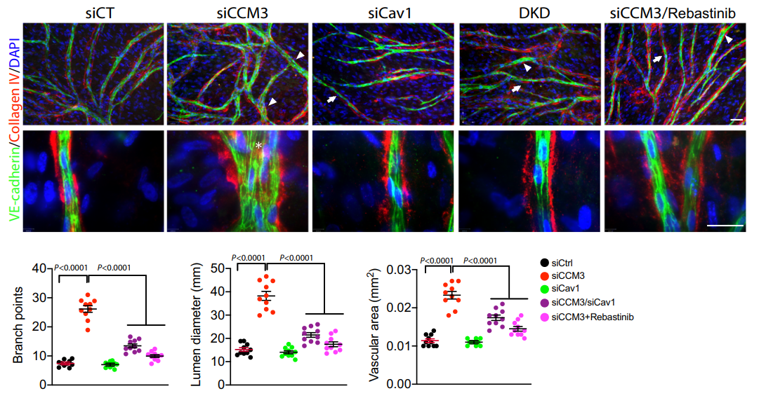
Rebastinib (1 μM) blunted increased EC sprouting and enlarged lumen formation induced by CCM3 knockdown.
Rebastinib purchased from MedChemExpress. Usage Cited in: Nat Commun. 2021 Jan 25;12(1):504. [Abstract]
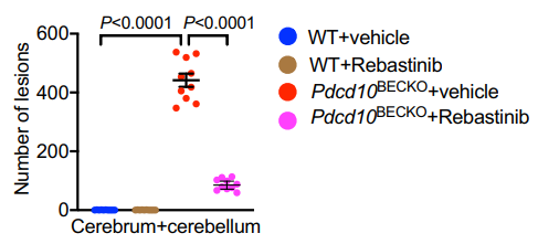
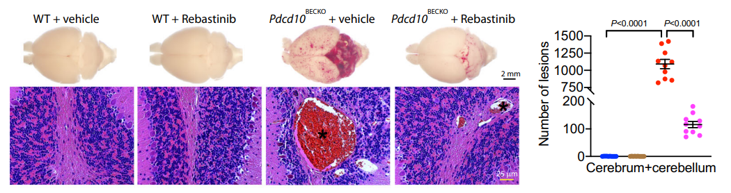
Rebastinib (10 μg/g; s.c.; 11 d) diminished the CCM lesions induced by Ccm3 deletion as visualized by whole-brain imaging and H&E staining compared to vehicle treatment in Pdcd10BECKO mice.
Rebastinib purchased from MedChemExpress. Usage Cited in: Nat Commun. 2021 Jan 25;12(1):504. [Abstract]
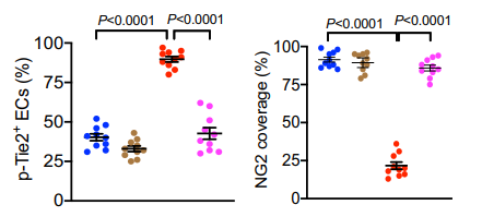
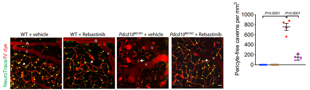
Rebastinib (10 μg/g; s.c.; once daily for 11 d) normalized the NG2+ pericyte coverage of CD31+ microvessels in Pdcd10BECKO mice.
Rebastinib purchased from MedChemExpress. Usage Cited in: Nat Commun. 2021 Jan 25;12(1):504. [Abstract]
Rebastinib (10 μg/g; s.c.; once daily for 11 d) caussed very fewer CCM lesions in brain of Pdcd10BECKO mice compared to vehicle group even at age of 2 months.
Rebastinib purchased from MedChemExpress. Usage Cited in: Nat Commun. 2021 Jan 25;12(1):504. [Abstract]
Rebastinib (10 μg/g; s.c.; 11 d) increased pericyte density and diminished pericyte-free caverns in Pdcd10BECKO mice.
-
Sci Transl Med
PP2A inhibition is a druggable MEK inhibitor resistance mechanism in KRAS-mutant lung cancer cells. [Abstract]2018 Jul 18;10(450):eaaq1093. PMID: 30021885 -
J Clin Invest
Vessels encapsulating tumor clusters promote noninvasive metastasis of hepatocellular carcinoma by shaping an immunosuppressive microenvironment. [Abstract]2026 Jan 6;136(4):e193758. PMID: 41493808 -
J Exp Clin Cancer Res
CDK16 promotes the progression and metastasis of triple-negative breast cancer by phosphorylating PRC1. [Abstract]2022 Apr 21;41(1):149. PMID: 35449080 -
Brain
Three-dimensional tissue engineered skeletal muscle modelling facioscapulohumeral muscular dystrophy. [Abstract]2025 May 13;148(5):1723-1739. PMID: 39556762 -
J Exp Med
2026 May 4;223(5):e20251374. PMID: 41891922 -
J Med Chem
Conformational analysis of the DFG-out kinase motif and biochemical profiling of structurally validated type II inhibitors. [Abstract]2015 Jan 8;58(1):466-79. PMID: 25478866 -
J Cell Sci
2026 May 28:jcs.264977. PMID: 42206576 -
Anticancer Drugs
2024 Jan 1;35(1):46-54. PMID: 37449977 -
-
-
-


溶剤 & 溶解度
体外:
DMSO : 50 mg/mL (90.32 mM; ultrasonic and warming and heat to 80°C; Hygroscopic DMSO has a significant impact on the solubility of product, please use newly opened DMSO)
Please refer to the solubility information to select the appropriate solvent. Once prepared, please aliquot and store the solution to prevent product inactivation from repeated freeze-thaw cycles.
Storage method and period of stock solution: -80°C, 2 years; -20°C, 1 year. When stored at -80°C, please use it within 2 years. When stored at -20°C, please use it within 1 year.
Please refer to the solubility information to select the appropriate solvent. Once prepared, please aliquot and store the solution to prevent product inactivation from repeated freeze-thaw cycles.
Storage method and period of stock solution: -80°C, 2 years; -20°C, 1 year. When stored at -80°C, please use it within 2 years. When stored at -20°C, please use it within 1 year.
濃度 (開始) × 体積 (開始) = 濃度 (終了) × 体積 (終了)
体内:
Select the appropriate dissolution method based on your experimental animal and administration route.
- For the following dissolution methods, please ensure to first prepare a clear stock solution using an In Vitro approach and then sequentially add co-solvents:
- To ensure reliable experimental results, the clarified stock solution can be appropriately stored based on storage conditions. As for the working solution for In Vivo experiments, it is recommended to prepare freshly and use it on the same day.
- The percentages shown for the solvents indicate their volumetric ratio in the final prepared solution. If precipitation or phase separation occurs during preparation, heat and/or sonication can be used to aid dissolution.
Add each solvent one by one: 10% DMSO 90% (20% SBE-β-CD in Saline)
Solubility: ≥ 2.08 mg/mL (3.76 mM); Clear solution
This protocol yields a clear solution of ≥ 2.08 mg/mL (saturation unknown).
Taking 1 mL working solution as an example, add 100 μL DMSO stock solution (20.8 mg/mL) to 900 μL 20% SBE-β-CD in Saline, and mix evenly.
Preparation of 20% SBE-β-CD in Saline (4°C, storage for one week): 2 g SBE-β-CD powder is dissolved in 10 mL Saline, completely dissolve until clear.
In Vivo Dissolution Calculator
Please enter the basic information of animal experiments:
-
-
-
-
Recommended: Prepare an additional quantity of animals to account for potential losses during experiments.
Please enter your animal formula composition:
-
%DMSO +
Recommended: Keep the proportion of DMSO in working solution below 2% if your animal is weak.
-
%+
-
+%Tween-80 + +
-
%Saline +
The co-solvents required include: DMSO, . All of co-solvents are available by MedChemExpress (MCE). , Tween 80. All of co-solvents are available by MedChemExpress (MCE).
Working solution concentration: 0.22 mg/mL
Method for preparing stock solution: mg drug dissolved in μL DMSO. Stock solution concentration: mg/mL.
1. Take μL DMSO stock solution;
2. Add μL .
μL , mix evenly;
3. Then add μL Tween 80, mix evenly;
4. Then add μL
Please ensure that the stock solution in the first step is dissolved to a clear state, and add co-solvents in sequence. You can use ultrasonic heating (ultrasonic cleaner, recommended frequency 20-40 kHz), vortexing, etc. to assist dissolution.


純度とドキュメンテーション
-
データシート (312 KB)
-
SDS (396 KB)
- English - EN (396 KB)
- Français - FR (396 KB)
- Deutsch - DE (396 KB)
- Norwegian - NO (396 KB)
- Español - ES (396 KB)
- Swedish - SV (396 KB)
- Italian - IT (396 KB)
- Korean - KR (396 KB)
- Portuguese - PT (396 KB)
-
取扱説明書 (2659 KB)
参考文献
[1]. Feng K, et al. The Suppressive Effect of Rebastinib on Triple-negative Breast Cancer Tumors Involves Multiple Mechanisms of Action. Anticancer research. 2023 Jun;43(6):2609-2624. [Content Brief]
[2]. Harney AS, et al. The Selective Tie2 Inhibitor Rebastinib Blocks Recruitment and Function of Tie2 Macrophages in Breast Cancer and Pancreatic Neuroendocrine Tumors. Molecular cancer therapeutics. 2017 Nov;16(11):2486-2501. [Content Brief]
[3]. Sato T, et al. Rebastinib inhibits FoxO1 activity and reduces dexamethasone-induced atrophy and its-related gene expression in cultured myotubes. The journal of physiological sciences : JPS. 2025 Mar;75(1):100012. [Content Brief]
[4]. Chan WW, et al. Conformational control inhibition of the BCR-ABL1 tyrosine kinase, including the gatekeeper T315I mutant, by the switch-control inhibitor DCC-2036. Cancer cell. 2011 Apr 12;19(4):556-68. [Content Brief]


Complete Stock Solution Preparation Table
Please refer to the solubility information to select the appropriate solvent. Once prepared, please aliquot and store the solution to prevent product inactivation from repeated freeze-thaw cycles.
Storage method and period of stock solution: -80°C, 2 years; -20°C, 1 year. When stored at -80°C, please use it within 2 years. When stored at -20°C, please use it within 1 year.
| Optional Solvent | Concentration Solvent Mass | 1 mg | 5 mg | 10 mg | 25 mg |
|---|---|---|---|---|---|
| DMSO | 1 mM | 1.8064 mL | 9.0320 mL | 18.0639 mL | 45.1598 mL |
| 5 mM | 0.3613 mL | 1.8064 mL | 3.6128 mL | 9.0320 mL | |
| 10 mM | 0.1806 mL | 0.9032 mL | 1.8064 mL | 4.5160 mL | |
| 15 mM | 0.1204 mL | 0.6021 mL | 1.2043 mL | 3.0107 mL | |
| 20 mM | 0.0903 mL | 0.4516 mL | 0.9032 mL | 2.2580 mL | |
| 25 mM | 0.0723 mL | 0.3613 mL | 0.7226 mL | 1.8064 mL | |
| 30 mM | 0.0602 mL | 0.3011 mL | 0.6021 mL | 1.5053 mL | |
| 40 mM | 0.0452 mL | 0.2258 mL | 0.4516 mL | 1.1290 mL | |
| 50 mM | 0.0361 mL | 0.1806 mL | 0.3613 mL | 0.9032 mL | |
| 60 mM | 0.0301 mL | 0.1505 mL | 0.3011 mL | 0.7527 mL | |
| 80 mM | 0.0226 mL | 0.1129 mL | 0.2258 mL | 0.5645 mL |


Rebastinib 関連分類
HY-13024 関連分類
 Powered by Bioz